Connecting groin pain, clinical assessment and everyday activities
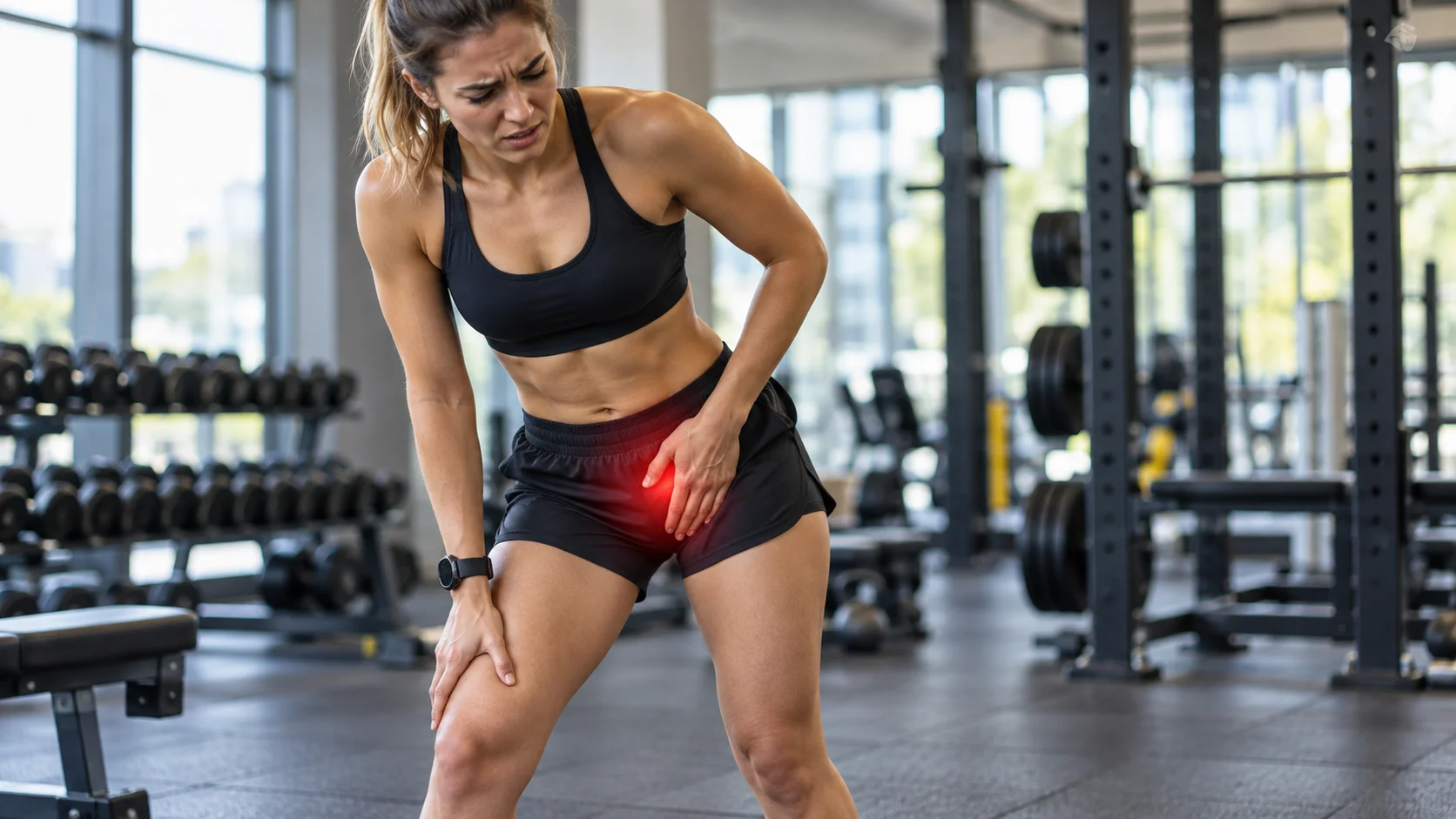
Deep groin pain, clicking during a pivot or discomfort when getting into a car may suggest a hip labral lesion. However, these symptoms are not specific. A click can be painless, several tissues can produce groin pain, and a tear can appear on MRI in someone with no symptoms [2, 3].
The task is to understand what limits the hip and determine whether the labrum actually contributes to the clinical picture. Imaging provides anatomical information. Diagnosis and treatment decisions also require a history, examination and assessment of capabilities. Recovery focuses on the movements the person wants to regain.
The labrum: one component of the joint
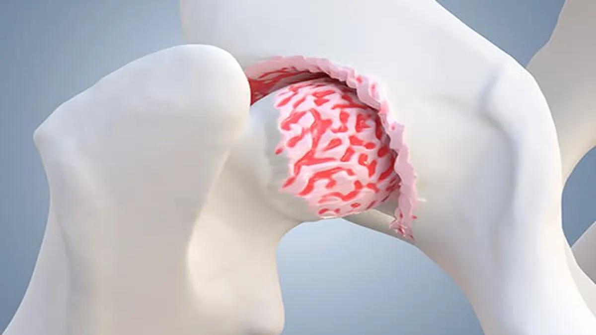
The acetabular labrum is a ring of fibrocartilage attached around the pelvic socket that receives the femoral head. It contributes to the joint’s seal, stability and distribution of loads. The capsule, bony structures, muscles and tendons also contribute to hip function [12].
The labrum should therefore not be imagined as a single part that explains every pain or noise. A lesion may interact with a particular bony shape, trauma or other changes. Its importance is assessed within that overall picture, rather than solely from the word “tear” in a report.
A traumatic tear, gradual change and an incidental finding do not necessarily create the same needs. Imaging descriptions should be specific enough to inform clinical reasoning, but cannot independently decide what someone must avoid or will be able to resume.
Bony shape and symptoms must be distinguished
Some shapes of the femur or acetabulum can alter contact in certain positions. Dysplasia can also affect coverage of the femoral head and joint loading. These characteristics do not automatically constitute a painful disease.
The Warwick Agreement defines femoroacetabular impingement syndrome through a combination of symptoms, clinical signs and imaging findings. Observed morphology alone is insufficient. This distinction avoids confusing a bony appearance with a diagnosis that necessarily explains every hip sensation [5].
Exercises do not change one bony shape into another. They can nevertheless improve strength, movement strategies and tolerance of certain tasks. Rehabilitation should define these goals rather than promise an anatomical correction that movement cannot produce.
An MRI finding can be asymptomatic
The review by Heerey and colleagues and Register’s prospective study show that intra-articular abnormalities, including labral lesions, occur in people without pain. Prevalence depends on the population and methods. These findings justify neither ignoring every lesion nor automatically attributing symptoms to every abnormality [2, 3].
The useful question is whether the findings agree. Do the location and course of the pain fit the clinical picture? Do the relevant movements reproduce the familiar symptom? Is another explanation plausible? Will the image change a decision? This approach gives a report its proper value without turning it into a complete prognosis.
Someone can improve their activities without a new MRI showing that the lesion has disappeared. Conversely, worsening pain warrants reassessment even if the report seems relatively reassuring. Clinical follow-up considers how the entire situation evolves.
Groin pain: considering several possibilities
In athletes, the adductors, iliopsoas, inguinal region, pubic region and hip can contribute to symptoms. The Doha Agreement provides clinical terminology for these presentations while recognising other causes. Several categories may coexist in the same person [9].
A femoral neck stress injury should be considered when exercise-related pain worsens, particularly when it subsequently occurs during walking. A delayed diagnosis can have important consequences. An MRI that also shows a labral lesion does not remove the need to investigate this possibility when clinically relevant [10].
| Possibility | Potential clue | What needs clarification |
|---|---|---|
| Intra-articular pain | Deep discomfort; painful flexion or rotation | Agreement between history, examination and imaging. |
| Adductor-related pain | Medial discomfort with some muscle efforts | Location, strength and appropriate tests. |
| Iliopsoas-related pain | Anterior discomfort, sometimes snapping | Relationship to effort and tendon movement. |
| Bone stress injury | Progressive exercise pain and decreasing tolerance of weight-bearing | Prompt assessment and protection from impact if suspected. |
| Inguinal or another cause | Pain with abdominal, urinary or regional symptoms | Consider a cause outside the joint. |
| Osteoarthritis or another hip condition | Pain, stiffness and reduced capacity | Examination, radiographs and health context. |
Signs overlap; no single clue identifies the cause on its own.

What clinical examination can contribute
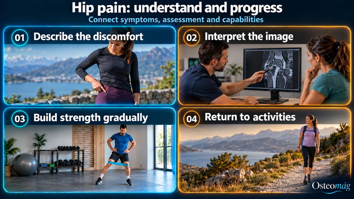
The consultation clarifies difficult movements, onset, trauma and the ability to bear weight. Examination observes walking, mobility, strength and relevant tasks. The lumbar region and nearby tissues can be considered according to the symptoms. This helps characterise the difficulty before choosing an additional investigation.
The FADIR test combines flexion, adduction and internal rotation. It can reproduce pain, but does not independently prove that a tear is symptomatic. The review by Reiman and colleagues highlights the limitations of clinical tests and their role within clinical reasoning, rather than as isolated anatomical confirmation [4].
A click also needs a precise description: painless or painful, occasional or reproducible, with or without genuine locking. A catching sensation does not always have the same meaning. Repeatedly performing a painful movement to obtain a noise on every attempt is not useful.
Radiographs and MRI: answering a specific question
The 2025 Canadian AAC–CASEM consensus recommends appropriate radiographic assessment when a symptomatic labral lesion is suspected. Radiographs assess osteoarthritis and bony characteristics, among other features. MRI, sometimes MR arthrography depending on the context, adds information about the labrum and other tissues [1].
The choice depends on the uncertainty to be resolved and its consequences for follow-up. Accumulating tests does not necessarily produce a more useful explanation. It should be possible to state what a result would change: ruling out a bone injury, clarifying an associated condition or preparing a specialist discussion.
Starting with an active approach when appropriate
The Canadian consensus considers nonoperative care a reasonable option for many patients. Activity adjustment and rehabilitation are individualised. This recommendation comes from expert agreement; it is not equivalent to a trial demonstrating that the same programme works for every lesion [1].
A particularly irritating activity can temporarily be reduced in range, duration or frequency. Comfortable movements can often be maintained according to the diagnosis. For someone troubled by deep flexion, beginning in a better-tolerated range and later discussing its reintroduction may help.
Hip muscle strength, lower-limb endurance and coordination are trained according to the needs identified. The programme does not aim to stitch the labrum back together through exercise. It aims to make the hip more capable in important movements, with symptoms compatible with progression.
Measuring progress in everyday life
Getting into a car, sitting at work, climbing stairs and returning to a walk are understandable goals. They can be followed without constantly repeating every pain-provocation test. The person then knows what they are trying to improve and can provide useful information between sessions.
The response during activity, afterwards and the following day helps adjust the programme. This does not directly measure the condition of the labrum. It mainly indicates whether the chosen dose suits current capabilities. Persistent worsening or reduced weight-bearing tolerance invites review of the strategy and, if necessary, the diagnosis.
Sporting demands differ: running in a straight line, pivoting in hockey and changing direction in tennis do not load the hip in the same way. Return to sport may rebuild endurance, strength, speed and specific movements separately. The timetable should not be determined solely by a click.
What do comparisons with surgery show?
The UK FASHIoN trial and Palmer’s trial mainly studied selected people with symptomatic femoroacetabular impingement. They reported an average advantage for arthroscopy in some outcomes, while people receiving conservative care could also improve. These trials do not apply to every isolated labral tear found on MRI [6, 7].
Average differences between groups do not guarantee an individual’s result. Selection criteria, the compared programmes, follow-up duration and complications need discussion. Arthroscopy has a role in certain situations, but its indication cannot rest on an image alone.
Martin and colleagues reported 24-month results in 97 patients older than 40 who were selected for limited radiographic osteoarthritis. Surgery combined with physiotherapy achieved better average outcomes on several measures. However, 32 people—71.1% of those initially assigned to physiotherapy alone—subsequently crossed over to arthroscopy [8].
This frequent crossover matters when interpreting the trial. It shows the complexity of a real-world comparison, not that 71% of everyone with a tear needs surgery. Conclusions remain tied to the studied population and its care criteria.
When specialist advice becomes relevant
A substantial limitation persisting despite appropriate rehabilitation may prompt a discussion of specialist assessment. Agreement between symptoms, signs and imaging, degenerative changes and possible dysplasia influences the options. The Canadian consensus emphasises this selection process [1].
The discussion should include expected benefits, limitations, risks and follow-up demands. Surgery does not remove the need for rehabilitation. Improvement through an active programme does not require proceeding to surgery simply because a tear remains recorded in the medical file.
The complementary role of osteopathy
Osteopathic support may help explore more comfortable movements and follow useful goals. The possible mechanisms of manual care are multiple; immediate improvement does not demonstrate labral repair or a lasting change in bony morphology [13].
The references presented do not establish that osteopathy restores the labrum. Its role must remain consistent with medical diagnosis, active rehabilitation and necessary referrals. A person does not need to learn to monitor every movement as a danger; they need understandable criteria for progression.
Signs that require a different priority
Significant pain after a fall, inability to walk or bear weight, loss of sensation after trauma, or a hot, painful hip with fever requires prompt assessment. Unusual sudden pain should also be reported [11].
In an athlete, worsening groin pain that becomes present during walking requires assessment to rule out a stress injury, among other possibilities. Persistent locking or rapidly deteriorating function also warrants reassessment. A known labral lesion must not automatically explain these changes [10].
The labrum may contribute to symptoms, but a person is more than their MRI. A useful approach connects symptoms with capabilities, distinguishes other causes and reassesses decisions as the situation evolves. Return to activity can then become gradual, chosen and guided by realistic expectations.
- [1] Matache BA et al., AAC-CASEM. Management of labral tears in the hip: a consensus statement. 2025.
- [2] Heerey JJ et al. Prevalence of imaging-defined intra-articular hip pathologies in people with and without pain. 2018.
- [3] Register B et al. Prevalence of abnormal hip findings in asymptomatic participants. 2012.
- [4] Reiman MP et al. Diagnostic accuracy of clinical tests for hip femoroacetabular impingement/labral tear. 2015.
- [5] Griffin DR et al. The Warwick Agreement on femoroacetabular impingement syndrome. 2016.
- [6] Griffin DR et al. UK FASHIoN: hip arthroscopy versus best conservative care. 2018.
- [7] Palmer AJR et al. Arthroscopic hip surgery compared with physiotherapy and activity modification. 2019.
- [8] Martin SD et al. Hip arthroscopy versus physical therapy in patients older than 40 years: 24-month randomized trial results. 2024.
- [9] Weir A et al. Doha agreement on terminology and definitions in groin pain in athletes. 2015.
- [10] Robertson GA, Wood AM. Femoral neck stress fractures in sport. 2017.
- [11] NHS. Hip pain in adults.
- [12] Groh MM, Herrera J. A comprehensive review of hip labral tears. 2009.
- [13] Bialosky JE et al. The mechanisms of manual therapy in musculoskeletal pain. 2009.