Midfoot pain: understand the navicular, adapt loading and recognise warning signs
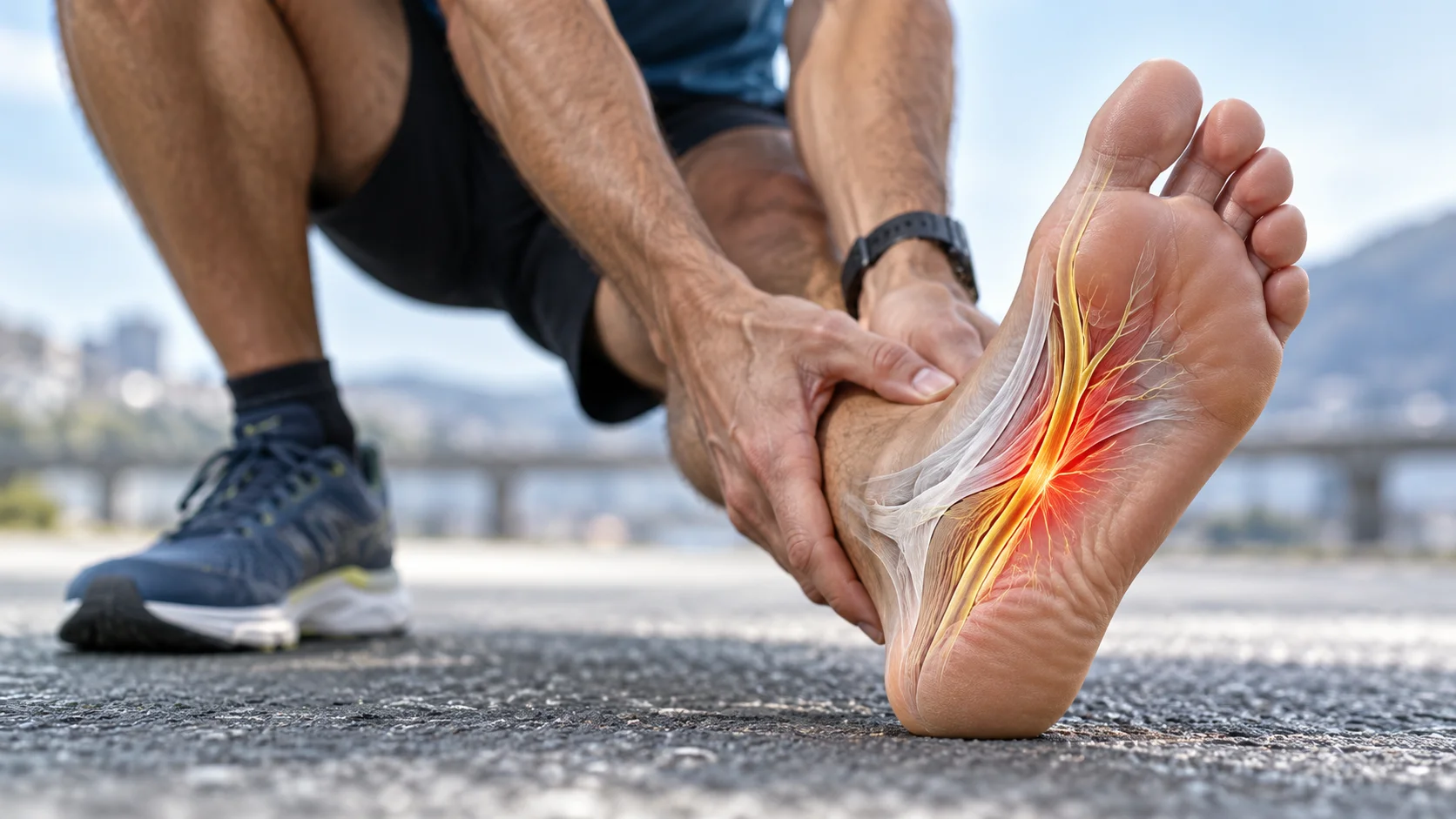
Persistent pain in the middle of the foot does not always come from an overworked tendon or arch. In some adults, deformity of the tarsal navicular is associated with altered loading and arthritis of neighbouring joints: Müller-Weiss disease. This uncommon and variable condition requires medical assessment and appropriate imaging.
The aim is to establish which structures are affected, maintain manageable walking and choose care proportionate to symptoms. A striking image does not automatically mean an operation is needed. Conversely, lasting bone pain or progressive deformity should not be treated indefinitely as a simple muscular tension. [1] [4]
Anatomy: the navicular at a junction of forces
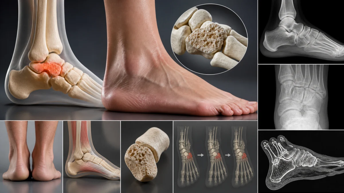
The navicular lies between the head of the talus behind it and the three cuneiforms in front. It forms part of the midfoot and contributes to the shape of the medial longitudinal arch. Its medial tuberosity receives an important attachment of the posterior tibial tendon. Ligaments, muscles and other joints also contribute to arch function.
During loading and push-off, forces pass through the talonavicular and naviculocuneiform joints. A change in navicular shape can alter their congruence and load distribution. Symptoms therefore depend on the whole foot rather than a single tender point. Pain may be medial, dorsal or sometimes felt more laterally. [2] [8]


What is known about the mechanisms?
Müller-Weiss disease was historically described as navicular osteonecrosis. Current publications discuss dysplasia, particular ossification patterns and asymmetric loading rather than one proven cause in every patient. Fragmentation should therefore not automatically be interpreted as the result of interrupted blood supply. [3] [9]
The lateral navicular may become compressed and deformed. Relative displacement of the talar head and medial navicular prominence then alter alignment. A flatfoot appearance with hindfoot varus has been described, but not everyone presents identically. The actual alignment must be examined instead of deriving a corrective plan from the disease name alone.
Both feet may be affected with different symptoms and deformities. The radiographic stage does not summarise pain or functional capacity. This variation explains why care needs to be individualised. Comparison of the two feet can inform assessment without assuming that the less painful side is structurally normal. [3] [4]
Symptoms and examination: what should guide assessment?
Common features include midfoot pain during walking, dorsomedial discomfort, stiffness and sometimes swelling. Push-off or prolonged standing may become difficult. Progressive arch changes and less comfortable footwear are also possible. These findings are not specific and cannot establish a diagnosis at home. [4]
The clinician asks about symptom onset, activity changes, injury, inflammatory disease and neuropathy. Assessment considers walking, skin condition, footwear and foot and hindfoot alignment. Pain location, tolerated movement and neurological or vascular findings add useful information. A change in how far someone can walk may matter more than a single pain rating.
A painful heel raise is not a compulsory test to repeat despite symptoms. When bone injury or a stress fracture is suspected, weight-bearing tests must be adapted. In the Quebec context, an osteopath describes functional restrictions and concerning findings; the practitioner does not make the medical diagnosis of Müller-Weiss disease. [1] [4]
Imaging: begin with a specific clinical question
Weight-bearing radiographs
Standing anteroposterior and lateral views of both feet generally form the basis of assessment. They show navicular shape, sclerosis or fragmentation, alignment and arthritic joints. Interpretation belongs to the medical team and radiologist. If standing is unsafe or impossible, the team adapts the investigations; weight-bearing should not be forced merely to obtain a standard view. [2]
CT, MRI and classification
CT clarifies bone structure and joint surfaces, particularly when surgery is being planned. MRI can help assess some early presentations or investigate another cause such as a stress fracture or infection. Every patient does not need all these investigations. The appropriate examination is the one most likely to answer a question that changes management. [8]
The Maceira classification describes five deformity stages. It facilitates communication, but does not independently determine treatment. Symptoms, the joints that are actually painful, alignment and the person’s goals must be considered together. An advanced stage is not an automatic indication for fusion. [6]
Which other causes of pain should be distinguished?
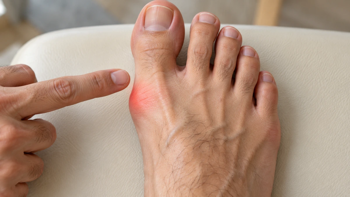
Medial pain may arise from the posterior tibial tendon or an accessory navicular. A navicular stress fracture needs consideration when focal bone pain follows impact loading, especially after increased training. Post-traumatic arthritis and inflammatory conditions can also resemble this presentation. Köhler disease affects children and should not be confused with adult Müller-Weiss disease. [1] [4]
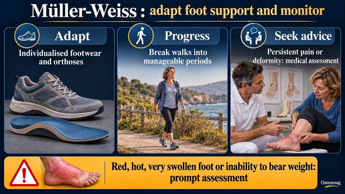
A red, hot, swollen foot in a person with diabetes and neuropathy requires particular consideration of active Charcot neuro-osteoarthropathy. Mild pain is not reassuring in that setting. Prompt assessment and foot protection are needed rather than waiting for a manual care appointment. [10]
| Possibility | Findings to assess | Useful next step |
|---|---|---|
| Müller-Weiss disease | Chronic pain, navicular deformity and adjacent arthritis | Medical assessment and weight-bearing radiographs when feasible |
| Stress fracture | Bone pain following impacts or training changes | Reduce loading and obtain medical advice |
| Posterior tibial tendon disorder | Pain along the tendon and arch changes | Adapted examination and imaging when indicated |
| Inflammatory arthritis | Prolonged stiffness and other affected joints | Broader medical assessment |
| Charcot foot | Warmth, redness and swelling with neuropathy | Prompt assessment and protection of the foot |
When is prompt or emergency assessment needed?
Severe pain after injury, a new inability to bear weight or an acute deformity needs prompt assessment. A cold, pale or blue foot, new sensory loss or sudden motor weakness requires emergency evaluation. Spreading redness, an infected wound or fever also warrants medical advice without delay.
A hot and markedly swollen foot in someone with neuropathy deserves special attention even without severe pain. Charcot guidance emphasises early recognition and protection of the limb when this condition is suspected. Waiting to see whether manual care helps can delay appropriate management. [10]
Outside an emergency, medical review is still necessary when pain persists, deformity progresses or footwear and walking become difficult. The clinician may refer to a foot and ankle specialist. There is no advantage in waiting for major loss of independence before discussing the problem. [4]
Conservative management: adapt support and loading
Nonoperative care aims to reduce painful stresses and preserve activity. A sufficiently stable shoe with enough space for the foot may help. A stiffer sole or rocker sole can reduce some painful movements during push-off. Selection depends on real-world comfort, balance and tolerance rather than a universal shoe prescription. [5]
Orthoses are fitted to alignment, deformity flexibility and pressure areas. A standard correction intended for every flatfoot may be inappropriate. The goal is improved load distribution, not moving the navicular back into place. New orthoses causing persistent pain need reassessment. Comfort should also be checked across the day and during the tasks that matter to the person.
Breaking walks into shorter periods, temporarily reducing impact and choosing better-tolerated activities can help maintain movement. A walking aid or greater protection may be prescribed when needed. Evidence specific to this rare condition remains limited, so functional progress should guide adjustments rather than claims that an insert will prevent every structural change. [5] [9]
Rehabilitation and a realistic role for osteopathic support
Rehabilitation depends on pain, foot stability and medical instructions. It may address walking, lower-limb strength, balance with safe support and comfortable movement in neighbouring regions. Exercises should not force a severely arthritic joint or load a bone whose condition has not been clarified. [6]
Osteopathic support may consider comfortable ankle, knee and hip movement and associated functional difficulties. Care should be gentle, consented and integrated with the overall plan. The person may decline a technique or stop a session. Immediate relief should be compared with walking and recovery between appointments.
The cited Müller-Weiss publications do not establish that a specific osteopathic treatment corrects the disease. Osteopathy does not reshape the navicular, reverse arthritis or replace imaging and indicated orthoses. Repeated flares after sessions or no useful functional improvement are reasons to review the strategy. Adjunctive care should have a practical purpose and should not delay specialist assessment. [1] [7]
When might surgery be discussed?
A foot and ankle surgeon may discuss an operation when pain and limitations remain substantial despite appropriate conservative management. Decisions consider painful joints, alignment, bone condition and patient goals. Radiographic stage alone is insufficient. Treatment preferences and the demands of work and daily life also inform the discussion. [6]
Arthrodesis fuses selected joints to reduce painful movement and stabilise the foot. Talonavicular or talonaviculocuneiform fusion may be considered depending on the case; other procedures address particular presentations. Expected benefits, lost movement, nonunion risk and recovery time should be discussed explicitly. An operation is not a guarantee that the foot will become entirely pain-free.
The systematic review published in a 2026 volume describes varied options without one universal gold standard. Evidence rests largely on studies with a low level of certainty. After surgery, loading and exercise progression depend on healing and the surgical team’s instructions, rather than a fixed schedule suitable for everyone. [7]
Follow-up: measure function rather than pursue perfect correction
Three useful measures are comfortable walking distance, footwear tolerance and recovery the following day. Increasing one demand at a time makes the response easier to interpret. Lasting pain escalation, new swelling or pain beginning after a shorter walk may justify adjustment and another medical review.
Prognosis varies. Some people remain functional with adaptations; others need more specialised management. The aim is useful independence with a manageable level of loading. Careful assessment, individualised footwear and coordinated care are more helpful than a promise of realignment or a decision based on imaging alone. Follow-up should support informed choices as needs and symptoms change. [5] [6] [7]
References
- [1] Hermena S, Francis M. Clinical Presentation, Imaging Features, and Management of Müller–Weiss Disease. Cureus. 2021;13(10):e18659. doi:10.7759/cureus.18659.
- [2] Carrascoso J, Monteagudo M, Llopis E, Jiménez M, Recio M, Maceira E. Imaging of Müller-Weiss Disease. Seminars in Musculoskeletal Radiology. 2023;27(3):293–307. doi:10.1055/s-0043-1766096.
- [3] Monteagudo M, Maceira E. Etiology, Epidemiology, and Pathogenesis of Müller-Weiss Disease—the Spanish Experience. Foot and Ankle Clinics. 2026;31(1):1–16. doi:10.1016/j.fcl.2024.08.007. Published online: 2025.
- [4] Cristofaro C, Park SS, Halai M. Clinical Features and Differential Diagnosis of Müller-Weiss Disease: The Canadian Experience. Foot and Ankle Clinics. 2026;31(1):17–28. doi:10.1016/j.fcl.2024.08.002. Published online: 2025.
- [5] Orejana Á, Monteagudo M. Nonoperative Treatment of Müller-Weiss Disease: The Spanish Experience. Foot and Ankle Clinics. 2026;31(1):65–80.
- [6] Angthong C, Younger ASE, Chuckpaiwong B, Harnroongroj T, Veljkovic A. A Novel Update on the Management of Müller-Weiss Disease: Presentation of a Treatment Algorithm. Cartilage. 2024;15(1):65–71. doi:10.1177/19476035231205684.
- [7] Busana M, Soares S, Garibaldi R, Tannast M, Seidel A. How to treat Müller-Weiss disease: A systematic review about the treatment algorithm. Orthopaedics & Traumatology: Surgery & Research. 2026;112(4):104551. doi:10.1016/j.otsr.2025.104551. Published online: 2025.
- [8] Imaging of Mueller-Weiss Syndrome: A Review of Clinical Presentations and Imaging Spectrum. American Journal of Roentgenology. 2016. PMID:27145453.
- [9] Mohiuddin T, Jennison T, Damany D. Müller-Weiss disease—review of current knowledge. Foot and Ankle Surgery. 2014;20(2):79–84.
- [10] International Working Group on the Diabetic Foot. Guidelines on the diagnosis and treatment of active Charcot neuro-osteoarthropathy in persons with diabetes mellitus (IWGDF 2023). Diabetes/Metabolism Research and Reviews. doi:10.1002/dmrr.3646.