Understanding vascular calf pain during exercise
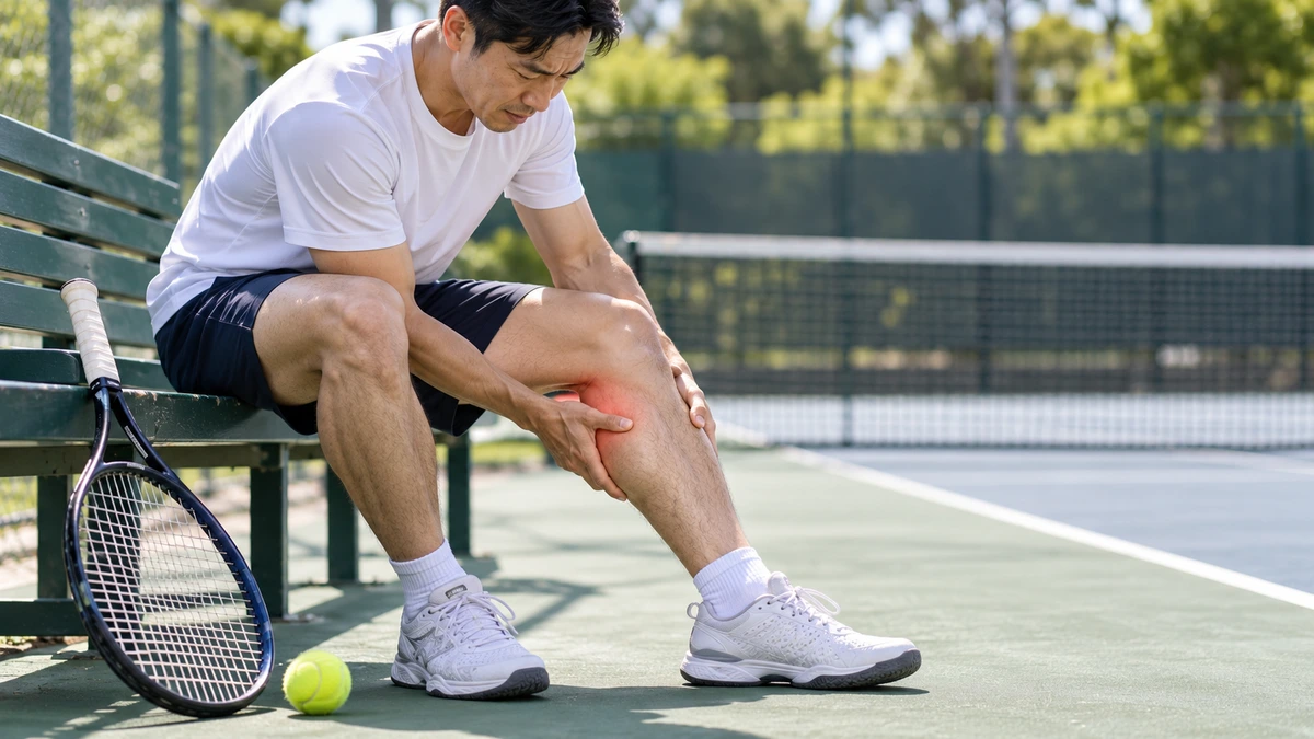
Calf pain that starts during running or intense exercise and settles at rest is often interpreted as muscular overload. That is a common explanation. However, in some people, particularly younger athletes without usual vascular risk factors, a rarer cause should be considered: popliteal artery entrapment syndrome, or PAES.
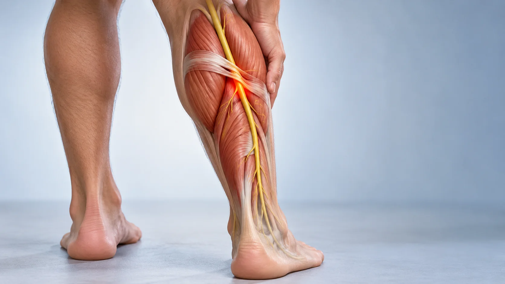
The popliteal artery runs behind the knee. In PAES, muscles, tendons or fibrous structures around it compress the artery. Compression can reflect an anatomical variation or occur mainly during muscle contraction. Blood flow towards the lower leg and foot may then decrease temporarily during exercise.
The syndrome is uncommon but important because delayed diagnosis may allow arterial wall injury, thrombosis or ischaemia to develop. Symptoms can resemble chronic exertional compartment syndrome, muscle injury, bone stress injury or neuropathy. Assessment combines the history, vascular examination and appropriate imaging, often with dynamic testing. [1] [2]
Where is the popliteal artery?

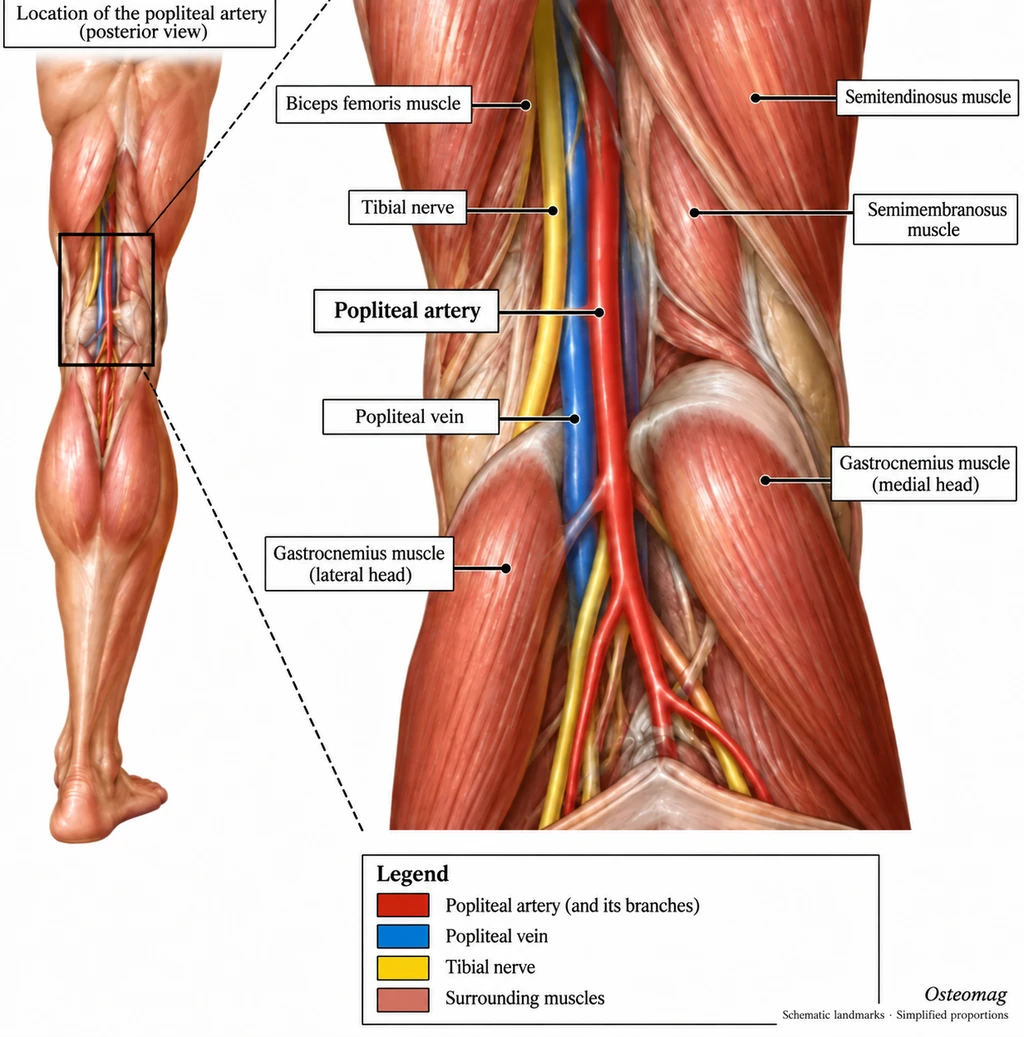
The popliteal artery continues from the femoral artery after passing through the adductor hiatus. It travels through the popliteal fossa behind the knee, then divides into vessels supplying the lower leg and foot.
Nearby structures include the gastrocnemius heads, popliteus, hamstrings, popliteal vein and tibial nerve. Variations in arterial course or muscle attachment may create a compression site. An anatomical drawing helps locate these relationships but cannot show whether a particular person has pathological compression. [7]
Anatomical and functional forms
Classical anatomical forms involve an unusual relationship between the artery and surrounding muscles. For example, the artery may take an atypical course relative to the medial gastrocnemius head. In functional forms, no major anatomical abnormality is apparent at rest. Compression appears during strong contraction or particular positions and may involve a well-developed calf musculature.
This distinction matters. Compression seen during a manoeuvre does not automatically establish disease: transient positional compression may also occur in asymptomatic people. Findings must agree with the person’s symptoms and vascular investigations. A positive provocative test alone should not determine treatment. [2] [6] [8]
Which symptoms deserve attention?
A characteristic presentation is calf pain, cramping or tightness at a relatively reproducible exercise level, decreasing after activity stops. Other features may include rapid leg fatigue, tingling or numbness, a cold sensation in the foot, pallor or colour change, temporary weakness and an exercise-related pulse change. Symptoms may involve both legs.
Examination at rest can be normal. A person may have a warm, normally coloured foot and palpable pulses during the appointment, while describing circulatory symptoms only during exercise. The timing, location and recovery pattern therefore matter. Patients should describe observed changes without repeatedly provoking symptoms or trying to diagnose themselves through pulse checks. [1]
Other causes of calf pain
Chronic exertional compartment syndrome
This can also produce reproducible running pain with marked tightness in a muscle compartment. It represents another possible mechanism, associated with changes in compartment pressure. Symptoms alone cannot reliably distinguish the conditions, which may coexist. [12]
Bone stress injury or muscle injury
A tibial or fibular stress fracture usually causes more localised pain. Initially it may occur only during exercise but can become persistent if loading continues. A gastrocnemius or soleus injury often follows an identifiable sprint, acceleration or sudden painful event. The history can differ from reproducible claudication, although no individual clue confirms the cause.
Deep vein thrombosis
A venous clot may cause pain, swelling, warmth or heaviness. It does not usually follow a reproducible exercise threshold. Suspected thrombosis requires urgent medical assessment. [11]
Peripheral arterial disease or nerve involvement
Atherosclerotic claudication is more common in older people or those with cardiovascular risk factors. PAES is particularly relevant in younger active people with an unusual presentation. Tibial nerve involvement, peripheral neuropathy or lumbar radiculopathy may cause pain, weakness and tingling. Exercise behaviour and circulatory findings help guide medical assessment. [7]
| Possible cause | Features to explore | Next step |
|---|---|---|
| Popliteal entrapment | Exercise pain, possible coldness or colour change | Dynamic vascular assessment |
| Exertional compartment syndrome | Reproducible tightness, possible sensory symptoms | Sports medicine assessment |
| Bone stress injury | Localised bone pain, reduced weight-bearing tolerance | Medical examination and indicated imaging |
| Muscle injury | Onset related to a movement, pain with contraction | Musculoskeletal assessment |
| Venous thrombosis | Unilateral swelling, warmth or persistent pain | Urgent medical assessment |
| Acute ischaemia | Suddenly cold or pale foot, new pain or deficit | Immediate emergency care |
What does clinical examination assess?
Assessment compares both limbs for temperature, colour, capillary refill, foot pulses, sensation, strength and swelling. The calf muscles, tibia, knee and lumbar region are examined as appropriate. Focal bone pain, neurological findings or swelling may redirect the investigation.
Some manoeuvres alter arterial compression, but must not be interpreted in isolation. A pulse changing in one position does not confirm PAES. Specialist tests aim to reproduce relevant symptoms under controlled conditions and measure circulation objectively. Protocol and interpretation matter; different positions and contraction intensities can influence findings. [4] [5]
What does the ankle-brachial index show?
The ankle-brachial index, or ABI, compares systolic blood pressure measured at the ankle with that measured at the arm. Measurements require appropriate cuffs and Doppler assessment according to a clinical protocol. The highest relevant ankle pressure is compared with the highest arm pressure when calculating the conventional index for each leg.

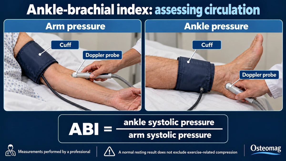
In some people with PAES, the resting value is normal but falls after exercise. ABI can provide a clue but does not establish the diagnosis alone. It is most useful alongside reproducible symptoms and dynamic vascular assessment. A normal resting result should not end the investigation when the clinical history remains suggestive. [9]
Duplex ultrasound and dynamic imaging
Duplex ultrasound visualises the artery and assesses blood flow. Testing may be performed at rest, during selected manoeuvres or after exercise. CT angiography and MR angiography show the artery’s course and neighbouring muscles, helping identify anatomical relationships and arterial wall damage.
A 2024 review emphasises interpreting the history, examination, ABI and static or dynamic imaging together. Work on ultrasound protocols also highlights the need for validation. Positional compression without matching symptoms is not equivalent to symptomatic PAES. The clinician chooses the investigation that can answer the relevant question rather than treating every image abnormality as disease. [1] [4]
Why timely assessment matters
Repeated compression may injure the arterial wall and contribute to narrowing, thrombosis or abnormal dilation. Once the artery is damaged, management becomes more complex than intermittent functional compression. Treatment depends on PAES type, arterial condition, symptom severity and anatomy.
A vascular specialist may consider decompression for appropriate symptomatic anatomical forms. An injured artery may require repair or reconstruction. Reviews report generally favourable outcomes in selected patients but heterogeneous studies and low-quality evidence limit conclusions about the best approach. The potential benefit, risks and uncertainty must be discussed individually. [3] [8]
A realistic role for osteopathic support
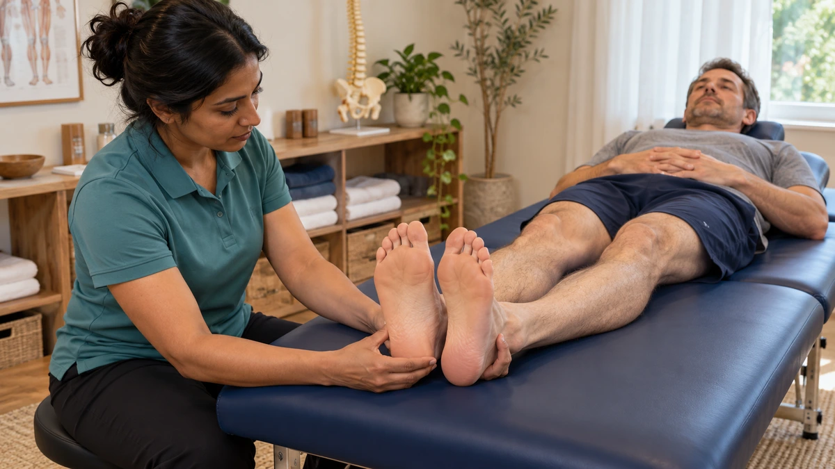
In Osteomag’s Quebec context, the osteopath does not make a medical diagnosis of PAES. Osteopathic care cannot permanently release an artery compressed by an anatomical abnormality or repair an injured arterial wall. Deep work in the popliteal fossa is not a treatment for PAES. The practitioner’s priority is recognising a presentation that may not be a simple muscle problem and arranging appropriate referral.
After medical diagnosis and specialist management, osteopathic support may contribute to general mobility and functional recovery within the treating team’s limits. Temporary comfort or reduced muscle tension does not demonstrate that circulation is safe. Support should encourage appropriate activity and never delay vascular assessment.
Warning signs
Immediate assessment is needed for a suddenly cold or pale foot, severe new leg pain, a newly absent distal pulse, substantial new weakness, rapidly worsening numbness, increasing rest pain or persistent colour change. Significant unilateral calf swelling also requires urgent assessment. These findings may indicate acute ischaemia or thrombosis. Do not wait for every sign to be present. [10] [11]
Unilateral calf swelling with breathlessness or chest pain requires calling emergency services: pulmonary embolism must be considered. [11]
When should you arrange a consultation without an emergency?
Recurrent calf pain over several sessions, symptoms beginning progressively earlier or pain that reduces normal activity deserves a medical appointment. Seek assessment when episodes include temperature or colour change, numbness or increasingly slow recovery, even if the foot appears normal at rest. The physician may arrange vascular medicine or vascular surgery review. Record the onset threshold, location and recovery time; do not deliberately provoke another episode to test circulation. [1] [7]
These criteria concern intermittent symptoms without acute signs. Severe sudden pain, persistent coldness or pallor, or new weakness requires urgent assessment rather than waiting for the scheduled appointment.
Returning to activity
Return to running or intense sport depends on the diagnosis and arterial condition. Improvement after rest does not confirm that the problem has resolved. Recurrent cramping with coldness, pallor or tingling requires stopping and reassessment. [3]
Progression criteria agreed with the team
Before return, the team specifies permitted activities, whether vascular follow-up is needed and which symptoms require stopping. Begin with a tolerated activity and duration, then change one variable at a time: duration, intensity or frequency. One pain-free session does not guarantee normal circulation. Progression also reflects arterial health, any procedure performed and follow-up findings.
Strength and endurance work do not replace treatment of symptomatic compression. They may form part of rehabilitation after medical agreement. Cramping with coldness, pallor, tingling or weakness means stopping exercise and contacting the team; persistent symptoms require urgent assessment. No universal return-to-running timeline applies to every form of PAES. [3] [8]
Key points
Reproducible calf pain should not automatically be attributed to muscles when circulatory signs accompany it. A normal resting examination cannot exclude dynamic compression. Medical referral, agreement between findings and a return approved by the specialist team matter more than promises to release tissues.
References
- [1] Neubauer TM, Chin JJ, Hill RD, Hu YE. Popliteal Artery Entrapment Syndrome: Updates for Evaluation, Diagnosis, and Treatment. Current Sports Medicine Reports. 2024;23(9):310–315. doi:10.1249/JSR.0000000000001194. Source
- [2] Gonzalez-Urquijo M, et al. Popliteal Artery Entrapment Syndrome: A Review of Current Concepts. The American Surgeon. 2026;92(3):991–998. doi:10.1177/00031348251391843. Source
- [3] Cui HJ, Wu YF. Popliteal Artery Entrapment Syndrome: A Systematic Review of Contemporary Management and Outcome Reporting. Journal of Endovascular Therapy. Published online 28 April 2026. doi:10.1177/15266028261439586. Source
- [4] Barrett DW, Carreira J, Bowling FL, Wolowczyk L, Rogers SK. Improving duplex ultrasound methods for diagnosing functional popliteal artery entrapment syndrome. Scandinavian Journal of Medicine & Science in Sports. 2024;34(3):e14592. doi:10.1111/sms.14592. Source
- [5] Altintas U, et al. Popliteal artery entrapment syndrome: ultrasound imaging, intraoperative findings, and clinical outcome. Vascular and Endovascular Surgery. 2013;47(7):513–518. doi:10.1177/1538574413495466. Source
- [6] Lane R, et al. Functional popliteal entrapment syndrome in the sportsperson. European Journal of Vascular and Endovascular Surgery. 2012;43(1):81–87. doi:10.1016/j.ejvs.2011.10.013. Source
- [7] Bradshaw S, et al. Popliteal artery entrapment syndrome. Cardiovascular Diagnosis and Therapy. 2021;11(5):1159–1167. doi:10.21037/cdt-20-186. Source
- [8] Shahi N, et al. Functional Popliteal Artery Entrapment Syndrome: A Review of Diagnosis and Management. Annals of Vascular Surgery. 2019;59:259–267. doi:10.1016/j.avsg.2018.12.105. Source
- [9] Aboyans V, et al. Measurement and interpretation of the ankle-brachial index: a scientific statement from the American Heart Association. Circulation. 2012;126(24):2890–2909. doi:10.1161/CIR.0b013e318276fbcb. Source
- [10] Björck M, et al. Editor’s Choice – European Society for Vascular Surgery (ESVS) 2020 Clinical Practice Guidelines on the Management of Acute Limb Ischaemia. European Journal of Vascular and Endovascular Surgery. 2020;59(2):173–218. doi:10.1016/j.ejvs.2019.09.006. Source
- [11] NHS. DVT (deep vein thrombosis). Updated 30 April 2026. Source
- [12] Vogels S, et al. Clinical Consensus on Diagnosis and Treatment of Patients with Chronic Exertional Compartment Syndrome of the Leg: A Delphi Analysis. Sports Medicine. 2022;52(12):3055–3064. doi:10.1007/s40279-022-01729-5. Source