Sudden pain on weight bearing may come from the bone beneath the cartilage
Knee pain appearing without major trauma may quickly be attributed to a meniscus or osteoarthritis. These diagnoses are common, but are not the only ones. A subchondral insufficiency fracture of the knee, often abbreviated SIFK in English-language literature, can produce a similar presentation, while its management may require greater protection of weight bearing. [1][2]
The important point is not to suspect a fracture with every discomfort. It is to recognize situations in which acute, unusual pain or pain marked on weight bearing deserves reassessment. A normal initial X-ray does not always exclude an early subchondral lesion. The history and examination determine whether further investigation is needed. [2]
What is a subchondral fracture?
Articular cartilage covers the surfaces that glide against each other. Immediately beneath it, subchondral bone helps bear loads. At the knee, the femoral condyles, tibial plateaus, menisci and other joint structures help distribute loads during walking and other activities.
An insufficiency fracture occurs when loads exceed the bone’s local capacity. Reduced strength, altered load distribution or several combined factors may be involved. SIFK alone does not demonstrate generalized osteoporosis. Distinguishing it from a fatigue fracture caused by unusual loads on otherwise normal bone forms part of medical assessment. [1][7]
The medial femoral condyle is frequently affected, but the tibial plateau or other sites may be involved. Lesions differ in size, subchondral condition and course. A diagnosis is therefore not enough to predict an identical recovery timetable for everyone. [3]
Why does the meniscus matter?
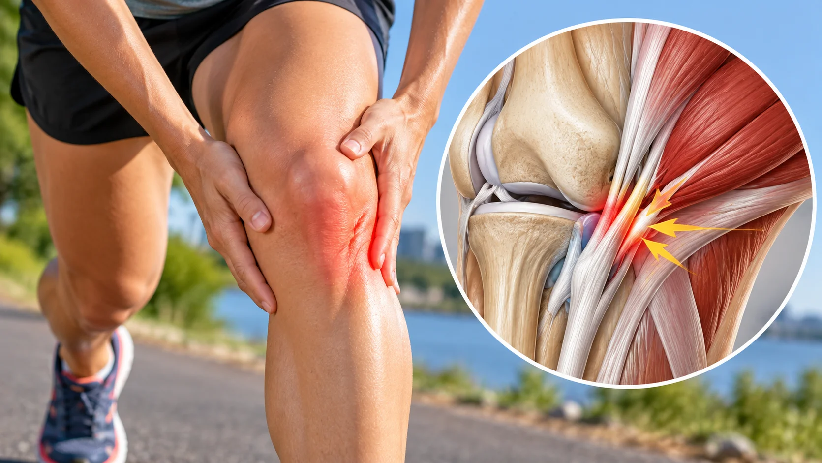
The menisci help distribute loads between the femur and tibia. A root tear or meniscal extrusion may change this function. Studies frequently find meniscal abnormalities associated with subchondral fractures, particularly in the medial compartment. [1][3]
This association does not establish that every tear causes a fracture or that every fracture requires a meniscal intervention. The knee must be considered as a whole: cartilage, bone, meniscus, alignment, function and clinical context. Treating a meniscal imaging finding without questioning unusual weight-bearing pain may miss another component of the problem.
In a study of 340 MRI examinations requested for a suspected meniscal problem with osteoarthritis, ten showed SIFK. This finding, close to 3%, concerns that selected population, not all painful knees. Nevertheless, it reminds us that the initial diagnosis may need revisiting before a weight-bearing exercise program. [4]

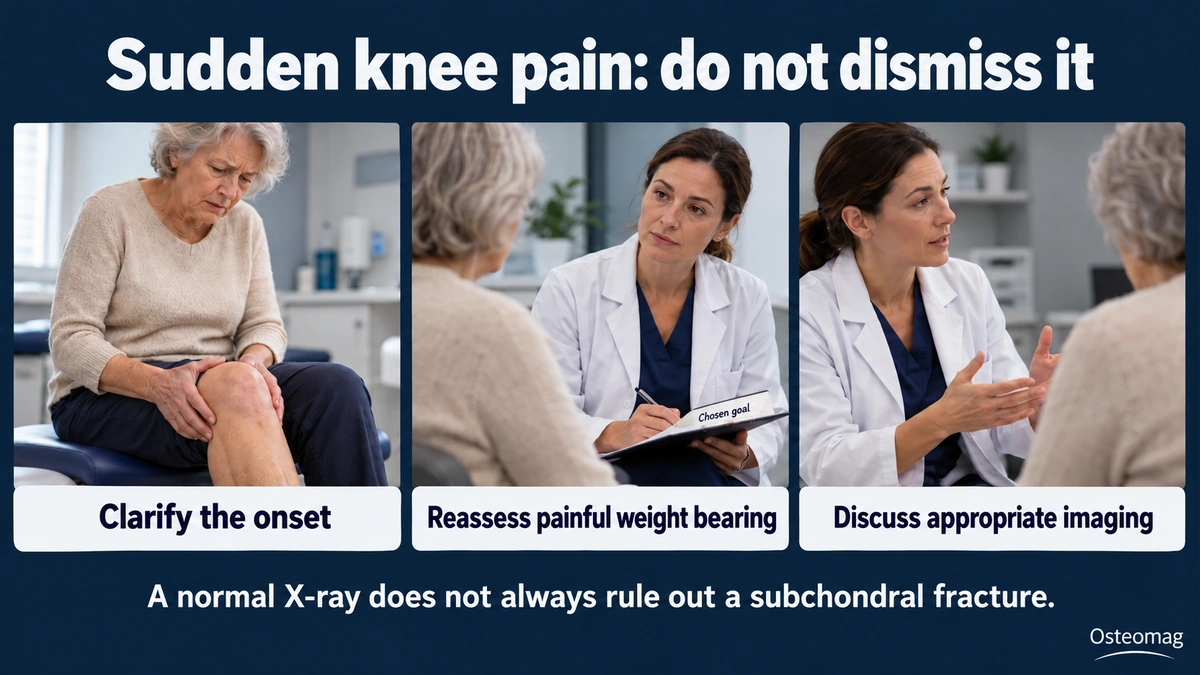
Which symptoms should prompt reconsideration of a simple explanation?
Pain can begin fairly abruptly, sometimes during an ordinary activity, then become significant when walking or standing. Night or rest discomfort, localized tenderness and an effusion may accompany the presentation. These signs are nonspecific: their presence does not allow self-diagnosis of a fracture. [1][6]
The clinician clarifies onset, progression, ability to bear weight, any trauma and recent activities. Pain very different from usual, clear worsening or recovery that does not follow the expected course warrants renewed assessment. Already having osteoarthritis does not protect against another condition.
Why can an X-ray be normal?
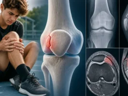
Initially, the lesion may be too subtle to be visible on radiographs. MRI can examine the subchondral region and marrow signal when clinical suspicion warrants it. This does not mean MRI should be requested for every knee pain: the indication depends on the presentation, severity and other hypotheses. [2][6]
A subchondral line and an edema-like marrow signal are among the features described. Edema alone remains nonspecific. The radiologist examines shape, location and other abnormalities to distinguish possible diagnoses. [1][7] The dimensions of subchondral abnormalities may be more informative for prognosis than the extent of edema alone. [8]
| Situation | What needs clarifying | Consequence |
|---|---|---|
| Recent pain very marked on weight bearing | Onset, trauma, walking ability | Do not dismiss it as simple stiffness |
| Reassuring initial X-ray | Progression and consistency with examination | Reassess if suspicion persists |
| Meniscal abnormality | Subchondral bone condition and clinical context | Do not assume it explains everything |
| Bone marrow edema on MRI | Subchondral line, size and morphology | Do not treat it as a stand-alone diagnosis |
What is the link with the former “spontaneous osteonecrosis”?
Terminology has evolved. Some presentations formerly called spontaneous osteonecrosis of the knee are now interpreted within the subchondral fracture spectrum. A histopathological study helped propose fracture as the initiating event in some of these lesions. [9] This does not mean that every knee osteonecrosis has the same cause or that every SIFK progresses to necrosis.
Contemporary reviews emphasize distinguishing forms that may progress favorably from those associated with collapse of the joint surface. [1][10] This possibility should be explained without presenting a sequence of images as an inevitable progression. An educational illustration cannot replace interpretation of real examinations.
Is the course always unfavorable?
No. Some lesions may improve with conservative management, while others progress to collapse and osteoarthritis. Size, subchondral morphology and cartilage or meniscal condition contribute to risk assessment. MRI classification studies remain tools for professionals, not individual calculators based on a symptom. [3][7]
In a retrospective cohort of 223 patients, a substantial proportion subsequently underwent surgery, including approximately one third receiving arthroplasty. This figure describes a group diagnosed and followed in a specialist setting. It does not represent the risk for everyone with recent knee pain and cannot predict the outcome of a particular case. [5]
When should prompt or emergency assessment be sought?
Inability to bear weight, significant acute pain, trauma or rapid worsening warrants medical assessment. A very swollen, hot and painful knee with fever or general illness requires urgent evaluation to avoid missing infection or another serious cause. Significant locking or an unusual change in function should also be reported.
Calf pain with unusual swelling must not automatically be attributed to the knee, especially when accompanied by shortness of breath or chest pain. Systemic or vascular signs change the priority. Temporary relief from contact or a position cannot exclude a condition needing different care.
Why is weight bearing sometimes protected?
Management does not necessarily follow that of uncomplicated osteoarthritis or an isolated meniscal lesion. Reduced or protected weight bearing may be indicated according to the lesion. The degree, walking aids and duration are defined with the medical team. An intensive walking or weight-bearing strengthening program must not be independently chosen to “test” knee strength. [4][6]
Optimal rehabilitation protocols remain insufficiently established by robust trials. [6] Imposing the same offloading duration or fixed return-to-sport timetable on everyone would therefore be misleading. Follow-up combines clinical progression and, when necessary, reassessment by imaging. Less pain is not equivalent to demonstrated bone healing.
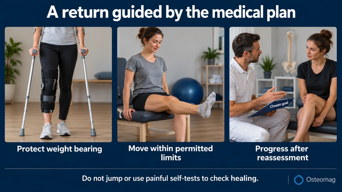
Returning gradually, within permitted limits
Rehabilitation may aim to maintain permitted movements, limit deconditioning and support a return to activities. Progression considers medical restrictions and does not depend on having to push through pain. Jumping, deep knee bends or repeated weight-bearing tests are not appropriate ways to check healing yourself.
A precise functional goal helps follow-up: necessary travel, independence at home or resuming an adapted task. Immediate tolerance and the subsequent response are discussed with the professional. If pain clearly increases or abilities decline, reassessment is the appropriate response, not an automatic increase in effort.
What role can osteopathy play?
The cited research does not demonstrate that osteopathic care heals the fracture, prevents collapse or restores bone circulation. Any support remains complementary and must respect protection instructions. Comfort in another region or adjustment of a position is not a causal treatment for the subchondral lesion.
The practitioner must recognize presentations requiring medical advice, communicate relevant information and avoid unverified reassuring explanations. Pain described as “mechanical” is not necessarily benign: an insufficiency fracture is itself influenced by mechanical loads.
Key points
With recent, unusual knee pain, the challenge is not to stop too quickly at the label of meniscus or osteoarthritis. SIFK is a possibility to consider in an appropriate context, even when an initial X-ray appears normal. Diagnosis, weight-bearing protection and return to activity must be individualized. Osteopathy replaces neither that assessment nor follow-up of the bone lesion.
References
- [1] Ochi J, Nozaki T, Nimura A et al. Subchondral insufficiency fracture of the knee: review of current concepts and radiological differential diagnoses. Jpn J Radiol. 2022;40(5):443-457. doi:10.1007/s11604-021-01224-3.
- [2] Jose J, Pasquotti G, Smith MK et al. Subchondral insufficiency fractures of the knee: review of imaging findings. Acta Radiol. 2015;56(6):714-9. doi:10.1177/0284185114535132.
- [3] Sayyid S, Younan Y, Sharma G et al. Subchondral insufficiency fracture of the knee: grading, risk factors, and outcome. Skeletal Radiol. 2019;48(12):1961-1974. doi:10.1007/s00256-019-03245-6.
- [4] Huizinga JL, Shah N, Smith SE et al. Prevalence of Undiagnosed Subchondral Insufficiency Fractures of the Knee in Middle Age Adults with Knee Pain and Suspected Meniscal Tear. Osteoarthr Cartil Open. 2020;2(4):100089. doi:10.1016/j.ocarto.2020.100089.
- [5] Pareek A, Parkes CW, Bernard C et al. Spontaneous Osteonecrosis/Subchondral Insufficiency Fractures of the Knee: High Rates of Conversion to Surgical Treatment and Arthroplasty. J Bone Joint Surg Am. 2020;102(9):821-829. doi:10.2106/JBJS.19.00381.
- [6] Musbahi O, Waddell L, Shah N et al. Subchondral Insufficiency Fractures of the Knee: A Clinical Narrative Review. JBJS Rev. 2023;11(10):e23.00084. doi:10.2106/JBJS.RVW.23.00084.
- [7] Buturoiu MM, Ghiea S, Weber MA. Subchondral insufficiency fractures: overview of MRI findings from hip to ankle joint. Rofo. 2024;196(11):1143-1154. doi:10.1055/a-2344-5337.
- [8] Lecouvet FE, van de Berg BC, Maldague BE et al. Early irreversible osteonecrosis versus transient lesions of the femoral condyles: prognostic value of subchondral bone and marrow changes on MR imaging. AJR Am J Roentgenol. 1998;170(1):71-7. doi:10.2214/ajr.170.1.9423603.
- [9] Yamamoto T, Bullough PG. Spontaneous osteonecrosis of the knee: the result of subchondral insufficiency fracture. J Bone Joint Surg Am. 2000;82(6):858-66. doi:10.2106/00004623-200006000-00013.
- [10] Zou Z, Zeng Y, Zeng J. [Research advances on subchondral insufficiency fracture and osteonecrosis of the knee]. Zhongguo Xiu Fu Chong Jian Wai Ke Za Zhi. 2026;40(3):482-486. doi:10.7507/1002-1892.202510036.