Measuring an anatomical reference while keeping function central
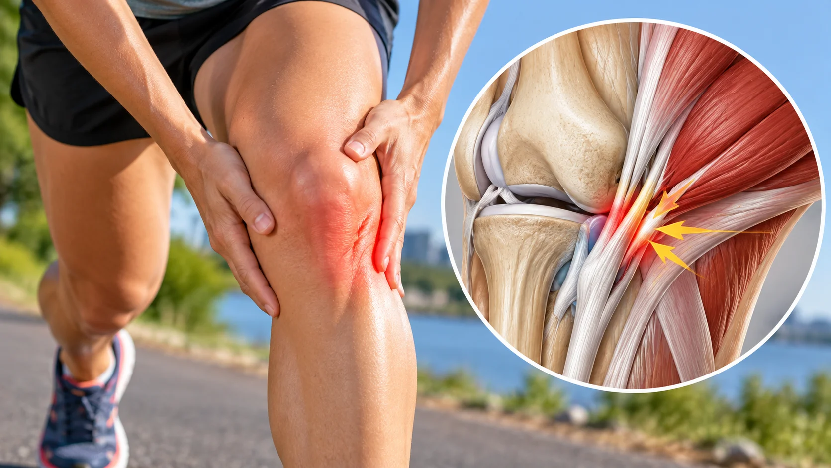
The Q angle is a geometric reference used to describe one aspect of lower-limb alignment. It links the pelvis, kneecap and tibial tuberosity. However, a large value alone cannot explain pain, diagnose a disease or predict injury. Measurement quality and the whole examination matter more than an isolated number. [1] [2]
For someone whose knee hurts on stairs, the practical problem is recovering movement capacity. Assessment should identify provocative activities, possible instability and signs suggesting another condition. Focusing only on normalising the angle can distract from these needs. Current recommendations for patellofemoral pain emphasise education and individualised exercise. [3] [4]
Which anatomical landmarks are used?
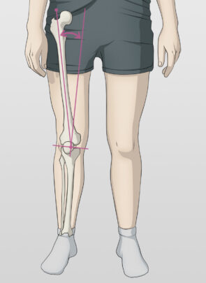
The first line runs from the anterior superior iliac spine, a bony landmark on the pelvis, to the centre of the patella. The second connects that centre to the tibial tuberosity, where the patellar tendon attaches. The angle between these directions offers a simplified representation of their relationship. It does not directly measure forces inside the knee. [1] [5]
The quadriceps generates force transmitted through the patella and patellar tendon. The shape of the femoral trochlea, supporting tissues and movements of the femur and tibia also contribute to joint function. Reducing this organisation to two lines drawn on the skin is insufficient. Pain can change while bony geometry changes very little.
Standing position, foot orientation, limb rotation and muscle activation affect the landmarks observed. A resting measurement is therefore not equivalent to analysing running or a jump landing. What is being measured must be specified before comparing results. [1] [6]

Measuring consistently and recognising uncertainty
A clinical measurement may use a goniometer after identifying the anatomical landmarks. Locating them by palpation involves some error. A systematic review found substantial disagreement about measurement reliability and validity, partly related to a lack of standardised procedures. Overly precise interpretations should therefore be avoided. [1]
When following one person, keep the position, instructions and, where possible, examiner consistent. Comparing a lying measurement with a standing measurement requires caution. A small difference does not demonstrate worsening alignment or a lasting treatment effect.
Historical alignment research and a more recent patellar-instability study highlight reproducibility limitations in some tests. Reliability means that a measurement can be repeated consistently; it does not prove that the measure identifies the cause of pain. These are separate questions. [6] [7]
Repeated photographs and daily self-measurements are not useful ways to monitor a knee. Camera perspective, position and landmark identification make comparisons uncertain. A video may help discuss movement with a professional, but an apparent angle does not become a diagnosis.
Association with pain: separating cause and consequence
A review comparing people with pain and controls found a greater average Q angle in some patellofemoral pain groups. However, an association measured at one point cannot establish whether the angle preceded pain, whether other factors contributed or whether participant selection explains part of the finding. [8]
Prospective studies ask a different question: does an initial characteristic predict future pain? A review of these studies and a later, larger synthesis did not identify the Q angle as a reliable predictor of patellofemoral pain. Telling an asymptomatic person that their measurement means they will become injured would therefore be misleading. [2] [9]
These findings do not make lower-limb observation useless. They clarify its limitations. A clinician can integrate several findings without giving one number more significance than the evidence supports. An unusual measurement with satisfactory function does not automatically justify preventive treatment.
Sex, body shape and individual variation
Published average values vary with populations and methods. Giving every man and woman different fixed limits, then labelling every exceedance abnormal, oversimplifies this variation. Body dimensions, chosen landmarks and measurement positions all contribute. The review of patellofemoral examination discusses these methodological issues. [5]
A wider pelvis does not condemn a woman’s knees to pain. Training, sporting demands, strength, load tolerance and previous problems deserve individual attention. Care based on an assumed universal fragility may instead increase worry and activity avoidance.
Static angle and dynamic movement
During squatting, stair use or landing, the lower limb moves in several planes. The knee may appear to move inwards because of combined hip, tibial and foot movements. This dynamic observation should not automatically be labelled an increased Q angle.
A professional can suggest variations of a task and explore their effects on discomfort, effort and control. The goal is not one perfect trajectory imposed on everyone. Research methods for patellar movement are themselves heterogeneous; a methodological review urges caution when comparing studies. [10]
Anterior knee pain: identifying the clinical presentation
Pain around or behind the kneecap, reproduced by activities loading a bent knee, may suggest patellofemoral pain. History and several consistent findings guide assessment. The Q angle alone cannot distinguish this from tendinopathy, osteoarthritis or instability. [3] [5]
The clinician asks when symptoms began, which loads increased and how the knee recovers. They check for swelling, locking, trauma or episodes of giving way. Movement, strength and tolerance to suitable tasks are examined before deciding whether further investigation is needed. Difficulty during a squat can arise from pain without indicating permanent structural weakness.
| Presentation | Clinical question | Useful response |
|---|---|---|
| Pain around the kneecap on stairs | Possible patellofemoral pain [3] [5] | Assess loading, strength and activity. |
| Giving way after dislocation or trauma | Instability or an injury needing clarification | Clinical assessment; do not focus only on the angle. |
| Locking, substantial swelling or inability to bear weight | Possible joint or traumatic injury | Prompt assessment according to the situation. |
| A large angle without symptoms | Variation in anatomy or measurement | No automatic treatment based on the number. |
Clinical pointers — Osteomag. This table does not replace individual assessment.
Distinguishing pain, injury and instability
Pain localised to the patellar tendon below the kneecap raises a different question from diffuse pain around it. An episode in which the kneecap moved out of place, true locking or a large effusion also requires separate assessment. Presentations sometimes overlap. History, examination and medically indicated investigations help clarify possible causes; the Q-angle value does not distinguish them. [3] [5]
An osteopath can help identify signs requiring referral and provide musculoskeletal support within their professional scope. They do not make a medical diagnosis. Suspected significant injury or inflammatory disease must not be reframed as a simple alignment defect to correct.
When is imaging relevant?
An X-ray or MRI is not requested simply because a clinical angle exceeds an average. Imaging answers a question: fracture after trauma, osteochondral injury, osteoarthritis, substantial instability or another suspected condition. Results are interpreted alongside symptoms and examination. [3] [5]
Some specialised measurements describe patellar structures using methods different from a goniometer. They are not interchangeable with the Q angle. An anatomical feature on an image does not prove that it explains all pain or requires intervention.
Adjusting loads to regain activities
For confirmed patellofemoral pain, education and knee strengthening, with hip exercises when appropriate, form the foundation of care. Complementary interventions depend on the presentation and preferences. The aim is better function without promising to change bony anatomy. [3] [4]
A starting point might involve a seated exercise, a sit-to-stand or knee bending through a tolerable range. Resistance and repetitions are individualised. An angle does not determine a programme: two people with the same measurement may have different abilities and goals.
Long descents, repeated deep knee bends or irritating jumps may be temporarily reduced while other tolerable activities continue. Complete rest is not a universal rule. Responses during activity, afterwards and the following day help guide progression with the professional.
Planning practical progression
Progression can prepare a specific task: climbing a flight of stairs, walking on a slope or returning to running. Change one variable at a time, such as duration before speed. This makes it easier to identify useful loading and demands that remain excessive.
New discomfort, worsening recovery or swelling calls for adjustment. Being pain-free at rest does not immediately permit every sporting load. Follow-up should also consider strength, confidence and the ability to repeat required movements.
A simple diary can record the chosen activity, discomfort and next-day function. This is more informative than a series of daily angle measurements. The programme needs to remain realistic alongside travel, work and other training.
An example focused on ability
For someone troubled by stairs, progress can be tracked through the number of steps tolerated and recovery afterwards. A strengthening exercise is chosen with the professional and adjusted according to response. Improvement in the task is meaningful even when the measured angle stays unchanged. Education and knee-targeted exercise, with or without hip exercise, remain the foundation of current recommendations. [4]
Temporary adaptations maintain achievable activities before gradually preparing for harder tasks. Return to sport considers symptoms and sport-specific capacity. No Q-angle value alone permits or prohibits that return. [3]
The role and limits of osteopathy
Osteopathic support can help discuss activity, assess musculoskeletal difficulties and offer individualised comfort care. It should not claim to permanently move the patella, correct a pelvis supposedly responsible for every symptom or normalise the Q angle in one session. Recommendations allow manual care as a possible individualised adjunct to active management. [4]
Benefit is judged through a concrete task over time. Temporary flexibility does not demonstrate anatomical correction. If sessions do not improve agreed goals, the plan needs review. Suggested links between the Q angle, migraine, the jaw and disc disease are not established explanations to offer a patient.
Signs requiring referral
Trauma with inability to bear weight, deformity or substantial locking requires prompt assessment. A red, hot, swollen knee with fever should be examined without waiting for comfort care. New weakness or loss of sensation also requires appropriate assessment. [11]
Persistent pain despite a coherent progression warrants reassessment. The question is not simply whether to repeat the measurement, but whether diagnosis, loading, possible injuries and goals need review. The Q angle can remain secondary information; function and safety guide decisions.
References
- [1] Smith TO et al. The reliability and validity of the Q-angle: a systematic review. 2008.
- [2] Neal BS et al. Risk factors for patellofemoral pain: a systematic review and meta-analysis. 2019.
- [3] Willy RW et al. Patellofemoral Pain: Clinical Practice Guidelines. JOSPT, 2019.
- [4] Neal BS et al. Best practice guide for patellofemoral pain. 2024.
- [5] Physical Examination and Patellofemoral Pain Syndrome: an Updated Review. 2021.
- [6] Patellofemoral alignment: reliability. 1996.
- [7] Common Physical Examination Tests for Patellofemoral Instability Demonstrate Weak Inter-Rater Reliability. 2021.
- [8] Lankhorst NE et al. Factors associated with patellofemoral pain syndrome: a systematic review. 2013.
- [9] Pappas E, Wong-Tom WM. Prospective Predictors of Patellofemoral Pain Syndrome. 2012.
- [10] A Word of Caution for Future Studies in Patellofemoral Pain: A Systematic Review With Meta-analysis. 2021.
- [11] NHS. Knee pain: signs requiring urgent assessment. View source.