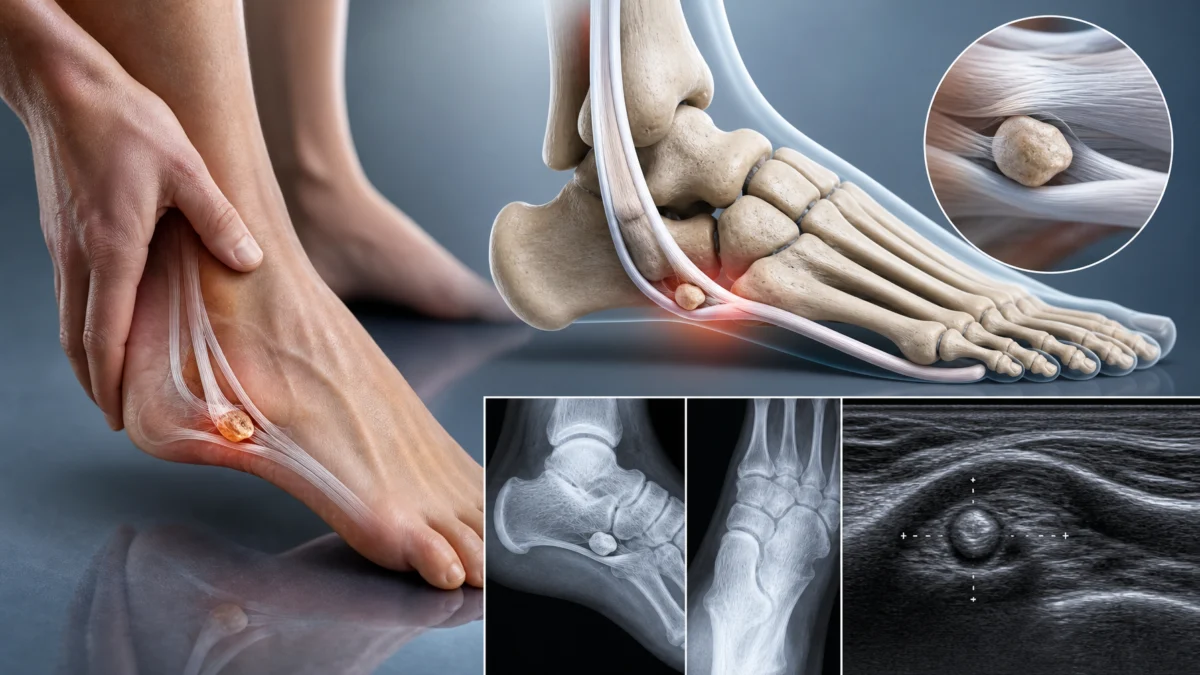
Pain along the plantar-lateral border of the foot is often attributed to an ankle sprain, the cuboid, the peroneal tendons, or the fifth metatarsal. A rarer cause deserves recognition: painful os peroneum syndrome.
The os peroneum is a small sesamoid bone located within the peroneus longus tendon near the cuboid. It is present in only part of the population and usually causes no symptoms. It becomes clinically important when fractured, separated, degenerative, or associated with injury to the peroneus longus tendon.
The diagnosis can be missed because this region is not always examined specifically. For the osteopath, recognizing the syndrome helps avoid attributing persistent lateral foot pain to a simple “blocked cuboid” when a sesamoid fracture or tendon injury may be present.
What Is the Os Peroneum?
The os peroneum is generally located within the peroneus longus tendon as the tendon curves around the lateral cuboid before crossing under the foot toward the base of the first metatarsal.
It can be ossified, cartilaginous, single, or multipartite. It is often bilateral.
Its presence is a normal anatomical variant, not a disease.
The Role of the Peroneus Longus
The peroneus longus contributes to eversion and plantar flexion. Its tendon passes behind the lateral malleolus, descends along the calcaneus, and changes direction around the cuboid.
This curve places substantial stress on the tendon during sport, propulsion, and sudden inversion movements.
What Is Painful Os Peroneum Syndrome?
The term covers several conditions described in the literature [1]:
- acute fracture of the os peroneum;
- separation of a multipartite os peroneum;
- partial peroneus longus tendon tear;
- complete tendon rupture;
- stenosing tenosynovitis;
- impingement around a hypertrophied peroneal tubercle;
- degenerative change or rare osteonecrosis.
The common feature is pain near the plantar-lateral cuboid region.
How Can a Fracture Occur?
Forced inversion or supination can suddenly tension the peroneus longus. The os peroneum may fracture or fragments of a multipartite ossicle may separate.
The tendon itself may also be injured.
Acute pain can be confused with a lateral ankle sprain or fifth-metatarsal fracture.
What Are the Symptoms?
- plantar-lateral midfoot pain;
- pain near the cuboid;
- pain with walking or push-off;
- tenderness along the peroneus longus;
- local swelling;
- pain with resisted eversion or plantar flexion;
- weakness or difficulty rising onto the forefoot.
In chronic cases, pain may be more diffuse and activity related.
Differential Diagnosis: Fifth-Metatarsal Fracture
A fracture at the base of the fifth metatarsal produces more distal and usually very focal bony pain.
The os peroneum is more proximal, near the calcaneocuboid region.
Radiographs often help distinguish the two.
Differential Diagnosis: Peroneal Tendinopathy
Peroneus brevis or longus tendinopathy can cause pain behind the lateral malleolus or along the lateral foot.
Painful os peroneum syndrome is often, in practice, a specific form of peroneus longus pathology.
Ultrasound and MRI can determine whether the tendon is torn, thickened, or surrounded by fluid.
Differential Diagnosis: Cuboid Syndrome
The term “cuboid syndrome” is used for lateral midfoot pain presumed to relate to cuboid mechanics.
Before attempting to manipulate the cuboid, fracture, tendon injury, and painful os peroneum syndrome should be excluded when significant trauma or pain is present.
Radiographs
Radiographs can show the os peroneum and identify fracture or unusual separation of fragments.
A normal multipartite ossicle generally has smooth, well-corticated, rounded borders. An acute fracture may show more irregular margins.
Comparison with the opposite foot can sometimes help.
Ultrasound
Ultrasound is particularly useful because it can examine the peroneus longus dynamically.
It may show:
- partial tendon tear;
- tenosynovitis;
- fragment displacement;
- irregularity around the os peroneum;
- dynamic impingement.
Classic radiological work has described the association between os peroneum fractures and peroneus longus tendon injuries [1,2].
MRI
MRI evaluates the ossicle, bone marrow, tendon, and soft tissues at the same time.
It is useful in chronic pain or when radiographs cannot distinguish a normal multipartite ossicle from a recent injury.
A 2021 pictorial review reminds clinicians that accessory ossicles can become symptomatic through fracture, inflammation, necrosis, or injury to neighbouring tissues [3].
Conservative Treatment
An injury without major tendon rupture can initially be managed with:
- relative rest;
- reduced running and jumping;
- appropriate footwear;
- temporary immobilization for some fractures;
- graded loading;
- calf and peroneal rehabilitation.
The plan depends on imaging findings and tendon stability.
The Role of Osteopathy
An osteopath can work on overall ankle and foot mobility, weight-bearing tolerance, and gradual return to movement.
A region where fracture or tendon rupture is possible should not be manipulated aggressively.
Acute pain after inversion with focal tenderness deserves a diagnosis first.
Peroneal Strengthening
After the painful phase, rehabilitation can include:
- band-resisted eversion;
- calf raises;
- single-leg balance;
- walking over varied surfaces;
- progression to running and changes of direction.
The peroneus longus should regain enough capacity to control the foot during propulsion.
The Role of the First Ray
The peroneus longus inserts near the base of the first metatarsal and medial cuneiform. It therefore contributes to dynamic forefoot stability.
Tendon pain can alter propulsion and transfer load elsewhere.
Rehabilitation should therefore address more than the local painful area.
When Is Surgery Considered?
Surgery may be required with:
- complete peroneus longus rupture;
- displaced fracture;
- persistent pain despite conservative treatment;
- substantial mechanical impingement;
- symptomatic non-union.
Options depend on the lesion and may include ossicle excision, tendon repair, fixation, or reconstruction.
Why Is Diagnosis Sometimes Delayed?
The condition is rare, the os peroneum is not present in everyone, and lateral foot pain has many possible causes.
In addition, radiographs may show the ossicle without its significance being immediately recognized.
Accurate diagnosis therefore requires correlation of imaging with the tendon course and symptoms.
When Should the Patient Be Referred?
Medical assessment is appropriate when:
- pain follows significant trauma;
- weight bearing is difficult;
- eversion strength is reduced;
- pain remains focal near the cuboid;
- radiographs show a displaced fragment;
- symptoms persist for several weeks despite good care.
Retesting
Functional measures can include walking, calf raises, single-leg stance, and resisted eversion.
Return to running should be gradual and assessed both during exercise and the following day.
What Osteopathy Cannot Claim
It cannot “put back” a fractured os peroneum, repair a torn tendon, or reliably distinguish a multipartite ossicle from a fracture by palpation alone.
It can support comfort, mobility, and functional return once important structural injury has been excluded.
Conclusion
Painful os peroneum syndrome is a rare but important cause of plantar-lateral foot pain. It represents a spectrum involving both the ossicle and the peroneus longus tendon.
Radiographs, ultrasound, and MRI may be needed to distinguish anatomical variation, fracture, and tendon injury.
For the osteopath, the essential point is not to reduce this pain to a “blocked cuboid.” Persistent or traumatic pain along the outside of the foot deserves structural assessment before aggressive intervention.
Acute Fracture or Normal Multipartite Ossicle?
This distinction is one of the main radiological pitfalls. A multipartite os peroneum can naturally have several fragments. Smooth, well-corticated borders favour an old variant, whereas new separation, irregular edges, marked displacement, or adjacent edema suggest a recent injury.
Comparison with the opposite foot and imaging of the peroneus longus can be particularly useful. Misinterpretation can lead either to unnecessary immobilization of a normal variant or failure to identify a tendon rupture.
Why Can Pain Occur Without a Fracture?
The peroneus longus changes direction sharply around the cuboid. Tenosynovitis, tendon thickening, or impingement against a prominent peroneal tubercle can irritate the os peroneum without a visible fracture. Chronic pain may therefore be mechanical and tendinous rather than purely bony.
In these situations, dynamic ultrasound can be especially useful for observing the tendon during foot movement.
References
- Sobel M, Pavlov H, Geppert MJ, et al. Painful os peroneum syndrome: a spectrum of conditions responsible for plantar lateral foot pain. Foot Ankle Int. 1994;15(3):112-124. PMID:7951939.
- Brigido MK, Fessell DP, Jacobson JA, et al. Radiography and US of os peroneum fractures and associated peroneal tendon injuries. Radiology. 2005;237:235-241.
- Kim HG, Choi HY, Park JS, et al. Clinical Features and Radiological Differential Diagnoses of Symptomatic Sesamoid Bones and Accessory Ossicles: A Pictorial Essay. J Korean Soc Radiol. 2021;82(1):82-98.