A clinical syndrome, not just a bone shape
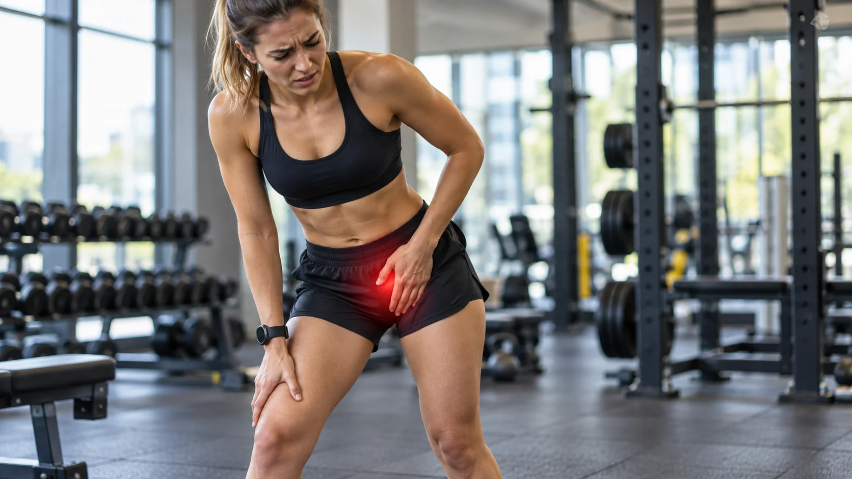
Groin pain during a squat, rotation, or prolonged sitting may suggest femoroacetabular impingement. However, anterior hip pain alone does not constitute this diagnosis. According to the Warwick international consensus, the syndrome combines compatible symptoms, clinical signs, and concordant imaging findings. Finding a particular bone shape in an asymptomatic person therefore does not amount to diagnosing painful impingement.[1]
The term “anterior impingement” often describes the region or position in which discomfort occurs. It does not automatically define a separate condition that affects only normally shaped hips. To understand the problem, it is more useful to clarify the situation: which activity causes the pain, which movements are limited, and which findings genuinely support the diagnosis? This approach avoids attributing all groin pain to bone-on-bone contact.
The key point
Cam or pincer morphology is not sufficient for a diagnosis. The risk of osteoarthritis varies between individuals, and improving symptoms does not guarantee that future osteoarthritis will be prevented.
The hip, cartilage, and labrum
The head of the femur articulates with the acetabulum, the socket in the pelvis. Cartilage covers the joint surfaces, and the labrum contributes to the function of the acetabular rim. In certain positions, the shape of the head-neck junction or acetabular coverage can alter contact and loading. These interactions are not simply a matter of constant rubbing with every step; they depend on anatomy and movement.[1] [3]
Cam morphology is a bony prominence or loss of sphericity at the femoral head-neck junction. Pincer morphology involves excessive acetabular coverage or a configuration that promotes contact with the rim. They may coexist. The word pincer conveys the idea of a clamp, not a “notch.” Insufficient coverage is a different issue, particularly dysplasia, and requires a different line of reasoning.[2] [3]

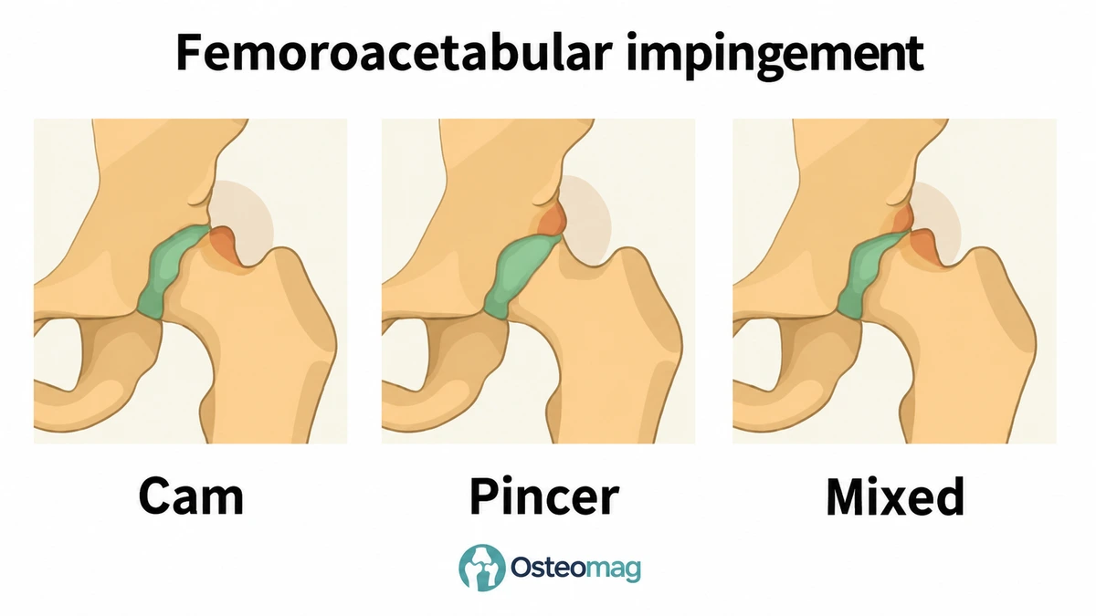
Why do some athletes have this morphology?
The Oxford consensus describes primary cam morphology as a bone variation that may develop during growth in relation to certain sporting loads. It is common in several athlete populations, but many people remain asymptomatic. The presence of this shape should therefore not be presented as a defect that makes the hip fragile or requires stopping sports.[2]
It is important to distinguish the existence of an anatomical variation from how well an activity is currently tolerated. An increase in load, repeated positions, or insufficient recovery can change symptoms without suddenly creating a new bone shape. Age, activities, and tissue condition also influence the situation. The cause of pain cannot be determined solely from the sport played or a photograph of posture.
Suggestive but nonspecific symptoms
Pain is often felt in the groin or at the front of the hip, sometimes extending toward the thigh or buttock. It may occur during deep flexion, pivoting, or after prolonged sitting. Restricted movement or mechanical discomfort may accompany the condition. An isolated click, especially if painless, is not enough to conclude that there is a labral injury or impingement.[1] [3]
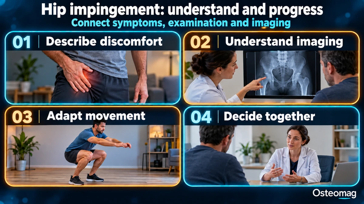
The history clarifies when the discomfort began, how it has evolved, recent loads, and which activities are difficult. Gradual pain while running does not have the same implications as severe pain after a fall or a very painful hip with fever. It is also useful to distinguish discomfort during activity from the response after exertion and the following day. These markers will then help guide progression.
Examination and imaging complement each other
The examination compares ranges of motion, strength, and representative tasks. The FADIR test, which combines flexion, adduction, and internal rotation, may reproduce hip pain. However, its accuracy does not allow it to be used as a stand-alone diagnostic test. A positive response must be interpreted alongside the other information, and a painful test does not justify forcefully repeating the movement to further confirm the result.[4]
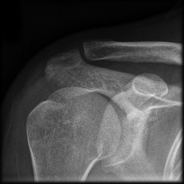
X-rays provide information about bone morphology, coverage, and signs of osteoarthritis. They do not directly show a labral tear. MRI may be used to investigate a question concerning the tissues or another condition; the choice depends on the context and the decision being considered. Abnormalities may also exist in asymptomatic people, which requires clinical interpretation rather than automatically equating an image with pain.[1] [6]
Considering other causes of groin pain
Pain in this region may arise from the adductors, iliopsoas, inguinal region, or a joint condition. Several contributing factors may coexist. The Doha agreement proposes clinical terminology that helps distinguish these situations, without claiming that a single test identifies every cause. Pain originating from the back or another region may also be considered when the signs warrant it.[5]
Bone pain that worsens with weight-bearing in a runner, particularly after a substantial increase in training, may prompt an investigation for a femoral neck stress fracture. This situation should not be treated as simply a stiff hip. Persistent pain that does not match the expected presentation requires reassessment and sometimes different investigations.[13] [14]
A table for interpreting the information
| Information | What it suggests | What it does not prove |
|---|---|---|
| Groin pain during deep flexion | A hip that is sensitive in certain positions | Definite femoroacetabular impingement |
| Cam morphology on an X-ray | A variation at the head-neck junction | A painful condition or inevitable osteoarthritis |
| Pincer morphology | Acetabular coverage or configuration requiring interpretation | An automatic indication for surgery |
| Painful FADIR | A clinical sign to integrate into the overall picture | A labral injury identified in isolation |
| Labral injury on MRI | A tissue abnormality to relate to the symptoms | The sole cause of all the pain |
| Improvement with exercises | Better functional tolerance | A change in bone shape |
| Progressive weight-bearing pain or fever | Another potentially important cause | A simple tightness to stretch |
The risk of osteoarthritis is not an individual destiny
Some longitudinal studies establish an association between cam morphology and the development of radiographic osteoarthritis. An individual participant data meta-analysis published in 2026 found a higher risk over several years in the populations studied. This finding concerns groups and changes visible on X-rays; on its own, it does not predict pain, limitations, or the need for a joint replacement in a specific individual.[7]
Osteoarthritis is a multifactorial condition. Bone shape, age, medical history, and other characteristics may contribute to its progression. Radiographic osteoarthritis must also be distinguished from its real-life consequences. A more pronounced imaging finding does not always correspond to a proportionate limitation. To guide care, osteoarthritis recommendations consider symptoms and function in particular.[12]
Adapting activities and building capacity
Nonsurgical management may include education, temporary modification of provocative positions, and an exercise program. The ISHA consensus supports an individualized approach that considers impairments and functional needs. This may include strengthening the hip and trunk, controlling a task, and gradually progressing activities. A consensus structures practice; it does not demonstrate that every component is essential.[8]
There is no need to force deep flexion that causes sharp pain in pursuit of an ideal range of motion. A shallower squat, a different sitting position, or a variation in weight-bearing may temporarily improve comfort. The aim is to return to a tolerable activity and then progress according to the response. These adaptations do not claim to reshape the bone, nor do they mean that a movement must be avoided forever.
Progression can begin with simple tasks and resistance levels compatible with recovery. The goals then become more specific: walking longer, cycling comfortably, resuming training, or tolerating certain work positions. Ease of activity, the response after exertion, and the response the following day are monitored. Some variability is possible, but repeated worsening should prompt a review of the parameters or the diagnosis.
When should surgery be discussed?
The UK FASHIoN trial and Palmer’s trial compared arthroscopy with nonsurgical care programs in selected patients with a diagnosed syndrome. Both groups improved, with average results favouring surgery for some outcomes. These trials concern a specific population and do not support recommending surgery for asymptomatic morphology or for every case of groin pain.[9] [10]
The decision takes into account persistent limitations, treatments already undertaken, the condition of the cartilage, any dysplasia, risks, and goals. A specialist consultation can clarify what surgery can reasonably achieve. Recovery requires rehabilitation and follow-up; immediate resolution of symptoms is not guaranteed. Significant osteoarthritis may also affect the appropriateness of arthroscopy.
Regarding the prevention of osteoarthritis, a review published in 2025 reports a favorable signal in a long-term comparison, but this was based on only two retrospective cohorts. It found no significant difference in the risk of hip replacement or resurfacing. These limitations mean that surgery cannot be presented as a definite means of preventing hip osteoarthritis.[11]
Role of osteopathy and situations requiring referral
The osteopath can help assess functional difficulties and, if the patient wishes, offer manual treatment for comfort. An improvement in how the patient feels does not prove that cam or pincer morphology has been corrected. The potential mechanisms of manual therapy are varied; its role should be linked to an activity goal and reassessment, as an adjunct to appropriate care.[15]
After a fall, an inability to walk or bear weight warrants urgent assessment. A very painful or hot hip, or hip pain accompanied by fever, also requires prompt referral. Progressive bone pain in an athlete requires investigation for a stress injury. In these situations, movements should not be forced and training should not be continued to test resilience.[13] [14]
Key takeaways
Femoroacetabular impingement syndrome combines symptoms, clinical signs, and imaging findings. Rehabilitation focuses on capacity and tolerance of activities. Surgery may be helpful in selected patients, but it guarantees neither complete resolution of pain nor prevention of osteoarthritis.
Scientific references and resources
- [1] Griffin et al. (2016). Warwick Agreement on femoroacetabular impingement syndrome.
- [2] Dijkstra et al. (2023). Oxford consensus on primary cam morphology, part 1.
- [3] AAOS. Femoroacetabular impingement.
- [4] Reiman et al. (2015). Diagnostic accuracy of clinical tests for hip impingement and labral tears.
- [5] Weir et al. (2015). Doha agreement on terminology and definitions in groin pain.
- [6] Register et al. (2012). Hip abnormalities in asymptomatic volunteers.
- [7] Cam morphology and radiographic hip osteoarthritis: individual participant data meta-analysis (2026).
- [8] Takla et al. (2021). ISHA physiotherapy agreement on assessment and treatment of FAIS.
- [9] Griffin et al. (2018). UK FASHIoN randomized trial: arthroscopy versus personalised hip therapy.
- [10] Palmer et al. (2019). Arthroscopy versus physiotherapy and activity modification: randomized trial.
- [11] Lameire et al. (2025). Impact of hip arthroscopy on osteoarthritis progression: systematic review and meta-analysis.
- [12] NICE. Osteoarthritis in over 16s, NG226.
- [13] Robertson and Wood (2017). Femoral neck stress fractures in sport.
- [14] NHS. Hip pain.
- [15] Bialosky et al. (2009). Mechanisms of manual therapy in musculoskeletal pain.