

Osteochondritis dissecans (OCD) is a condition where part of the joint cartilage surface dies due to lack of circulation. Necrosis of the subchondral bone forms. A necrotic bone fragment can break away from the joint surface and end up in the joint as a foreign body and damage the joint capsule. Such a fragment usually has a diameter of 1 to 2 cm.
Synonym: Osteochondrosis
Osteochondrosis can affect the knee, hip, elbow, foot and also the spine, but it most often affects the knee (85% are in the medial femoral condyle). When it affects the knee, it is called König’s disease or epiphyseal osteochondritis of the lateral femoral condyle. It is an osteonecrosis mainly of the medial condyle of the knee. This lesion is usually located on the posterior lateral aspect of the medial femoral condyle (70% of them).
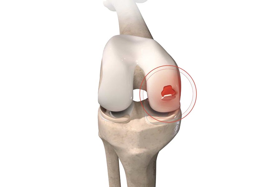
This osteochondritis becomes dissecting, as a separation of a fragment of bone and cartilage from the rest of the articular surface, occurs in a progressive way, to end up detaching completely, walking freely in the joint.
Risk factors
The exact cause has not yet been found, but possibly is a set of circumstances brought together by the following factors:
- When the bone under the cartilage is injured, the blood supply can be restricted (ischemia); this can lead to the death of bone tissue (necrosis), causing the cartilage to fragment and dislodge.
- Hereditary and endocrine factors
- Avascular necrosis (loss of blood flow)
- Lack of vitamin D
- Minor trauma
- Rapid growth
- Deficiencies and imbalances in the calcium/phosphorus ratio
- Bone formation deciency
Symptoms
- The nature of the pain depends on the pressure exerted on the hip and, in the presence of a bone fragment, it can be instantaneous and paralyzing. This fragment of cartilage that is free in the joint capsule will cause a lot of damage and inflammation in its passage.
- Pain related to activity that develops gradually.
- Feel the joint that catches, blocks, that makes popping noises
- Restriction of range of motion
Sports that can damage the cartilage surface of the hip
- Gymnastic
- Soccer
- Basketball
- The Crosse
- Football
- Tennis
- Squash
- Baseball
- Weightlifting
Screening test
Wilson test
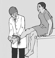
The Wilson test aids in the diagnosis of osteochondritis dissecans of the medial femoral condyle. The examiners place the knee at 90 degrees of flexion and internally rotate the tibia. When the knee is extended, the patient feels pain at approximately 30 degrees of flexion. External rotation of the tibia relieves pain.
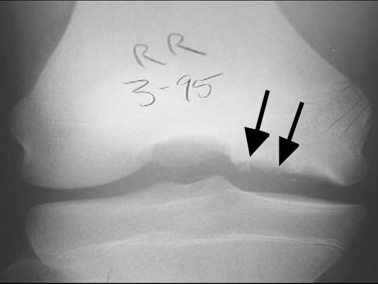
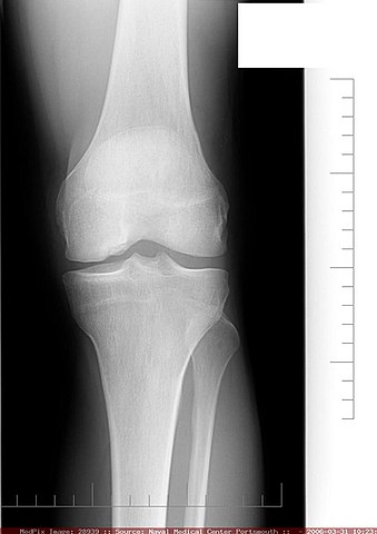
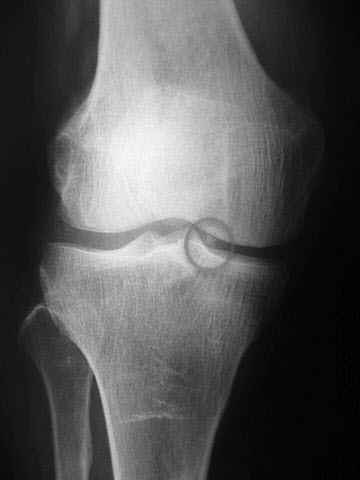
Radiography
References
- Edmonds EW, Polousky J. A review of knowledge in osteochondritis dissecans: 123 years of minimal evolution from König to the ROCK study group. Clin Orthop Relat Res. 2013 Apr;471(4):1118-26. [PMC free article] [PubMed]
- Crawford DC, Safran MR. Osteochondritis dissecans of the knee. J Am Acad Orthop Surg. 2006 Feb;14(2):90-100. [PubMed]
- Kocher MS, Tucker R, Ganley TJ, Flynn JM. Management of osteochondritis dissecans of the knee: current concepts review. Am J Sports Med. 2006 Jul;34(7):1181-91. [PubMed]
- König F. The classic: On loose bodies in the joint. 1887. Clin Orthop Relat Res. 2013 Apr;471(4):1107-15. [PMC free article] [PubMed]
- Lindén B. The incidence of osteochondritis dissecans in the condyles of the femur. Acta Orthop Scand. 1976 Dec;47(6):664-7. [PubMed]
- Cruz AI, Shea KG, Ganley TJ. Pediatric Knee Osteochondritis Dissecans Lesions. Orthop Clin North Am. 2016 Oct;47(4):763-75. [PubMed]
- Andriolo L, Crawford DC, Reale D, Zaffagnini S, Candrian C, Cavicchioli A, Filardo G. Osteochondritis Dissecans of the Knee: Etiology and Pathogenetic Mechanisms. A Systematic Review. Cartilage. 2020 Jul;11(3):273-290. [PMC free article] [PubMed]
- Credit in part: Bryce Mohr; John D. Baldea.


 Weakness: Causes, Implications, and Solutions")


