

What is Chondromalacia Patella?
Chondromalacia patellae, often referred to as patellofemoral syndrome, is a medical condition characterized by softening or wearing away of the cartilage located under the kneecap of the knee. This condition primarily affects the joint area between the kneecap and the femur. Chondromalacia patellae often occurs in young adults, particularly teenagers and people in their twenties, although cases can also be seen in older individuals.
The main contributing factor to chondromalacia patellae is abnormal pressure on the cartilage of the patella, usually due to abnormalities in knee mechanics. When the patella does not move properly along the groove of the femur during knee movements, this can lead to uneven distribution of pressure on the cartilage, leading to softening.
Common symptoms of chondromalacia patellae include pain located at the front of the knee, often exacerbated by activities such as going up or down stairs, sitting for long periods of time, or squatting. Pain may also be felt when participating in sports activities, especially those involving repetitive knee movements.
Diagnosis of chondromalacia patellae is often based on a clinical evaluation, a thorough medical history, and imaging tests such as X-rays and MRIs. These tests help assess the condition of the cartilage under the kneecap and determine how severe the disease is.
Management of chondromalacia patellae involves several approaches. Initially, conservative measures such as rest, icing, knee elevation, and anti-inflammatory medications may be recommended to relieve pain and reduce inflammation. Osteopathy is also often prescribed to strengthen the surrounding muscles, improve knee stability, and correct muscle imbalances that may contribute to the condition.
In more severe or persistent cases, surgical interventions may be considered. These may include procedures such as debridement surgery, where fragments of damaged cartilage are removed, or performing realignment surgery to correct joint mechanics.
It is essential to emphasize that the management of chondromalacia patellae should be personalized according to the severity of symptoms and the response to different interventions. Regular follow-up with a healthcare professional is necessary to adjust the treatment plan as the patient progresses in his recovery. In addition, preventing overuse of the knee and adopting appropriate techniques during physical activities can play a crucial role in the long-term management of this condition.
Knee Biomechanics: Understanding Chondromalacia
Chondromalacia patellae, when examined from a biomechanical perspective, reveals complex interactions between the articular structures of the knee, primarily the patella and femur. Joint biomechanics play a crucial role in the development of this condition, involving specific forces, movements, and pressures that affect the cartilage beneath the patella.
The biomechanical mechanism of chondromalacia patellae often begins with abnormalities in the natural trajectory of the patella during knee movements. Normally, the patella glides smoothly along the groove of the femur during knee flexion and extension. However, muscular imbalances or anatomical factors can disrupt this trajectory, generating uneven pressure on the cartilage.
A muscle imbalance, where some muscle groups are weaker or tighter than others, can change the path of the kneecap. For example, weakness in the quadriceps muscles can allow the kneecap to deviate from its normal path, causing uneven pressure distribution on the cartilage. This abnormal pressure can cause it to soften and wear out.
The biomechanics of chondromalacia patellae may also be influenced by anatomical factors, such as the shape of the patella or the slope of the femur. A flatter patella or a different femoral slope can create uneven forces during knee movements, contributing to cartilage degradation.
Physical activities, especially those involving repetitive knee movements, can exacerbate the effects of altered biomechanics. Activities such as running, jumping, or playing certain sports can increase pressure on the kneecap and accelerate the process of cartilage softening.
Accurate diagnosis of chondromalacia patellae from a biomechanical perspective often involves advanced imaging studies, such as MRI, to assess patella trajectory, cartilage status, and possible anatomical abnormalities. Understanding these biomechanical aspects is essential to developing an effective treatment plan.
Biomechanical management of chondromalacia patellae is based on correcting muscle imbalances and improving joint stability. Physiotherapy plays a central role by strengthening the knee muscles, restoring muscle balance and correcting biomechanical deficiencies. Specific rehabilitation techniques can be applied to realign the patella trajectory and minimise uneven forces on the cartilage.
Underlying mechanism
- Excessive pressure on the patella: Chondromalacia patellae is often the result of excessive pressure on the kneecap during knee movements. This can result from a variety of factors such as misalignment of the patella, weakened quadriceps muscles, joint instability, or repeated trauma.
- Cartilage Degeneration: Excessive pressure and abnormal movements can lead to degeneration of the cartilage in the patella. Initially, the cartilage may become softened and lose its normal resilience, and then it may crack and deteriorate over time.
- Inflammation and inflammatory response: Degeneration of cartilage can trigger an inflammatory response in the knee joint. The resulting inflammation can cause pain, swelling, and stiffness.
- Impaired patella function: Damaged cartilage no longer effectively performs its role as a shock absorber between the patella and the femur. This can lead to impaired normal patella function during knee movements.
- Progression to more advanced stages: Without adequate treatment, chondromalacia patellae can progress to more advanced stages, involving more severe cartilage damage and even exposure of the underlying bone.
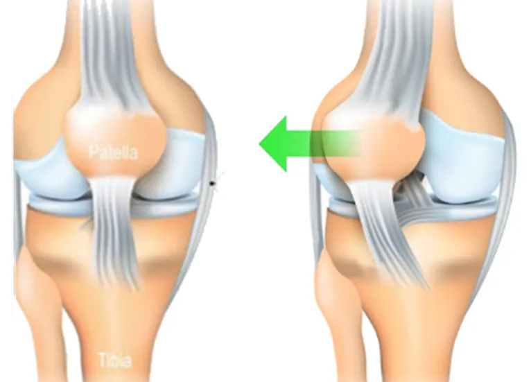
On the right, a condition is seen where the kneecap has moved laterally, a condition known as patellar dislocation or subluxation. This lateral displacement is often due to a muscular or ligamentous imbalance, or an anatomical abnormality. The quadriceps ligament and patellar tendon are stretched and no longer adequately hold the kneecap in its natural axis. This condition can lead to pain, knee instability, and an increased risk of repetitive injury.
The image clearly illustrates the difference between a healthy knee and one with lateral displacement of the patella, highlighting the importance of proper alignment for optimal function of the knee joint.
Anatomy of the Knee: The Structure of the Patella and Its Role
The anatomy of the knee is complex, involving a sophisticated interconnection of ligaments, tendons, muscles, and bones. At the heart of this structure is the kneecap, or patella, a triangular bone located at the front of the knee. Its crucial role in knee biomechanics makes it a key element in joint stability and force transmission during movement.
The patella plays a vital role in improving knee mechanics by acting as a lever for the quadriceps muscles. These muscles attach to the patella via the quadriceps tendon, and by pulling on the patella, they facilitate knee extension. This action helps stabilize the patella during movement, ensuring optimal alignment along the groove of the femur.
The structure of the patella consists of several important parts. The posterior surface of the patella is covered with articular cartilage, which ensures smooth articulation with the femur during flexion and extension of the knee. This cartilage is essential for minimizing friction and distributing forces evenly across the articular surface.
The kneecap is also surrounded by the patellar tendon, which connects the kneecap to the tibial tubercle. This tendon, often used during jumping or activities requiring rapid knee extension, is crucial for knee stability and function.
In terms of its functional role, the patella acts as a lever mechanism that increases the mechanical efficiency of the quadriceps during knee extension. As the knee extends, the patella moves into the groove of the femur, facilitating movement and reducing pressure on the quadriceps tendon.
However, this anatomical complexity makes the patella susceptible to various conditions, including the aforementioned chondromalacia patella. Abnormalities in the patella’s trajectory, muscular imbalances, or anatomical factors can cause uneven pressure on the cartilage, causing it to soften and degrade.
Diagnosis and management of patella-related problems requires a thorough understanding of its anatomy and function. Imaging tests, such as X-rays or MRIs, are often used to assess the condition of the patella, detect abnormalities, and guide the treatment plan.
The Femoral-Rotellar Joint: Center of Chondromalacia
The patellofemoral joint, playing a crucial role, fulfills two main functions: acting as an anatomical pulley to provide mechanical advantage to the extensors and reducing friction between the extensor mechanism and the femur. The patella, shaped like an inverted triangle, integrates harmoniously into the quadriceps tendon, giving it the status of the largest sesamoid bone in the body. Distally, it attaches to the tibial tubercle via the patellar tendon.
The posterior articular surface of the patella is divided into two distinct facets, medial and lateral, separated by a vertical ridge. In 30% of the population, a third facet, the unpaired facet, the most medial, completes this unique configuration.
The patella engages with the trochlear groove of the anterior femur, presenting corresponding articular surfaces: patellar, lateral and medial. It should be noted that the lateral trochlear articular surface is generally distinguished by a greater prominence than its medial part. During movements of the knee joint, from extension to flexion, the articular surface of the patella comes into contact with the variations of the femur.
At full extension, the patella has little to no contact with the trochlear groove, potentially placing it in an unstable position. Between 10 and 20 degrees of flexion, the patella engages the trochlear groove, with the contact area being the lower portion of the medial and lateral facets. As the knee progresses to greater flexion, the contact surface moves more proximally on the patella. Only beyond 90 degrees of flexion does the unpaired facet contact the medial femoral condyle, contributing to load sharing with the lateral facet. Additionally, at this degree of flexion, the quadriceps tendon itself engages the proximal trochlear groove, actively participating in force distribution.
Why Chondromalacia Patella Develops: Contributing Factors
Chondromalacia patellae, often referred to as patellofemoral syndrome, has its origins in a variety of factors, and understanding its causes is crucial for diagnosis and the development of an appropriate treatment plan. This joint condition, characterized by softening of the cartilage located under the kneecap, can be influenced by anatomical, mechanical, muscular, and even lifestyle factors.
One of the major contributing factors to chondromalacia patellae is misalignment of the patella during knee movements. An abnormal patella trajectory can cause uneven pressure on the cartilage, promoting its softening. Anatomical abnormalities, such as a flat patella or a specific femoral slope, can predispose to this misalignment and contribute to the development of chondromalacia.
Muscle imbalances also play a crucial role. The quadriceps muscles, which attach to the kneecap via the quadriceps tendon, are essential for maintaining optimal trajectory. Any weakness or imbalance in these muscles can compromise the stability of the kneecap, increasing the risk of chondromalacia.
Repeated trauma or microtrauma, often related to intense physical activities or repetitive knee movements, can also contribute to the development of chondromalacia patellae. These repeated stresses on the kneecap and its cartilage can gradually cause it to soften and wear out.
Lifestyle factors should not be overlooked. Being overweight, for example, puts increased pressure on the joints, including the kneecap, which can contribute to the development of chondromalacia. Similarly, poorly adapted physical activities, lack of muscle flexibility, or inappropriate footwear can negatively influence knee biomechanics.
People with chronic joint instability or congenital knee malformations may also be more likely to develop chondromalacia patellae. Genetic factors may thus play a role in predisposition to this condition.
Accurately diagnosing the causes of chondromalacia patella often involves imaging tests, such as X-rays or MRIs, to assess the condition of the cartilage and identify possible structural abnormalities. A thorough evaluation of medical history, physical activities, and lifestyle is also essential to determine contributing factors.
Management of chondromalacia patellae involves correcting muscle imbalances, reeducating the patellar trajectory, and managing modifiable risk factors, such as excess weight. Physical therapy plays a central role in these approaches, aiming to strengthen muscles, improve joint stability, and teach appropriate movement techniques.
- Poor Knee Alignment: Poor knee alignment, such as bowed legs (genu varum) or knocked legs (genu valgum), can cause pressure to be distributed poorly on the cartilage of the patella, causing premature wear.
- Trauma: Knee injuries, such as sprains, fractures, or dislocations, can damage cartilage and contribute to the development of chondromalacia patellae.
- Knee Instability: Chronic knee instability, often due to ligament damage, can cause abnormal pressure distribution across the kneecap, leading to cartilage wear.
- Overuse or High-Impact Activities: Regular participation in high-impact activities, such as running or jumping sports, can contribute to wear and tear on the cartilage of the kneecap, especially if combined with poor technique or overuse.
- Muscle Dysfunction: A muscular imbalance between the quadriceps and hamstring muscles can lead to poor stabilization of the knee, thus promoting the development of chondromalacia.
- Congenital Anatomical Factors: Some individuals may have congenital anatomical abnormalities, such as a malpositioned patella, that increase their predisposition to chondromalacia patella.
- Age: Although chondromalacia patellae can affect individuals of any age, it is often associated with age-related degenerative changes, particularly in older individuals.
- Gender: Women appear to be more likely to develop chondromalacia patellae than men, which may be attributed to differences in knee biomechanics and hormonal fluctuations.
- Joint Inflammation: Inflammatory conditions such as arthritis can contribute to deterioration of the cartilage in the kneecap.
Chondromalacia patellae can result from a combination of several of these factors.
Why Chondromalacia Patella Develops: Contributing Factors
The symptoms of chondromalacia patella are varied and can affect the quality of life of affected individuals. This joint condition, characterized by softening of the cartilage under the kneecap, usually manifests itself with specific signs related to knee mobility, causing a range of noticeable symptoms.
Pain is one of the most common symptoms of chondromalacia patellae. This pain is often located in the front of the knee, behind the kneecap, and may be felt as diffuse or focused. The pain may be exacerbated by specific activities, such as going up or down stairs, sitting for long periods, or squatting. It may also be triggered by repetitive knee movements, such as running or jumping.
A cracking or crepitating sensation may accompany the pain in some people with chondromalacia patellae. This audible sound often results from abnormal friction between the kneecap and femur due to softened cartilage. Crepitation may be perceived as an audible symptom or felt by the patient during knee movements.
Chondromalacia patellae can also present with knee instability. Affected individuals may experience a feeling of “give” or weakness in the knee, which can compromise their ability to perform physical activities without discomfort. This instability may be associated with muscle imbalances that disrupt the normal trajectory of the kneecap.
Knee swelling is a common symptom of chondromalacia patellae. Inflammation often results from irritation of damaged cartilage, leading to an inflammatory response in the joint area. Swelling can contribute to knee stiffness, limiting its range of motion and worsening pain.
- Patella Pain: Pain is often felt directly below the kneecap, especially during activities such as going up or down stairs, running, or after prolonged sitting.
- Crepitus: Some individuals may experience a crepitus or rubbing sensation when moving the kneecap, usually associated with cartilage deterioration.
- Knee Flexion Pain: Pain may increase when bending the knee, especially when the knee is flexed for a long period of time.
- Pain When Sitting: Some people experience pain after sitting for an extended period of time, which can put extra pressure on the kneecap.
- Swelling: Mild swelling or a feeling of warmth may sometimes accompany chondromalacia patella, especially if inflammation occurs.
- Muscle Weakness: Weakness in the muscles around the knee, especially the quadriceps, can develop due to avoidance of activities that cause pain.
- Difficulty Bending or Extending the Knee: Some individuals may have difficulty fully bending or extending the knee due to pain or stiffness associated with chondromalacia.
Identify the Risk Factors for Chondromalacia Patella
Several risk factors can contribute to the development of chondromalacia patella. Some of these factors include:
- Trauma or Knee Injury: Previous knee injuries, such as sprains, fractures, or dislocations, can damage the cartilage under the kneecap, promoting chondromalacia.
- Knee Instability: Chronic knee instability, often due to ligament damage, can lead to abnormal pressure distribution on the cartilage and contribute to the development of chondromalacia.
- Poor Knee Alignment: Anatomical problems such as bowed legs (genu varum) or knocked legs (genu valgum) can cause pressure to be distributed improperly across the cartilage, increasing the risk of chondromalacia.
- Overuse: Overuse or intense physical activities, especially those involving repetitive knee movements, can contribute to premature cartilage wear.
- Muscle Dysfunction: Muscle imbalance around the knee, particularly between the quadriceps muscles and hamstring muscles, can lead to poor knee stability, increasing the risk of chondromalacia.
- Anatomical Factors: Some individuals may have congenital anatomical abnormalities that increase their predisposition to chondromalacia patella.
- Age: Although chondromalacia patellae can occur at any age, it is often associated with age-related degenerative changes.
- Gender: Women appear to be more likely to develop chondromalacia patellae than men, which may be related to differences in knee biomechanics.
- High-Impact Activities: Participating in high-impact activities, such as running on hard surfaces, can increase stress on cartilage and contribute to the development of chondromalacia.
It is important to note that chondromalacia patellae can result from a combination of several of these risk factors.
Exploring the Differential Diagnosis of Chondromalacia Patella
- Knee Osteoarthritis: Chondromalacia patellae and knee osteoarthritis share symptoms such as pain and stiffness, but they affect different parts of the knee. Osteoarthritis involves wear and tear of articular cartilage throughout the joint, while chondromalacia patellae focuses specifically on the kneecap.
- Patellar tendonitis: Inflammation of the patellar tendon can cause similar symptoms, such as pain during physical activity and bending the knee.
- Iliotibial band syndrome: This condition involves inflammation of the iliotibial band, often causing pain on the outside of the knee. It may be confused with chondromalacia patellae due to similar symptoms.
- Prepatellar bursitis: Inflammation of the prepatellar bursa can cause pain in the front of the knee, similar to that of chondromalacia patellae.
- Meniscal injuries: Meniscal injuries can cause pain and discomfort similar to chondromalacia patellae. A thorough evaluation is necessary to differentiate these conditions.
- Patellofemoral Syndrome: This condition, also known as “patellofemoral pain syndrome,” shares similarities with chondromalacia patellae in terms of anterior knee pain.
- Ligament injuries: Ligament injuries, such as anterior cruciate ligament (ACL) or medial collateral ligament (MCL) sprains, can cause similar symptoms, including knee pain and stiffness.
The Outerbridge Classification: An Essential Tool for Assessing Chondromalacia
The classification of chondromalacia patellae may vary depending on the severity of the lesions observed. The most commonly used classification system for chondromalacia patellae is the Outerbridge classification, which divides lesions into four stages:
- Stage I: Changes are limited to softening of the cartilage without any tears. There may be softening or thinning of the cartilage, but no cracking is present.
- Stage II: Superficial cartilage cracks are observed, but they do not penetrate to the underlying bone. These cracks can be considered minor to moderate.
- Stage III: The cracks reach the underlying bone, causing more extensive cartilage damage. This can lead to loss of cartilage substance.
- Stage IV: This stage is characterized by significant loss of cartilage substance, with exposure of the underlying bone. More severe degenerative changes are present, and the joint may show signs of advanced osteoarthritis.
The Outerbridge classification helps determine the severity of chondromalacia patellae and often guides treatment decisions. It is frequently used by healthcare professionals, including orthopedists, to assess the extent of cartilage damage and plan treatment accordingly.
It is important to note that the Outerbridge classification is an arthroscopic assessment, meaning it is based on what the surgeon observes during an arthroscopy, a procedure that allows the inside of the knee joint to be inspected using a camera.

Prevention of Chondromalacia Patella
Prevention of chondromalacia patella typically involves strategies to strengthen the muscles around the knee, improve joint stability, and minimize risk factors. Here are some recommendations to prevent the development of chondromalacia patella:
- Muscle strengthening:
- Quadriceps: Targeted strengthening exercises for the quadriceps muscles can help stabilize the kneecap. This can include knee extensions and squats.
- Hamstrings: Strengthening the hamstrings can balance muscle strength around the knee, reducing pressure on the kneecap.
- Stability Exercises:
- Balance and Proprioception: Exercises that improve balance and proprioception, such as standing on one leg, can help with knee joint stability.
- Gliding Exercises: Controlled gliding movements can be incorporated to strengthen stabilizer muscles.
- Stretching:
- Quadriceps and Hamstrings: Regular stretching of the quadriceps and hamstrings can help maintain muscle flexibility and reduce stress on the kneecap.
- TFL (Tensor Fasciae Latae): Stretching the TFL can be beneficial in releasing tension on the kneecap.
- Weight Control:
- Maintaining a healthy body weight is crucial, as excess weight can increase pressure on the knees, increasing the risk of chondromalacia patellae.
- Moderation in High Impact Activities:
- Avoiding excessive stress on the knees by limiting high-impact activities, such as running on hard surfaces, can help prevent premature wear of the cartilage.
- Suitable Shoes:
- Wearing appropriate shoes for physical activity, with adequate support, can help absorb shock and protect joints.
- Correction of Biomechanical Imbalances:
- For some people, biomechanical imbalances may contribute to chondromalacia patellae. Regular evaluations by healthcare professionals can help detect and correct these imbalances.
- Proper Warm-Up and Cool-Down:
- Before any physical activity, a proper warm-up and gentle stretching can prepare muscles and joints, reducing the risk of injury.
Chondromalacia Treatment: What You Need to Know
Chondromalacia patellae, also called patellofemoral syndrome, is a condition in which the cartilage under the kneecap (patella) becomes softened or damaged, often resulting in knee pain, especially when bending or extending. Treatment for chondromalacia patellae may involve conservative approaches aimed at relieving pain and improving knee function. Some common treatment options include:
- Osteopathy:
- An osteopathic program may be prescribed to strengthen the muscles around the knee, with an emphasis on the quadriceps and hamstrings.
- Stretching exercises can help improve muscle flexibility and reduce stress on the kneecap.
- Drugs :
- Nonsteroidal anti-inflammatory drugs (NSAIDs), such as ibuprofen, may be recommended to reduce inflammation and relieve pain.
- Analgesics may also be used for pain relief.
- Weight Control:
- Maintaining a healthy body weight can reduce pressure on the knee, thereby relieving symptoms.
- Orthotics:
- Orthotics, including orthotic insoles, may be used to correct biomechanical abnormalities that contribute to chondromalacia patellae.
- Reduction of High Impact Activities:
- Avoiding or reducing high-impact activities, such as running on hard surfaces, can help prevent symptoms from worsening.
- Injections :
- Corticosteroid injections may be considered to reduce inflammation.
- Viscosupplementation injection can help improve joint lubrication.
- Therapeutic Exercises:
- Some specific exercises, such as isometric contraction quadriceps exercises, may be included in the rehabilitation program.
- Surgery :
- In severe and persistent cases, when conservative treatments do not work, surgery may be considered. This may include cartilage debridement surgery, patella realignment, or other procedures depending on the specific situation.
Radiographic Signs
Radiographic signs of chondromalacia patellae can be identified using imaging tests such as X-rays. These signs provide information about the condition of the cartilage under the kneecap. Some of the radiographic signs associated with chondromalacia patellae include:
- Cartilage Thinning: X-rays may reveal thinning or decreased opacity of the cartilage under the kneecap. This may indicate softening or wearing away of the cartilage, which is characteristic of chondromalacia.
- Cartilage Surface Irregularities: X-ray images may show irregularities in the surface of the cartilage under the kneecap, including cracks, rough areas, or cartilage defects. These changes indicate cartilage damage.
- Joint Space Narrowing: The joint space, the space between the kneecap and the femur, may appear narrowed on x-rays due to loss of cartilage height.
- Osteophytes: Osteophytes, also called bone spurs, can form on the edges of bone surfaces in the patella region. These bony growths are a response to cartilage wear.
- Patella Subluxation: In some cases, x-rays may show a subluxation of the patella, indicating poor positioning of the patella relative to the femur.
- Severity Stage: Certain classifications can be used to determine the severity stage of chondromalacia patellae based on radiographic findings, ranging from mild to severe.
It is important to note that X-rays may not always reveal all the details of chondromalacia patellae because the cartilage is not directly visible on X-ray images. In some cases, more advanced imaging tests, such as MRI (magnetic resonance imaging), may be recommended to provide a more detailed assessment of the cartilage and surrounding structures.
The final diagnosis and determination of the treatment plan are usually based on a complete evaluation, including clinical information, medical history, physical examination, and imaging results.

Conclusion
In conclusion, chondromalacia patella is a knee condition characterized by softening or wearing away of the cartilage beneath the kneecap, often resulting in pain and discomfort. Treatment for this condition may involve various approaches depending on the severity of symptoms and the individual characteristics of the patient.
Treatment options range from conservative approaches, such as physiotherapy or osteopathy, to medical interventions, including the use of anti-inflammatory drugs, injections, or even surgery in severe and resistant cases.
It is imperative to consult a healthcare professional for an accurate diagnosis and a treatment plan tailored to each situation. Multidisciplinary treatment, combining different therapeutic approaches, can often provide the best results for the effective management of chondromalacia patellae, improving mobility, relieving pain, and promoting healing.
References
- Kelly MA, Insall JN. Historical perspectives of chondromalacia patellae. Orthop Clin North Am. 1992 Oct;23(4):517-21. [PubMed]2.
- Gordon HM. Chondromalacia patellae. Aust J Physiother. 1977 Sep;23(3):103-6. [PubMed]3.
- Bączkowicz D, Kręcisz K, Borysiuk Z. Analysis of patellofemoral arthrokinematic motion quality in open and closed kinetic chains using vibroarthrography. BMC Musculoskelet Disord. 2019 Jan 31;20(1):48. [PMC free article] [PubMed]4.
- Franco BAFM, Sadigursky D, Daltro GC. Patellar position in patients with patellofemoral syndrome as characterized by anatomo-radiographic study. Rev Bras Ortop. 2018 Jul-Aug;53(4):410-414. [PMC free article] [PubMed]5.
- Aysin IK, Askin A, Mete BD, Guvendi E, Aysin M, Kocyigit H. Investigation of the Relationship between Anterior Knee Pain and Chondromalacia Patellae and Patellofemoral Malalignment. Eurasian J Med. 2018 Feb;50(1):28-33. [PMC free article] [PubMed]6.
- Resorlu M, Doner D, Karatag O, Toprak CA. The Relationship between Chondromalacia Patella, Medial Meniscal Tear and Medial Periarticular Bursitis in Patients with Osteoarthritis. Radiol Oncol. 2017 Dec;51(4):401-406. [PMC free article] [PubMed]7.
- Eckenrode BJ, Kietrys DM, Parrott JS. Effectiveness of Manual Therapy for Pain and Self-reported Function in Individuals With Patellofemoral Pain: Systematic Review and Meta-analysis. J Orthop Sports Phys Ther. 2018 May;48(5):358-371. [PubMed]8.
- Outerbridge RE, Dunlop JA. The problem of chondromalacia patellae. Clin Orthop Relat Res. 1975 Jul-Aug;(110):177-96. [PubMed]9.
- Zhang H, Kong XQ, Cheng C, Liang MH. A correlative study between prevalence of chondromalacia patellae and sports injury in 4068 students. Chin J Traumatol. 2003 Dec;6(6):370-4. [PubMed]10.
- Marega T. [Post-traumatic chondromalacia of the knee-cap]. Fracastoro. 1968 Jan-Feb;61(1):83-9. [PubMed]11.
- OUTERBRIDGE RE. The etiology of chondromalacia patellae. J Bone Joint Surg Br. 1961 Nov;43-B:752-7. [PubMed]12.
- Bentley G, Dowd G. Current concepts of etiology and treatment of chondromalacia patellae. Clin Orthop Relat Res. 1984 Oct;(189):209-28. [PubMed]13.
- Leslie IJ, Bentley G. Arthroscopy in the diagnosis of chondromalacia patellae. Ann Rheum Dis. 1978 Dec;37(6):540-7. [PMC free article] [PubMed]14.
- Elias DA, White LM. Imaging of patellofemoral disorders. Clin Radiol. 2004 Jul;59(7):543-57. [PubMed]15.
- Pihlajamäki HK, Kuikka PI, Leppänen VV, Kiuru MJ, Mattila VM. Reliability of clinical findings and magnetic resonance imaging for the diagnosis of chondromalacia patellae. J Bone Joint Surg Am. 2010 Apr;92(4):927-34. [PubMed]16.
- Özgen A, Taşdelen N, Fırat Z. A new MRI grading system for chondromalacia patellae. Acta Radiol. 2017 Apr;58(4):456-463. [PubMed]17.
- Tuna BK, Semiz-Oysu A, Pekar B, Bukte Y, Hayirlioglu A. The association of patellofemoral joint morphology with chondromalacia patella: a quantitative MRI analysis. Clin Imaging. 2014 Jul-Aug;38(4):495-498. [PubMed]

 Weakness: Causes, Implications, and Solutions")


 injury of the knee")


