

Discover the fascinating world of peroneal tendons, from their structure to their crucial role in ankle stability. This text offers you keys to understanding, preventing and taking care of these essential elements. Immerse yourself in enriching reading for a better understanding of your musculoskeletal health.
Introduction
An essential aspect in the preventive management of peroneal tendon disorders lies in understanding the risk factors. Common causes of these disorders include trauma and acute injuries, excessive or repetitive physical activities, biomechanical abnormalities, inappropriate footwear, sports-related injuries, anatomical abnormalities, inflammatory or rheumatic diseases, aging, deficits muscle, and other medical conditions.
Warming up before exercise plays a crucial role in preparing muscles and tendons for physical activity, thereby reducing the risk of injury. Muscle strengthening exercises, with emphasis on the peroneal muscles, help stabilize the ankle and prevent disorders. Working on stability and balance is also key to strengthening the ankle support structures.
Choosing appropriate footwear that provides adequate ankle support and matches your foot type is a crucial part of prevention. Regular stretching, with emphasis on the calf and ankle muscles, maintains tendon flexibility and prevents stiffness. Weight management helps reduce pressure on the tendons, minimizing the risk of disorders.
Particular attention should be paid to signs of overwork and muscle fatigue. Periods of adequate rest are essential to allow optimal recovery of muscles and tendons. Additionally, in the event of an injury or pain, early consultation with a healthcare professional is essential to prevent the progression of tendon disorders.
Additionally, correcting biomechanical issues, such as excessive pronation, can be done with the help of medical professionals such as podiatrists or osteopath. These experts can recommend specific orthotics or exercises to correct these underlying problems.
In summary, prevention of peroneal tendon disorders involves a holistic approach, incorporating factors such as understanding potential causes, proper warm-up, muscle strengthening, stability and balance, choosing proper footwear, stretching regular routines, weight management, adequate rest, and correction of biomechanical problems. By adopting these practices, it is possible to significantly reduce the risk of peroneal tendon disorders and promote long-lasting ankle health.
Anatomy of the Peroneal Tendons
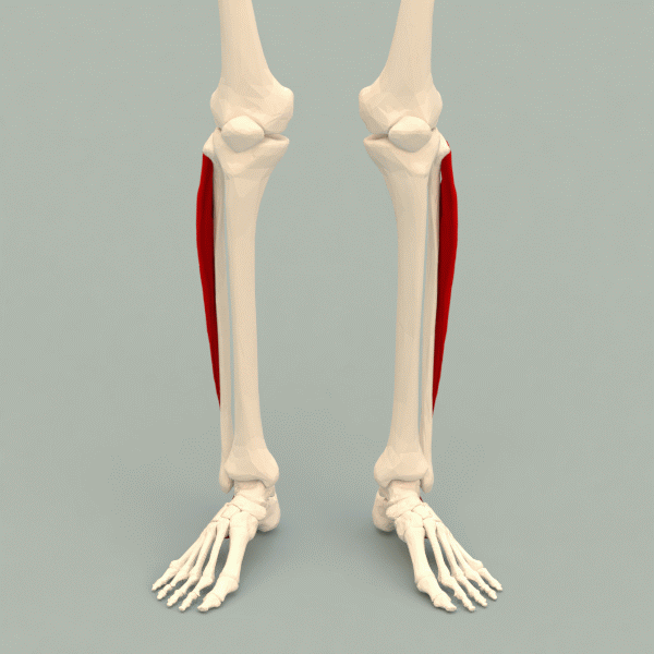
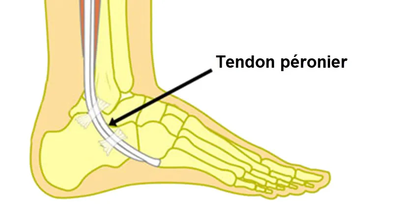
The peroneal tendons include two main tendons, the short tendon of the peroneus brevis muscle and the long tendon of the peroneus longus muscle. These muscles, also known as the peroneal muscles, are located in the lower leg region and have their origins at the fibula.
Short Fibular Muscle (Peronaeus brevis):
- Origin: Lateral head of the fibula.
- Path: Descends obliquely towards the back of the ankle.
- Insertion: Attaches to the base of the fifth metatarsal.

Long Fibular Muscle (Peronaeus longus):
- Origin: Proximal head of the fibula.
- Path: Goes down the leg and forms the long tendon.
- Insertion: Attaches to the first metatarsal and medial cuneiform.
Structure of the Peroneal Tendons
Peroneal tendons are strong fibrous structures that connect muscles to bones. The structure of tendons includes several components:
- Connective Tissue:
- Tendons are primarily composed of collagen, forming bundles that provide strength and tensile strength.
- Tendon Sheaths:
- The peroneal tendons are surrounded by tendon sheaths that provide protection and facilitate movement by reducing friction.
- Vascularization and Innervation:
- The peroneal tendons are vascularized and innervated, receiving a blood supply and nerve signals to maintain their health and function.
Function of the Peroneal Tendons
The peroneal tendons play an essential role in several functions related to the movement and stability of the ankle:
- Ankle Stabilization:
- The peroneal tendons contribute to lateral stabilization of the ankle, helping to prevent excessive inversion of the foot.
- Foot Eversion:
- The peroneal muscles, particularly the peroneus longus, are responsible for eversion of the foot, that is, turning the sole of the foot outward.
- Support for the Arch of the Foot:
- The peroneal tendons help maintain the longitudinal arch of the foot by supporting the outer edge of the foot.
- Participation in Plantar Flexion:
- When walking and running, the peroneal tendons participate in plantar flexion, contributing to the downward movement of the foot.
Common Peroneal Tendon Disorders
Peroneal tendon disorders refer to various conditions affecting the peroneal tendons located on the lateral (outer) side of the ankle. The peroneal tendons are responsible for stabilizing the foot and ankle, especially when walking and running. Here are some common peroneal tendon disorders:
- Peroneal Tendonitis:
- Description: Inflammation of the peroneal tendons.
- Causes: Overexertion, repetitive activities, trauma or inappropriate shoes.
- Symptoms: Pain, swelling and tenderness on the outer side of the ankle.
- Subluxation/Dislocation of the Peroneal Tendons:
- Description: The peroneal tendons may displace or subluxate from their normal position behind the lateral malleolus.
- Causes: Trauma, anatomical variations or overwork.
- Symptoms: Snapping or popping sensation, pain, instability and swelling.
- Peroneal Tendon Tears:
- Description: Partial or complete tear of the peroneal tendons.
- Causes: Acute injury, chronic overwork or degeneration.
- Symptoms: Pain, weakness, swelling and sometimes feeling unsteady.
- Tenosynovitis:
- Description: Inflammation of the synovial sheath surrounding the tendon.
- Causes: Overexertion, repetitive activities or inflammatory conditions.
- Symptoms: Pain with movement, swelling, and a crunching or grinding sensation.
- Subluxation of the Peroneal Tendons:
- Description : Tendons can move out of their normal groove behind the lateral malleolus.
- Causes : Trauma, anatomical factors or overwork.
- Symptoms: Instability, pain, and a feeling that the tendons are moving or coming out of place.
- Chronic Lateral Ankle Instability:
- Description: Weakened or stretched ligaments, including those supporting the peroneal tendons, leading to instability.
- Causes: Previous ankle sprains or injuries.
- Symptoms: Frequent ankle sprains, feeling of weakness and pain on the outer side of the ankle.
Causes of Peroneal Tendon Disorders
Peroneal tendon disorders encompass a range of conditions that affect the peroneal tendons, which are located on the outside of the ankle and play a crucial role in stabilizing the foot and ankle during movement. These disorders can result from various causes, including anatomical factors, biomechanical issues, overuse or repetitive stress, trauma, and underlying medical conditions.
Anatomical factors can predispose individuals to peroneal tendon disorders. For example, individuals with a high arch or cavus foot deformity may experience increased tension and stress on the peroneal tendons due to the altered biomechanics of the foot and ankle. Similarly, those with a tight Achilles tendon or a varus hindfoot alignment may also be at higher risk of developing peroneal tendon disorders due to increased strain on the tendons during movement.
Biomechanical issues, such as abnormal foot pronation or supination, can also contribute to peroneal tendon disorders. Excessive pronation (inward rolling) or supination (outward rolling) of the foot can place uneven stress on the peroneal tendons, leading to inflammation, irritation, or even tears. These biomechanical abnormalities may result from muscle imbalances, improper footwear, or faulty walking or running mechanics.
Overuse or repetitive stress is a common cause of peroneal tendon disorders, particularly in athletes or individuals engaged in activities that involve repetitive ankle movements or excessive loading of the foot and ankle. Activities such as running, jumping, or participating in sports that require rapid changes in direction can place significant strain on the peroneal tendons, increasing the risk of overuse injuries such as tendonitis or tendonosis.
Trauma, such as ankle sprains or fractures, can also lead to peroneal tendon disorders. Ankle sprains, in particular, can cause stretching or tearing of the peroneal tendons, especially if the injury is severe or if the tendons are already weakened due to other factors. Fractures or dislocations of the ankle or foot can also damage the peroneal tendons directly or disrupt their normal functioning, leading to instability or dysfunction.
Underlying medical conditions, such as rheumatoid arthritis, gout, or inflammatory diseases, can contribute to the development of peroneal tendon disorders. These conditions can cause inflammation, degeneration, or weakening of the tendons, making them more susceptible to injury or dysfunction. Additionally, certain systemic diseases or metabolic disorders, such as diabetes or obesity, can impair blood flow and nutrient delivery to the tendons, further increasing the risk of tendon disorders.
List of causes
- Overuse and Microtrauma:
- Repeated exposure to specific movements, such as dorsiflexion and foot inversion, can result in cumulative microtrauma to the lateral peroneal tendons, leading to tendinopathy.
- Sudden Changes in Activity:
- A sudden increase in the intensity or duration of physical activity, especially activities involving foot movements, can put excessive strain on the tendons, promoting the development of tendinopathy.
- Anatomical Anomalies of the Foot:
- The presence of anatomical abnormalities, such as a flat foot or excessive pronation, can create uneven stress on the lateral peroneal tendons, contributing to their irritation and problems.
- Inappropriate Footwear:
- Wearing inappropriate shoes, especially those that do not provide adequate support, can increase stress on the tendons and promote the development of tendinopathy.
- Trauma or Acute Injuries:
- Acute injuries, such as an ankle sprain or direct impact to the lateral peroneal region, can damage the tendons and lead to long-term problems.
- Mechanical Factors:
- Muscular imbalances, poor biomechanics or ankle instability can lead to uneven distribution of forces on the tendons, contributing to the development of tendinopathy.
- Age and Tissue Degeneration:
- The natural aging process can lead to degeneration of tendon tissue, increasing vulnerability to tendinopathy.
- Inflammatory Diseases:
- Inflammatory medical conditions, such as rheumatoid arthritis, can affect the lateral peroneal tendons and contribute to the development of tendinopathy.
- Bad Training Technique:
- Improper training technique, especially when exercising, can overwork the tendons and lead to injury.
- Congenital Structura
- Some individuals may have congenital abnormalities of tendons or surrounding structures, increasing the risk of tendinopathies.
- Metabolic Factors:
- Metabolic conditions such as diabetes can influence tendon health and increase the risk of tendinopathy.
- Overweight:
- Excess weight puts additional pressure on the tendons, increasing stress and the likelihood of developing tendinopathy.
- Nervous System Dysfunction:
- Neurological disorders or imbalances in the nervous system can affect muscle regulation and contribute to tendinopathy.
- Dehydration and Nutrition:
- Insufficient hydration and nutritional deficiencies can compromise the health of connective tissues, including tendons.
- Hormonal Conditions:
- Hormonal fluctuations, such as those associated with the menstrual cycle in women, can influence tendon sensitivity.
- Repetitive Activities:
- Certain occupations or sporting activities that involve repetitive foot movements can contribute to the development of tendinopathy.
- Vascular Conditions:
- Vascular problems, such as blood circulation disorders, can compromise tendon health.
- Emotional Stress:
- Chronic emotional stress can contribute to physiological imbalances that can influence tendon health.
- Systemic Inflammatory Response:
- Systemic inflammatory conditions, even of non-local origin, can have an impact on tendon health.
- Poor Recovery:
- Inadequate recovery after strenuous physical activity may increase susceptibility to tendinopathy.
- Genetics and Genetic Factors:
- Genetic predispositions may influence susceptibility to lateral peroneal tendinopathy.
- Anatomical Variations:
- Individual anatomical differences, such as the shape or position of the peroneals, may contribute to tendinopathy.
- Inadequate Shoes:
- Wearing inappropriate shoes, especially during physical activities, can increase stress on the peroneals.
- Exposure to Toxic Agents:
- Prolonged exposure to toxic substances can have implications for general health, including tendon health.
- Inadequate Stretching Practices:
- Incorrect or insufficient stretching practices can affect the flexibility and resilience of the peroneals.
- Ineffective Body Mechanics:
- Poor body mechanics during physical activities can lead to overload of the peroneals.
- Age and Aging Process:
- Natural aging of the body can make tendons more prone to degeneration and tendinopathy.
- Specific Professional Activities:
- Certain jobs involving specific foot movements can increase the risk of tendinopathy.
- Posture Problems:
- Postural problems, such as flat foot, can influence the distribution of weight on the feet and affect the tendons.
- Local or Systemic Infections:
- Infections, whether local to the foot or systemic, can contribute to tendon problems.
Symptoms of Peroneal Tendon Disorders
Peroneal tendon disorders include a variety of conditions affecting the peroneal tendons, crucial structures positioned along the outer aspect of the ankle, which play a vital role in stabilizing the foot and ankle during movement.
These disorders can manifest with a variety of symptoms, ranging from mild discomfort to severe pain and functional limitations. Common symptoms of peroneal tendon disorders include pain, swelling, instability, weakness, and altered gait mechanics.
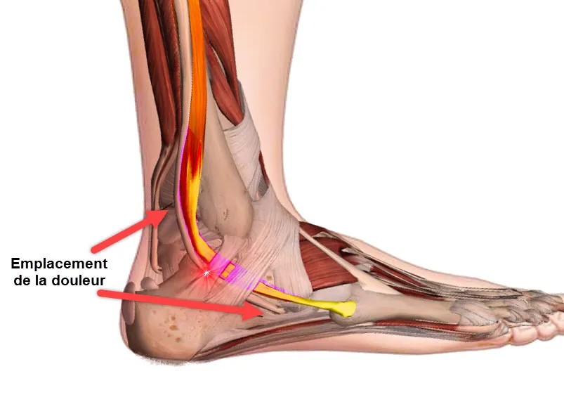
Pain is one of the hallmark symptoms of peroneal tendon disorders and is typically localized to the outside of the ankle or behind the lateral malleolus (the bony prominence on the outer aspect of the ankle). The pain may be dull or sharp and can worsen with activity, particularly movements that involve ankle dorsiflexion (lifting the foot upward) or eversion (turning the foot outward). Individuals may also experience pain during palpation or manual manipulation of the peroneal tendons.
Swelling is another common symptom of peroneal tendon disorders and is often associated with inflammation or irritation of the tendons. Swelling may be localized to the area around the peroneal tendons or may extend along the outside of the ankle. In some cases, swelling may be accompanied by warmth or redness, indicating an inflammatory response.
Instability or a feeling of “giving way” in the ankle is frequently reported by individuals with peroneal tendon disorders. This instability may be due to weakening or stretching of the peroneal tendons, which can compromise their ability to stabilize the ankle joint during weight-bearing activities. As a result, individuals may feel unsteady or insecure when walking, running, or participating in sports.
Weakness in the ankle and foot muscles, particularly those responsible for eversion and dorsiflexion, is another common symptom of peroneal tendon disorders. Weakness may manifest as difficulty lifting the foot upward or turning it outward, leading to decreased control and coordination during movement. Weakness in the peroneal muscles can also contribute to functional deficits such as difficulty maintaining balance or propelling the body forward during walking or running.
Altered gait mechanics are often observed in individuals with peroneal tendon disorders as a compensatory response to pain, instability, or weakness. These alterations may include changes in foot strike pattern, decreased push-off power, or an uneven distribution of weight during stance phase. Over time, altered gait mechanics can lead to secondary musculoskeletal issues such as overuse injuries, joint pain, or muscle imbalances.
List of symptoms
- Pain :
- Pain is one of the most common symptoms. It may be felt along the outer side of the ankle, lower leg, or foot.
- The pain can be acute, chronic or occur during certain activities.
- Swelling :
- Swelling around the peroneal tendon may occur due to inflammation or irritation.
- Touch Sensitivity:
- The area around the peroneal tendons may be tender to the touch.
- Crackling or Cracking:
- Some patients may experience a cracking or crackling sensation when moving the ankle.
- Muscular weakness :
- Weakness of the peroneal muscles may manifest, affecting ankle stability.
- Ankle Instability:
- Certain peroneal tendon disorders can lead to a feeling of instability in the ankle, increasing the risk of falls or twists.
- Difficulty Standing on Tiptoes:
- Some patients may have difficulty standing on their tiptoes due to pain or weakness in the peroneal tendons.
- Changes in the Arch of the Foot:
- Biomechanical abnormalities or disorders of the peroneal tendons can cause changes in the arch of the foot, such as its sagging.
- Numbness or Tingling:
- In some cases, numbness or tingling sensations may be felt along the outer side of the leg or foot.
- Deformation or Displacement of Tendons:
- In more severe cases, deformities or visible displacement of the peroneal tendons may be observed.

Pathophysiology of Peroneal Tendon Disorders
The pathophysiology of peroneal tendon disorders involves a complex interplay of mechanical stress, anatomical factors, inflammation, and degenerative changes within the peroneal tendons and surrounding structures. Understanding these underlying mechanisms is crucial for accurate diagnosis and effective management of these conditions.
Mechanical stress plays a central role in the pathophysiology of peroneal tendon disorders. Repetitive or excessive stress on the peroneal tendons, often due to activities such as running, jumping, or walking on uneven surfaces, can lead to microtrauma and overuse injuries. These injuries may result in inflammation, swelling, and structural changes within the tendons, predisposing them to further damage and dysfunction.
Anatomical factors also contribute to the development of peroneal tendon disorders. Variations in foot and ankle anatomy, such as high arches, flat feet, or abnormal foot pronation (rolling inward) or supination (rolling outward), can alter the biomechanics of the foot and ankle. These alterations can increase the strain on the peroneal tendons and other supporting structures, leading to instability, inflammation, and degeneration over time.
Inflammation plays a significant role in the pathophysiology of peroneal tendon disorders. In response to mechanical stress or injury, the body’s immune system initiates an inflammatory response, characterized by the release of pro-inflammatory mediators and recruitment of immune cells to the affected area. While inflammation is a normal part of the healing process, chronic or excessive inflammation can contribute to tissue damage and impair tendon healing. In peroneal tendon disorders, inflammation may occur within the tendons themselves (tendonitis) or in the surrounding tissues (peritendonitis), leading to pain, swelling, and functional impairment.
Degenerative changes within the peroneal tendons are common in individuals with chronic or recurrent peroneal tendon disorders. Over time, repeated episodes of mechanical stress and inflammation can lead to structural alterations within the tendons, including collagen degeneration, fibrosis, and neovascularization (the formation of new blood vessels). These changes can weaken the tendons and compromise their ability to withstand mechanical loads, increasing the risk of further injury and dysfunction.
In addition to changes within the tendons themselves, peroneal tendon disorders can also affect the surrounding soft tissues and bony structures. Chronic inflammation and mechanical stress can lead to secondary changes in the ligaments, joint capsules, and bony prominences around the ankle, contributing to instability, joint stiffness, and pain. In some cases, structural abnormalities such as bone spurs or accessory bones may exacerbate tendon pathology by impinging on the peroneal tendons during movement.
List
- Repetitive Strain Injuries: Lateral peroneal tendinopathy is often caused by repetitive strain injuries resulting from repeated or excessive movements of the ankle. These microtraumas can occur in sporting activities, professional activities or even in daily life.
- Overuse of the tendons: The lateral peroneal muscles, the peroneus longus and the peroneus brevis, are heavily used during plantar flexion and inversion movements of the ankle. Overuse of these muscles, especially during frequent changes of direction or lateral movements, can cause excessive strain on the tendons, contributing to the development of tendinopathy.
- Lack of vascularization: Tendons have relatively limited vascularization compared to other tissues. Insufficient blood supply can make tendons more susceptible to injury and degenerative processes. Repetitive microtrauma can lead to decreased vascularity in the lateral peroneal region, further compromising tendon health.
- Inflammation and degeneration: Repetitive microtrauma can trigger an inflammatory response in the tendons, characterized by a release of inflammatory mediators. However, unlike acute inflammation, tendinopathy can progress to tendon degeneration, with structural alterations such as collagen fragmentation, decreased cell number, and scar tissue formation.
- Abnormal cellular response: Tendon cells may exhibit an abnormal metabolic response due to repeated stress. This can lead to changes in the regulation of proteins and enzymes, contributing to the degeneration of tendon tissue.
- Anatomical and biomechanical factors: Certain anatomical factors, such as irregularities in the structure of the ankle, may contribute to the development of lateral peroneal tendinopathy. Additionally, biomechanical issues, such as muscular imbalances or ankle instability, can increase susceptibility to tendon injuries.
- Age: Lateral peroneal tendinopathy tends to be more common in older individuals. The natural aging process can cause the quality of collagen in tendons to decline, making them more prone to injury.
- Genetic factors: Some individuals may be genetically predisposed to developing tendon problems. Genetic variations can influence collagen structure, inflammatory response, and other aspects involved in tendon health.
- Inappropriate Footwear: Wearing shoes that do not properly support the ankle can contribute to inappropriate movements, increasing stress on the lateral peroneal tendons. Shoes that do not provide adequate stability can increase the risk of injury.
- Specific sporting activities: Certain sporting activities, such as basketball, soccer, or other sports involving frequent lateral movements, may increase the risk of lateral peroneal tendinopathy due to specific biomechanical demands.
Diagnostic
- History:
- The doctor will begin by taking a detailed medical history, including the symptoms you are experiencing, triggering factors, duration of symptoms, and any associated recent activities or injuries.
- Physical examination :
- A physical exam will be performed to assess the painful area, ankle mobility, muscle strength, stability, and the presence of swelling or deformities.
- Stress Tests:
- Certain stress tests may be performed to evaluate the stability of the ankle and the reaction of the tendons to specific movements.
- Medical imaging :
- Imaging studies may be ordered to confirm the diagnosis and assess the severity of the condition. Options may include:
- X-rays: To rule out bone problems such as fractures.
- Ultrasound: To visualize the tendons and assess their condition.
- MRI (Magnetic Resonance Imaging): To obtain detailed images of tendons and soft tissues.
- Bone scintigraphy: To detect possible metabolic or inflammatory abnormalities.
- Imaging studies may be ordered to confirm the diagnosis and assess the severity of the condition. Options may include:
- Electromyography (EMG):
- In some cases, an EMG may be performed to assess the nerve and muscle function associated with the peroneal tendons.
- Differential diagnosis :
- The doctor can also rule out other conditions that might present with similar symptoms by performing a differential diagnosis, including joint problems, nerve damage, or systemic pathologies.
- Specialized Consultation:
- Depending on the test results, the doctor may refer the patient to a specialist, such as an orthopedist or orthopedic surgeon, for further evaluation or intervention if necessary.
Role of osteopathy in treatment
Osteopathy is a complementary health care approach that focuses on the manual manipulation of musculoskeletal tissues to improve the patient’s overall health. Although osteopathy is not always the first line of treatment for peroneal tendon disorders, it can play a beneficial role as a complementary approach as part of an overall treatment plan. Here is how osteopathy can be used in the treatment of peroneal tendon disorders:
Principles of osteopathy
- Holism:
- Osteopathy views the body as an interconnected whole, recognizing the importance of balance and coordination between different anatomical structures.
- Mobility and Fluidity:
- Osteopathy aims to improve the mobility and fluidity of tissues, including joints, muscles, tendons and ligaments, to promote better function.
- Self-regulation of the Body:
- The central idea is that the body has the ability to self-regulate and self-heal. The osteopath seeks to facilitate this process by eliminating obstacles to health.
- Structure and Function:
- Osteopathy posits that the structure and function of the body are interdependent. Structural dysfunctions can affect function, and vice versa.
Role of osteopathy in the treatment of peroneal tendon disorders
- Global evaluation :
- The osteopath carries out a global assessment of the patient, taking into account posture, ankle mobility, and other factors influencing the peroneal tendons.
- Reduction of Tensions and Blockages:
- Using manual techniques, the osteopath can work on reducing muscle tension, joint blockages and movement restrictions that could affect the peroneal tendons.
- Improved Joint Mobility:
- Osteopathy aims to improve joint mobility, which can have a positive impact on the function of surrounding tendons and muscles.
- Correction of Postural Imbalances:
- The osteopath can work on correcting postural imbalances that could contribute to peroneal tendon disorders.
- Facilitation of Blood and Lymphatic Circulation:
- By improving blood and lymphatic circulation, osteopathy promotes the transport of nutrients and the elimination of waste, thus contributing to the healing process.
- Integration with Other Approaches:
- Osteopathy can be integrated with other treatment approaches, such as physical therapy, chiropractic, and medical treatments, to create a comprehensive care plan.
Limitations and Considerations
- Osteopathy is not suitable for all cases, and it is essential to consult a healthcare professional to determine if this approach is appropriate.
- In cases of severe peroneal tendon disorders or serious injuries, medical or surgical intervention may be necessary.
- The choice of a treatment approach, including osteopathy, should be discussed with the healthcare team and based on a comprehensive assessment of the individual condition.
Osteopathy may be a viable option in addition to other treatments for some patients, but it does not necessarily replace conventional medical interventions. The choice of this approach will depend on the specific nature of the peroneal tendon disorder and the individual needs of the patient.
Prevention of peroneal tendon disorders
- Warm up before exercise: Before any physical activity, make sure you warm up properly. This may include dynamic stretching, ankle rotations, and light exercises to activate the leg muscles.
- Muscle strengthening: Strengthen the leg muscles, especially the peroneal muscles. Exercises such as heel raises, resisted ankle rotations, and dorsiflexion exercises can help strengthen the peroneal tendons.
- Stability and balance: Working on stability and balance can help prevent injuries. Single-leg exercises, such as the single-leg stand, can improve ankle stability.
- Appropriate Footwear: Choose appropriate footwear for your activity. Shoes that provide good ankle support and match your foot type can help prevent peroneal tendon disorders.
- Regular stretching: Incorporate regular stretching into your routine, focusing on your calf and ankle muscles. This can help maintain tendon flexibility.
- Weight Management: Maintaining a healthy body weight reduces pressure on tendons and joints, helping to prevent peroneal tendon disorders.
- Adequate rest: Be sure to allow your muscles and tendons to recover adequately by including rest days in your training program.
- Immediate treatment of injuries: If you experience injury or pain, consult a healthcare professional as soon as possible. Early treatment can prevent tendon disorders from progressing.
- Correcting biomechanical problems: If you have biomechanical problems, such as excessive pronation, consult a podiatrist or osteopath. They may recommend orthotics or specific exercises to correct these problems.
- Gradual progression of intensity: If you are starting a new exercise program, be sure to progress gradually in intensity. Avoid sudden changes that could overload the tendons.
Preventative stretching
Preventative stretching plays a vital role in preventing peroneal tendon disorders and maintaining overall ankle health. Here are some specific stretches that can be incorporated into a preventative routine:
- Calf stretch against the wall:
- Stand facing a wall with one leg bent in front of you and the other straight behind you.
- Keep your heels on the floor and gently lean forward to feel the stretch in the calf of the straight leg.
- Achilles Tendon Stretch:
- Stand upright, place one foot forward and the other back, slightly bend your front knee.
- Keep the back heel on the ground and lean forward, feeling the stretch along the Achilles tendon.
- Stretching the lateral peroneal muscle:
- Sitting or standing, cross the leg to be stretched over the other knee.
- Turn the foot slightly downward to target the lateral peroneus muscle, then gently lean forward.
- Stretching the deep peroneal muscle:
- Sit on your heels with your knees apart.
- Slowly lean forward with your arms extended in front of you, feeling the stretch in the deep peroneus muscle.
- Tibialis anterior stretch:
- Stand with one foot behind you and bend your knee slightly.
- Gently bend the front knee while keeping the heel on the ground, feeling the stretch in the tibialis anterior.
- Stretching the gastrocnemius muscle:
- Standing, place one foot forward and the other back.
- Flex the front knee while keeping the back heel on the ground, feeling the stretch in the gastrocnemius muscle.
- Iliotibial Band Stretch:
- Stand with one leg crossed behind the other.
- Lean slightly toward the opposite side, stretching the iliotibial band to the side of the hip.
- Stretching the dorsal muscles of the foot:
- Sit on the floor with your legs extended in front of you.
- Use your hands to pull your toes toward you, stretching the dorsal muscles of the foot.
It is important to warm up before doing stretches and hold them for at least 15 to 30 seconds to allow for optimal effectiveness. These stretches can be incorporated into a regular routine to help prevent peroneal tendon disorders and maintain ankle flexibility. Before beginning a stretching program, it is recommended to consult a healthcare professional, particularly if you have a history of tendon problems or other medical conditions.
Preventive exercises
Here are some specific exercises that can help strengthen the peroneal tendon muscles and prevent associated disorders. Before beginning any exercise program, be sure to consult a healthcare professional to ensure that these exercises are appropriate for your individual physical condition and needs.
- Heel raises (Calf Raises):
- Standing, slowly lift your heels off the floor while standing on your tiptoes.
- Hold the position for a few seconds, then slowly lower your heels.
- Repeat the movement to strengthen your calf muscles.
- Ankle rotation with resistance:
- Sit in a chair with your foot flat on the floor.
- Tie a resistance band around the front of your foot and hold the other end in your hands.
- Slowly turn your foot outward against resistance, then return it to the starting position.
- Repeat the movement to strengthen the peroneal muscles.
- Back flexion with elastic band:
- Sit with your legs straight and an elastic band tied around the top of the foot.
- Slowly flex your toes toward you against the resistance of the band, then return to the starting position.
- This exercise targets the dorsal ankle muscles.
- Single-Leg Stand:
- Raise one leg and hold the position for 30 seconds to 1 minute.
- Be sure to stand near stable support if necessary.
- Alternate between both legs to improve balance and stability.
- Calf Stretch:
- Stand up and place your hands against a wall.
- Step one leg back while bending the other leg slightly forward.
- Hold the position to stretch your calf muscles.
- Repeat with the other leg.
- Ankle inversion and eversion:
- Sitting or standing, do inversion (inwards) and eversion (outwards) movements of the ankle.
- These movements can be performed without resistance to improve mobility and strength of the peroneal muscles.
- Band Resistance Exercises for Feet:
- Tie a resistance band around your feet and spread them slightly apart.
- Perform dorsi and plantar flexion movements against the resistance of the band to strengthen the ankle muscles.
- Stair climbs:
- Going up and down stairs can strengthen leg muscles, including the peroneal tendon muscles.
Differential diagnosis
- Ankle Sprain: An ankle sprain, especially a peroneal ligament sprain, can cause pain similar to that of lateral peroneal tendinopathy. Clinical evaluation and imaging studies may be necessary to differentiate between these two conditions.
- Ankle Fracture: An ankle fracture can have similar symptoms, such as pain and swelling. X-rays may be used to rule out a fracture.
- Tarsal tunnel syndrome: This condition is characterized by compression of the posterior tibial nerve in the ankle, causing similar pain and tingling sensations. Specific tests and imaging studies may be needed for diagnosis.
- Retrocalcaneal bursitis: Inflammation of the bursa located at the back of the heel can cause pain in the ankle area. Clinical assessment and imaging tests can help distinguish bursitis from tendinopathy.
- Plantar fasciitis: Plantar fasciitis, characterized by inflammation of the plantar fascia at the arch of the foot, can cause heel and ankle pain. A thorough clinical evaluation and imaging studies can help differentiate these conditions.
- Ankle Arthritis: Inflammatory or degenerative forms of arthritis can cause pain in the ankle area. Blood tests and imaging studies may be needed to make a diagnosis of arthritis.
- Iliotibial band syndrome: This syndrome often presents with pain on the outside of the knee, but it can also affect the ankle area. Accurate clinical assessment and specific tests can help distinguish this condition from lateral peroneal tendinopathy.
- Morton’s neuroma: Compression of the nerve between the toes can cause burning sensations and pain in the ankle. Specific tests and imaging studies may be needed to confirm the diagnosis.
Conclusion
In conclusion, preventive management of peroneal tendon disorders is of critical importance to maintain ankle health and functionality. Preventive stretching plays a key role in this, helping with flexibility, mobility and injury prevention. The integration of osteopathy as a treatment alternative offers a holistic approach, considering the body as a whole.
Engaging in a regular routine of targeted stretching can not only prevent peroneal tendon disorders, but also improve athletic performance, reduce muscle tension and promote better blood circulation. Stretching, combined with other preventive measures such as muscle strengthening, correcting biomechanical imbalances and a healthy lifestyle, forms a complete package to promote lasting health of the peroneal tendons.
In addition, the integration of osteopathy into treatment offers a complementary approach, focused on restoring the structural and functional balance of the body. Osteopathic professionals can help identify and treat imbalances, thereby promoting recovery and prevention of peroneal tendon disorders.
In short, the combination of preventive maintenance, appropriate stretching and osteopathy offers a comprehensive approach to maintaining peroneal tendon health, allowing individuals to enjoy an active and balanced life. It is always recommended to consult health professionals for personalized advice and care tailored to each individual.
References
- Davda K, Malhotra K, O’Donnell P, Singh D, Cullen N. Peroneal tendon disorders. EFORT Open Rev. 2017 Jun;2(6):281-292. [ PMC free article ] [ PubMed ]2.
- Philbin TM, Landis GS, Smith B. Peroneal tendon injuries. J Am Acad Orthop Surg. 2009 May;17(5):306-17. [ PubMed ]3.
- Zammit J, Singh D. The peroneus quartus muscle. Anatomy and clinical relevance. J Bone Joint Surg Br. 2003 Nov;85(8):1134-7. [ PubMed ]4.
- Sobel M, Geppert MJ, Olson EJ, Bohne WH, Arnoczky SP. The dynamics of peroneus brevis tendon splits: a proposed mechanism, technique of diagnosis, and classification of injury. Foot Ankle. 1992 Sep;13(7):413-22. [ PubMed ]5.
- Sobel M, Geppert MJ, Warren RF. Chronic ankle instability as a cause of peroneal tendon injury. Clin Orthop Relat Res. 1993 Nov;(296):187-91. [ PubMed ]6.
- Selmani E, Gjata V, Gjika E. Current concepts review: peroneal tendon disorders. Foot Ankle Int. 2006 Mar;27(3):221-8. [ PubMed ]7.
- Dombek MF, Lamm BM, Saltrick K, Mendicino RW, Catanzariti AR. Peroneal tendon tears: a retrospective review. J Foot Ankle Surg. 2003 Sep-Oct;42(5):250-8. [ PubMed ]8.
- Brandes CB, Smith RW. Characterization of patients with primary peroneus longus tendinopathy: a review of twenty-two cases. Foot Ankle Int. 2000 Jun;21(6):462-8. [ PubMed ]9.
- Roster B, Michelier P, Giza E. Peroneal Tendon Disorders. Clin Sports Med. 2015 Oct;34(4):625-41. [ PubMed ]10.
- Safran MR, O’Malley D, Fu FH. Peroneal tendon subluxation in athletes: new examination technique, case reports, and review. Med Sci Sports Exercise 1999 Jul;31(7 Suppl):S487-92. [ PubMed ]11.
- Grant TH, Kelikian AS, Jereb SE, McCarthy RJ. Ultrasound diagnosis of peroneal tendon tears. A surgical correlation. J Bone Joint Surg Am. 2005 Aug;87(8):1788-94. [ PubMed ]12.
- Lamm BM, Myers DT, Dombek M, Mendicino RW, Catanzariti AR, Saltrick K. Magnetic resonance imaging and surgical correlation of peroneus brevis tears. J Foot Ankle Surg. 2004 Jan-Feb;43(1):30-6. [ PubMed ]13.
- Dallaudière B, Pesquer L, Meyer P, Silvestre A, Perozziello A, Peuchant A, Durieux MH, Loriaut P, Hummel V, Boyer P, Schouman-Claeys E, Serfaty JM. Intratendinous injection of platelet-rich plasma under US guidance to treat tendinopathy: a long-term pilot study. J Vasc Interv Radiol. 2014 May;25(5):717-23. [ PubMed ]14.
- Steinböck G, Pinsger M. Treatment of peroneal tendon dislocation by transposition under the calcaneofibular ligament. Foot Ankle Int. 1994 Mar;15(3):107-11. [ PubMed ]


