

Rheumatoid arthritis is a disease in which the immune system decides to attack the human cell instead of the foreign one. In the joint, it is the surface of the inner synovial membrane that is targeted. This attack activates the inflammatory system causing fusiform soft tissue swelling and juxta-articular osteoporosis. This liquid has the ability to break down cartilage and bone. This loss of cartilage and bone will eventually cause misalignment and uniform narrowing of the joint space. Erosions begin near the joint margins (“bare areas”) and usually show no repair. Once the surfaces are devoid of cartilage, they can coalesce (joint ankylosis).
- Rheumatoid arthritis is a systemic autoimmune disease.
- It can aggressively affect a wide variety of body systems:
- The skin
- The eyes
- Lungs
- The heart
- The blood vessels
- The function of the immune system is to defend the body against infection.
- However, in people with rheumatoid arthritis, the immune system is confused and mistakenly attacks the tissues of the body.
- One of its targets is the inner lining of the joint capsule.
- This will cause an inflammatory response:
- Painful swelling
- Exudate has a proteolytic property (1), (the fluid in the capsule breaks down the proteins, which are the foundation of the capsular membrane)
- The capsular mucosa is affected by the synovial fluid
- Bone erosion
- RA primarily affects the small joints of the hands and wrists.
- Foot involvement may occur early.
- In more advanced cases, RA affects the following areas:
- Cervical spine
- Knees
- Shoulders
- Hips
Radiographic features of RA include
- Marginal erosions:
- Occur first at the intracapsular joint margins in the “bare zone.”
- The bare zone is an area of exposed bone just inside the joint capsule that is not covered by thick cartilage.
- Soft tissue swelling.
- Diffuse and symmetrical joint space narrowing.
- Periarticular osteopenia.
- Joint subluxations.
Initial radiographic findings
- The first radiographic findings are a non-specific periarticular soft tissue swelling with a fusiform configuration
- The fluid in the joint capsule is overproducing and is composed of
- Edema
- Synovial hypertrophy (pannus formation)
- Tenosynovitis (and/or)
- Periarticular osteopenia.
Marginal erosion
- Occurs in the area of the joint that does not have articular contact
- That is, where the bone is not covered by cartilage in the joint capsule
- Hypertrophic synovium is called pannus
- It is the pannus that destroys the bone and cartilage.
- Erosions are visible on plain X-rays as breaks in the bone surface.
- Bone erosion is the result of increased osteoclasts and inhibition of osteoblast-mediated bone repair
Erosions typically begin at the intracapsular joint margins – an area of exposed bone between the joint capsule and the beginning of the cartilage surface. This area of exposed bone is called the bare zone and is devoid of thick cartilage. These areas are the first sites of erosion in RA.
Late radiographic findings
- Joint swelling will eventually cause poor joint biomechanics.
- The dislocations will now be subluxations
- Once subluxation sets in, joint contact will no longer be cartilage-cartilage, but rather bone-bone, contact will now be on the side of the joint
- In fact, erosion progresses from cartilage-cartilage to bone-bone.
- This erosion will then be present throughout the joint.
- Joint fusion or ankylosis is a late complication that can occur after a complete loss of cartilage.
- Poor joint mechanics will result in secondary osteoarthritis.
- Indeed, this can lead to the formation of:
- Osteophytes
- Subchondral sclerosis
- Subchondral cysts
Ankylosis
- As the destruction of cartilage and bone progresses, the joint space can be completely destroyed.
- Ankylosis is present when there is complete loss of cartilage
- This stage is the terminal phase of rheumatoid arthritis
- This stage is most often found in: (order of importance)
- Carpus
- Tarsus
- Metacarpophalangeal joint
- Proximal interphalangeal joint
Rheumatoid arthritis of the hand and wrist
- The hands are frequently affected in patients with RA.
- The typical joints involved are the
- MCP
- PIP
- Intercarpal joints.
- The PIPs are usually spared

- The first radiographic changes in RA are
- Soft tissue swelling
- Periarticular osteopenia
- Erosions occur early in the disease
- Joint subluxations are present in more advanced disease and lead to several common deformities:
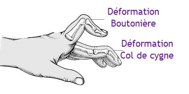
- Buttonhole deformity (PIP flexion and DIP hyperextension).
- Gooseneck deformity (PIP hyperextension and DIP flexion).
- Ulnar subluxation of the fingers at the MCP.

Rheumatoid arthritis of the spine
- The cervical spine is involved in up to 70% of patients.
- Involvement increases with more severe and long-standing disease.
- The thoracic and lumbar spine is almost never involved.
- The general pattern of rheumatoid arthritis in the cervical spine includes:
- Multi-level subluxation
- Osteopenia
- Erosions
- Odontoid
- Facet joints
- Vertebral plates
- Spinal apophyses.
- Unlike osteoarthritis, there is no bone production
Atlantoaxial subluxation in rheumatoid arthritis
- Atlantoaxial subluxation (C1 – C2) is a characteristic sign of rheumatoid arthritis.
- Atlantoaxial subluxation can occur in the following directions:
- Anterior (most common)
- Caused by inflammation of the adjacent bursa and resulting laxity of the transverse ligament.
- May not be apparent if flexion radiographs are not obtained
- Present if the Atlanta-dental interval (ADI) is > 2.5 mm (> 5 mm in children)
- Posterior
- Vertical (atlantoaxial impaction)
- Erosion and collapse of the C1 – C2 facets, leading to protrusion of the ordinate through the foramen magnum.
- May compress the midbrain.
- Rotation
- Lateral
- Anterior (most common)
- Atlanto-dental interval is the distance between the anterior aspect of the dens and the posterior aspect of the anterior ring of C1, measured at the inferior aspect of the C1 – C2 joint.


 Weakness: Causes, Implications, and Solutions")


