


Introduction
This protrusion can subsequently trigger painful bursitis, a condition characterized by inflammation of the bursa, a fluid-filled sac located between the tendon and the bone.
It was initially described in 1927 by a Swedish orthopedic surgeon Patrick Haglund.

Bursitis results from constant irritation of the bursa, which can cause inflammation, redness and tenderness in the affected area. In addition to the characteristic bony bump, Haglund’s disease can cause persistent heel pain, especially when walking or using shoes that put pressure on the affected area.
Risk factors associated with Haglund’s disease often include anatomical problems, such as retroversion of the foot or abnormal gait, as well as frequent wearing of shoes that fit poorly or have a stiff back. Activities involving repetitive foot movements, such as running, can also aggravate the condition.
Treatment for Haglund’s disease generally aims to relieve pain and reduce inflammation. Non-invasive methods, such as the use of adapted shoes, heel lifts, icing and anti-inflammatory medications, are often recommended as first line treatment. In more severe cases or when symptoms persist, treatment options such as physiotherapy or surgical procedures may be considered to reduce irritation and correct the bone abnormality.
It is crucial to consult a healthcare professional for an accurate diagnosis and a treatment plan tailored to each individual, in order to minimize the impacts of Haglund’s disease on quality of life and mobility.
What is Haglund’s Syndrome?
Definition and Key Characteristics
Haglund’s syndrome, also known as “pump bump” or “retrocalcaneal bursitis,” is a painful orthopedic condition affecting the back of the heel. It is characterized by the formation of a bony prominence, or exostosis, on the posterior aspect of the calcaneus (heel bone). This condition is often accompanied by inflammation of the Achilles tendon and the retrocalcaneal bursa, a fluid-filled sac that cushions the tendon from the heel bone.
The hallmark features of Haglund’s syndrome include:
- A noticeable, painful bump on the back of the heel.
- Redness, swelling, and tenderness around the affected area.
- Pain that worsens with walking, running, or wearing shoes with rigid backs.
- Restricted ankle mobility due to pain and inflammation.
Haglund’s syndrome is often referred to as a triad condition, encompassing:
- Haglund’s deformity: The bony protrusion on the back of the calcaneus.
- Achilles tendinitis: Inflammation of the Achilles tendon due to repetitive friction.
- Retrocalcaneal bursitis: Inflammation of the bursa between the Achilles tendon and the heel bone.
While it can affect anyone, Haglund’s syndrome is particularly common among individuals who engage in activities involving repetitive heel motion, such as running or wearing improperly fitted footwear.
Anatomy of the Heel
Understanding the anatomy of the heel is essential to grasp the underlying mechanisms of Haglund’s syndrome. The heel is a complex structure composed of bones, tendons, and soft tissues that work together to provide stability, shock absorption, and mobility.
Key components of the heel include:
- Calcaneus (Heel Bone):
- The calcaneus is the largest bone in the foot, forming the foundation of the heel. It serves as the attachment site for the Achilles tendon and various ligaments. The posterior aspect of the calcaneus is where the bony prominence in Haglund’s syndrome forms.
- Achilles Tendon:
- The Achilles tendon connects the calf muscles (gastrocnemius and soleus) to the calcaneus. It facilitates movements like walking, running, and jumping. In Haglund’s syndrome, repetitive irritation of the Achilles tendon exacerbates inflammation and pain.
- Retrocalcaneal Bursa:
- A fluid-filled sac located between the Achilles tendon and the calcaneus. Its role is to reduce friction and cushion the tendon from the bone. Chronic irritation can lead to retrocalcaneal bursitis, a key feature of Haglund’s syndrome.
- Plantar Fascia:
- Though not directly involved in Haglund’s syndrome, this connective tissue supports the arch of the foot and influences the biomechanics of the heel.
- Heel Pad:
- A thick layer of fatty tissue under the calcaneus that absorbs shock during weight-bearing activities. While it doesn’t directly contribute to Haglund’s syndrome, its integrity can influence overall heel health.
Symptoms of Haglund’s disease
Haglund’s disease has a characteristic set of symptoms that may vary in intensity from person to person, but are generally similar in presentation. These symptoms are mainly localized in the heel region and can have a significant impact on the mobility and quality of life of those affected.
One of the most common symptoms of Haglund’s disease is a painful bump on the back of the heel. This bump, often described as a “Haglund’s bump,” is a bony exostosis that forms as a result of chronic irritation of the Achilles tendon and heel bone. The bump may be visible and palpable, and it is usually painful to the touch.
Another common manifestation of Haglund’s disease is pain in the heel, especially when walking, running, or any other activity that involves flexing or stretching the foot. This pain, often described as a shooting or burning pain, may be located at the back of the heel, around Haglund’s bump, and may radiate down the foot or along the Achilles tendon.
Symptoms of Haglund’s disease may also include inflammation of the Achilles tendon, called Achilles tendinitis. This inflammation can lead to increased stiffness and pain in the heel, especially in the morning upon waking or after a prolonged period of immobility. Movements that stretch or strain the Achilles tendon, such as climbing stairs or running uphill, can make symptoms worse.
Some patients with Haglund’s disease may also experience redness or swelling around the heel area, especially if there is severe inflammation of the Achilles tendon. This swelling may be associated with increased sensitivity to touch and may limit the range of motion of the foot and ankle.
Additionally, Haglund’s disease can cause deterioration in posture and gait, as individuals often attempt to relieve pain by changing the way they walk or stand. This alteration in posture can lead to additional muscle tension in other parts of the body, such as the knees, hips and lower back, thus worsening the symptoms of the condition.
- Pain at the back of the heel: Pain is one of the characteristic symptoms of Haglund’s disease. It is usually felt at the back of the heel, where the Achilles tendon attaches to the heel bone.
- Swelling: Inflammation of the soft tissues around the heel can lead to visible or palpable swelling in the affected area.
- Redness: The skin around the heel may become red due to inflammation, especially in cases of associated retrocalcaneal bursitis.
- Sensitivity to touch: The affected area may be sensitive to touch. Light pressure or wearing shoes can trigger discomfort.
- Noticeable bony bump: Haglund’s deformity, a posterosuperior calcaneal exostosis, may be palpable on the back of the heel.
- Increased pain when using shoes: Pain may become worse when wearing shoes, especially those that have a rigid back or that put pressure on the heel area.
- Possible limitation of foot mobility: Some individuals may experience a limitation of foot mobility, particularly when dorsiflexion of the foot.
- Symptoms Worsened by Activity: Pain and discomfort may intensify during activities such as walking, running, or climbing stairs.
Causes of Haglund’s disease
Haglund’s disease, also known as “posterior heel protrusion” or “posterior calcaneal exostosis,” is a painful orthopedic condition that affects the heel region. This condition is characterized by a painful bony bump on the back of the heel, often associated with inflammation of the Achilles tendon and pain when walking or running.
One of the main causes of Haglund’s disease is the anatomy of the foot and ankle. People with a particular foot shape, such as pes cavus or a foot with an excessive tilt angle, may be more likely to develop this condition. This anatomy can cause increased pressure on the back of the heel, promoting the formation of the characteristic bony bump.
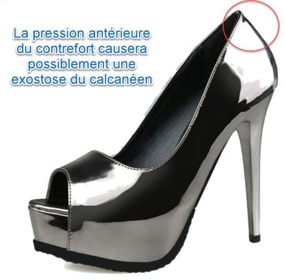
Additionally, inappropriate footwear can contribute to the development of Haglund’s disease. Shoes with a stiff heel or a design that puts excessive pressure on the back of the heel can worsen symptoms and encourage the bony bump to form. Shoes that rub or irritate the heel area can also cause inflammation of the Achilles tendon, making the condition worse.
Repetitive physical activities and repeated trauma to the heel may also play a role in the development of Haglund’s disease. Sports that involve flexion and extension movements of the foot, such as running or jumping, can put excessive pressure on the Achilles tendon and promote inflammation and bony bump formation.
Genetic factors may also contribute to predisposition to Haglund’s disease. Some people may have a genetic predisposition to developing bone abnormalities or orthopedic conditions, making them more likely to develop this condition.
Finally, factors such as age and body weight can also influence the development of Haglund’s disease. Older people, due to loss of flexibility and decreased tissue recovery capacity, may be more prone to orthopedic problems such as Haglund’s disease. Additionally, excess body weight can place additional pressure on the heel, exacerbating symptoms of the condition
- Foot Anatomy: Some individuals have foot anatomy that predisposes them to Haglund’s disease. This may include an outward tilt of the heel (retroversion) or other anatomical variations that increase pressure on the heel region.
- Abnormal Biomechanics: Biomechanical abnormalities, such as excessive pronation of the foot, can contribute to overload of the Achilles tendon and irritation of surrounding soft tissues.
- Inappropriate Footwear: Frequently wearing high-heeled shoes, especially those with a stiff back, can put constant pressure on the heel region, thereby promoting the development of Haglund’s disease.
- High Impact Activities: Participating in high impact activities, such as running, can increase stress on the Achilles tendon and contribute to tissue irritation.
- Heel Spurs: The presence of heel spurs, bony growths on the heel, may be associated with Haglund’s disease and contribute to the triad of symptoms.
- Genetic Factors: There may be a genetic component to the predisposition to Haglund’s disease, although research in this area is still ongoing.
- Overweight or Obesity: Excess weight can increase the load on the Achilles tendon and contribute to the development of Haglund’s disease.
- Predisposition to Inflammation: Some individuals have a predisposition to inflammation, which can worsen the inflammatory response in the heel area.
- Previous Trauma: Previous injuries, especially those involving the heel, can increase the risk of developing Haglund’s disease.
- Gender: Although Haglund’s disease can affect both sexes, some studies suggest it is more common in women.
Pathophysiology
The pathophysiology of Haglund’s disease involves a complex combination of anatomical, mechanical, and inflammatory factors that contribute to the formation of the characteristic bony bump and associated symptoms. This orthopedic condition typically begins with chronic irritation of the Achilles tendon and heel bone, leading to a cascade of pathological responses.
At the heart of the pathophysiology of Haglund’s disease is the particular anatomy of the heel region. In some people, the shape of the heel and Achilles tendon may be such that there is increased pressure on the back of the heel, allowing chronic irritation and inflammation to develop. This excess pressure can be exacerbated by factors such as excessive foot pronation, high heel tilt angle, or being overweight.
Chronic irritation of the Achilles tendon and heel bone can lead to a local inflammatory response, characterized by the release of pro-inflammatory cytokines and chemical mediators. This inflammation contributes to the pain, swelling, and stiffness associated with Haglund’s disease. Additionally, it can stimulate excessive bone growth, leading to the formation of the bony bump at the back of the heel, called exostosis.
The formation of this bony bump can lead to a cycle of friction and continued irritation of the Achilles tendon, worsening inflammation and pain. Repeated friction between Haglund’s hump and the Achilles tendon can also cause additional injury and microtrauma to the tendon, compromising its structure and function.
Alongside these local mechanisms, Haglund’s disease may also involve alterations in the immune system and systemic inflammatory response. Studies have suggested a genetic predisposition to inflammation and excessive bone formation in some people, which may increase their susceptibility to developing Haglund’s disease.
Finally, certain external factors, such as inappropriate shoes or repetitive physical activities, can worsen Haglund’s disease symptoms by putting additional pressure on the heel area. Shoes with a stiff heel or a design that puts excessive pressure on the back of the heel can worsen Achilles tendon irritation and encourage bony bump formation.
The pathophysiology of Haglund’s disease can be understood by examining key steps in the process. Here is a step-by-step description of the pathophysiology of this condition:
- Predisposing Anatomy: Haglund’s disease is often associated with particular foot anatomy, such as an outward tilt of the heel (retroversion) or other anatomical variations. These anatomical features may create predisposing conditions.
- Mechanical Stress: Frequent use of inappropriate shoes, especially those with a rigid back such as high heels, generates excessive mechanical stress on the heel region.
- Achilles Tendon Irritation: Shoes cause constant friction between the heel and the shoe, irritating the soft tissues near the Achilles tendon. This can trigger inflammation and irritation.
- Retrocalcaneal Bursitis: Repeated irritation can lead to retrocalcaneal bursitis, characterized by inflammation of the bursa between the Achilles tendon and the heel bone.
- Achilles tendinopathy: Persistent inflammation and repeated mechanical stress can lead to Achilles tendinopathy, marked by degenerative changes in the tendon.
- Generalized Inflammatory Response: The pathophysiology of Haglund’s disease involves a generalized inflammatory response, affecting the surrounding soft tissues, including the Achilles tendon and bursa.
- Calcaneal Exostosis Formation: In response to continued traction of the Achilles tendon, a Haglund’s deformity forms in the form of a posterosuperior calcaneal exostosis, a bony bump at the junction of the Achilles tendon and the heel.
- Cycle of Damage and Repair: Persistence of mechanical stress and inflammation creates a cycle of damage and repair, with degenerative tissue changes that can be self-perpetuating.
Haglund’s triad
Haglund’s triad, is an orthopedic condition characterized by a combination of three main clinical features: Haglund’s hump, Achilles tendinitis, and retrocalcaneal bursitis. This triad of symptoms is often associated with chronic irritation of the Achilles tendon and heel bone, leading to a cascade of inflammatory responses and significant pain in the heel region.
The first component of Haglund’s triad is Haglund’s hump, a bony exostosis that forms at the back of the heel in response to chronic irritation of the Achilles tendon and heel bone. This bump, often described as a bony protrusion, can be visible and palpable, and is usually painful to the touch. It is caused by excessive bone growth in response to persistent inflammation and irritation.
The second component of Haglund’s triad is Achilles tendonitis, an inflammation of the Achilles tendon that manifests as pain, swelling, and stiffness in the heel. This inflammation is usually caused by repeated friction between the Achilles tendon and Haglund’s hump, thus worsening the symptoms of the disease.
The third component of Haglund’s triad is retrocalcaneal bursitis, an inflammation of the bursa located between the Achilles tendon and the heel bone. This bursitis develops in response to irritation and friction between the inflamed Achilles tendon and Haglund’s hump. It can lead to additional pain and swelling in the heel area, thus worsening the symptoms of the condition.
Haglund’s triad is often associated with activities that involve repetitive movements of the foot, such as running, jumping, or walking on hard surfaces. These activities exacerbate irritation of the Achilles tendon and promote the formation of Haglund’s hump, thus leading to worsening of the symptoms of the disease.
Diagnosis of Haglund’s triad usually relies on a thorough clinical examination, as well as imaging techniques such as x-rays, ultrasound or MRI to assess the extent of lesions and anatomical changes in the region of the heel.
Diagnosis
Accurate diagnosis is the cornerstone of effective management for Haglund’s syndrome. Osteopaths play a crucial role in identifying this condition through a comprehensive approach that includes clinical examination and advanced imaging techniques. Understanding these methods ensures an accurate diagnosis and helps tailor interventions to reduce pain, improve mobility, and enhance overall well-being.
Clinical Examination
The diagnostic process for Haglund’s syndrome begins with a detailed clinical assessment. Osteopaths emphasize a holistic evaluation, considering not only the localized symptoms but also the patient’s overall biomechanics and lifestyle factors.
- Patient History
- A thorough history-taking is crucial. Patients often report a painful bump on the back of the heel, exacerbated by walking, running, or wearing rigid-backed shoes.
- Questions about footwear habits, physical activities, and prior injuries help identify contributing factors.
- Chronic conditions, such as Achilles tendinitis or retrocalcaneal bursitis, may also surface during the history.
- Physical Examination
- Palpation: Gentle pressure on the back of the heel reveals tenderness and swelling around the bony prominence.
- Visual Inspection: A visible bump, redness, or signs of inflammation are commonly observed.
- Functional Assessment: The osteopath assesses foot and ankle mobility, including dorsiflexion and plantarflexion, to identify movement restrictions or compensatory patterns.
- Gait Analysis: Subtle changes in posture or walking mechanics, often due to pain avoidance, are critical for holistic treatment planning.
- Biomechanical Evaluation
- The osteopath evaluates foot alignment, arch structure, and weight distribution. Conditions like excessive pronation or high arches (pes cavus) can predispose individuals to Haglund’s syndrome and must be addressed in treatment.
Imaging Techniques
When clinical findings suggest Haglund’s syndrome, imaging confirms the diagnosis and assesses the extent of tissue damage. Osteopaths collaborate with radiologists or other healthcare professionals to incorporate imaging results into their comprehensive care plans.
- X-rays
- X-rays are a primary tool for identifying the hallmark bony prominence (Haglund’s deformity) on the posterior calcaneus.
- They also reveal associated conditions, such as heel spurs or calcification at the Achilles tendon insertion.
- MRI (Magnetic Resonance Imaging)
- MRI is used to assess soft tissues, including the Achilles tendon and retrocalcaneal bursa.
- It helps detect inflammation, tendinitis, or partial tendon tears that may not appear on X-rays.
- Ultrasound
- Ultrasound is a dynamic, cost-effective option for evaluating the Achilles tendon and bursae in real-time.
- It identifies soft tissue swelling, fluid accumulation, and structural abnormalities.
Osteopathy’s Perspective
Osteopaths integrate clinical examination and imaging findings into a holistic framework, addressing underlying causes like biomechanical imbalances and improper footwear. This approach not only alleviates symptoms but also prevents recurrence, enhancing the patient’s overall quality of life.
Differential diagnoses
- Plantar Fasciitis: Plantar fasciitis is an inflammation of the plantar fascia, the tissue that connects the heel to the toes. It can cause heel pain similar to that of Haglund’s disease.
- Isolated Heel Spur: A heel spur, a bony growth in the heel, may be independent of Haglund’s triad and cause similar symptoms.
- Posterior Compartment Syndrome: This condition involves increased pressure in the posterior compartment of the calf, which can cause heel pain and symptoms similar to Haglund’s disease.
- Heel Arthritis: Certain types of arthritis, such as rheumatoid arthritis, can affect the joints of the foot, leading to heel pain.
- Achilles Tendinopathy: Achilles tendinopathy can manifest with similar symptoms, although Haglund’s triad is specific to this condition.
- Isolated Retrocalcaneal Bursitis: Inflammation of the retrocalcaneal bursa, without the presence of Haglund’s triad, can also cause heel pain.
- Tarsal Tunnel Syndrome: This condition involves compression of the nerves in the ankle, which can cause heel pain and similar symptoms.
- Calcaneus Stress Fracture: A stress fracture of the heel can cause painful symptoms similar to those of Haglund’s disease.
- Posterior Pregnant Syndrome: This condition is characterized by pain at the back of the ankle, often involving the Achilles tendon.
- Upper Heel Bone Spur: A distinct bony growth in the heel can cause similar symptoms without the specific triad of Haglund’s disease.
Prevention Tips for Haglund’s Syndrome: A Holistic Osteopathic Approach
Preventing Haglund’s syndrome involves addressing its root causes and minimizing factors that contribute to heel irritation and inflammation. Osteopathy emphasizes a proactive approach to prevention, focusing on proper footwear, biomechanics, and exercises that promote foot health. These tips not only reduce the risk of developing Haglund’s syndrome but also support overall musculoskeletal wellness.
Choosing the Right Footwear
Footwear plays a pivotal role in preventing Haglund’s syndrome. Ill-fitting shoes with rigid backs can exacerbate heel friction, leading to irritation of the Achilles tendon and retrocalcaneal bursa. Osteopaths recommend the following footwear tips:
- Opt for Supportive Shoes:
- Choose shoes with a soft, padded heel counter to reduce friction and pressure on the back of the heel.
- Shoes with a slightly raised heel can help decrease tension on the Achilles tendon.
- Ensure Proper Fit:
- Avoid shoes that are too tight, particularly around the heel. Tight shoes can rub against the heel bone, worsening inflammation.
- Shoes that are too loose can also cause the foot to slide, increasing irritation.
- Replace Worn-Out Shoes:
- Old or damaged shoes may lose their cushioning and support, contributing to poor biomechanics.
- Specialized Orthotics:
- Custom orthotics or heel lifts can help redistribute pressure and support proper foot alignment.
Maintaining Healthy Biomechanics
Biomechanics—the way your body moves—plays a significant role in preventing Haglund’s syndrome. Poor alignment or gait patterns can increase stress on the heel. Osteopaths focus on identifying and correcting these issues.
- Gait Analysis:
- Have your walking and running patterns evaluated to identify potential biomechanical imbalances, such as overpronation or high arches.
- Postural Awareness:
- Poor posture can affect weight distribution and increase pressure on the heels. Osteopaths can guide patients in maintaining proper alignment through personalized advice and exercises.
- Weight Management:
- Maintaining a healthy weight reduces excessive stress on the feet and heels.
- Activity Modifications:
- Avoid repetitive high-impact activities on hard surfaces. Incorporate cross-training to reduce strain on the Achilles tendon.
Treatment Options for Haglund’s Syndrome: An Osteopathic Perspective
Treating Haglund’s syndrome requires a personalized approach that addresses the underlying causes and provides long-term relief. Osteopathy emphasizes conservative and holistic measures, focusing on non-invasive treatments to improve mobility, reduce pain, and restore balance to the musculoskeletal system. However, in more severe cases, medical and surgical interventions may be necessary. Here’s a detailed look at the treatment options for Haglund’s syndrome.
Conservative Approaches
Osteopathy often begins with conservative treatments that aim to reduce inflammation and alleviate pressure on the affected area. These approaches emphasize natural healing and functional restoration.
- Rest and Ice Therapy
- Rest: Reducing weight-bearing activities allows inflamed tissues to heal. Avoid running, jumping, or wearing shoes that irritate the heel.
- Ice Therapy: Applying ice packs to the affected area for 15-20 minutes several times a day can help reduce inflammation and swelling. Osteopaths may recommend specific cryotherapy techniques to target the inflamed bursa or Achilles tendon.
- Physical Therapy
- Osteopaths use manual therapy techniques, such as soft tissue mobilization and myofascial release, to improve circulation, reduce inflammation, and alleviate pain.
- Stretching exercises for the Achilles tendon and calf muscles help reduce tension on the heel, while strengthening exercises improve foot stability and biomechanics.
- Appropriate Footwear and Orthotics
- Switching to shoes with a soft, padded heel counter and a slightly elevated heel reduces friction and strain on the Achilles tendon.
- Custom orthotics or heel lifts, often recommended by osteopaths, redistribute pressure and support proper foot alignment. These adjustments can significantly improve gait and reduce heel irritation.
Medical Interventions
When conservative measures fail to provide adequate relief, medical interventions may be considered. These treatments focus on managing inflammation and pain to facilitate healing.
- Anti-inflammatory Medications
- Nonsteroidal anti-inflammatory drugs (NSAIDs) such as ibuprofen or naproxen are commonly prescribed to reduce pain and inflammation. Osteopaths may incorporate these medications as a complementary measure to manual therapy.
- Topical anti-inflammatory gels can also target localized inflammation without systemic side effects.
- Corticosteroid Injections
- In severe cases, corticosteroid injections may be administered to reduce inflammation in the retrocalcaneal bursa or surrounding tissues.
- While effective for temporary relief, osteopaths caution against frequent injections, as they can weaken the Achilles tendon and increase the risk of rupture.
Surgical Treatments
When conservative and medical treatments fail, and the pain significantly impacts daily activities, surgical intervention may be necessary.
- Exostectomy
- This procedure involves removing the bony prominence (Haglund’s deformity) on the calcaneus to eliminate friction and irritation.
- Surgeons may also repair or debride the Achilles tendon if it has been significantly damaged.
- Bursa Removal
- If retrocalcaneal bursitis persists, the inflamed bursa may be surgically removed to alleviate pain and improve function.
- Post-Surgical Osteopathic Care
- Post-surgical rehabilitation often includes osteopathic techniques to restore mobility, reduce scar tissue formation, and improve overall biomechanics.
- Gentle manual therapy, coupled with progressive exercises, ensures a smooth recovery and reduces the risk of recurrence.
Exercises for Relief
Heel Raises

Stand behind a chair. Place both hands on the backrest to balance yourself. Place your feet hip-width apart. Lift your heels off the floor, so your weight is on your toes. Do 10 repetitions. Frequency: 2 sets of 10 to 15 repetitions. Hold the weight in your hands, if it becomes too easy.
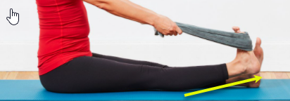
Achilles Tendon Stretching
Sit with your legs extended and your knees straight. Place a towel around your foot, just under the toes. Hold each end of the towel in each hand, with your hands above your knees. Pull the towel back so your foot stretches toward you. Hold this position for at least 15 to 30 seconds
Radiographic Features
- Calcaneal Exostosis: One of the most obvious signs of Haglund’s disease on x-rays is the presence of a calcaneal exostosis, a bony bump on the back of the heel. This growth forms at the junction of the Achilles tendon and the heel bone.
- Haglund deformity: Calcaneal exostosis is often associated with a Haglund deformity, which is a posterosuperior bony outgrowth. This deformity is generally visible on lateral radiographs.
- Heel Spur: In some cases, heel spurs may be present, which manifests as additional bony growths.
- Abnormal Joint Space: X-rays may show signs of an abnormal joint space between the calcaneus and the posterior malleolus of the fibula.
- Widening of the Bony Cover: An x-ray may reveal a widening of the bony cover at the junction between the Achilles tendon and the heel bone.
- Assessment of Alignment: Radiographs also help assess the overall alignment of the foot and ankle, which can provide additional information about the pathophysiology of Haglund’s disease.


Conclusion
In conclusion, Haglund’s disease is a complex and debilitating orthopedic condition characterized by the presence of a painful bony bump on the back of the heel, often accompanied by Achilles tendonitis and retrocalcaneal bursitis. This condition can lead to persistent pain, inflammation and impaired function of the foot, impacting the quality of life of those affected.
The pathophysiology of Haglund’s disease involves a complex interplay between anatomical, mechanical, inflammatory, and genetic factors, contributing to the formation of Haglund’s hump and the associated triad of symptoms. Inappropriate shoes, such as those with high heels, stiff heels, or those with insufficient support, can make symptoms worse by increasing pressure on the back of the heel and causing irritation of the Achilles tendon.
Early recognition of Haglund’s disease symptoms and appropriate management are key to relieving pain, reducing inflammation, and preventing long-term complications. Treatment options may include conservative measures such as rest, ice, stretching, wearing appropriate shoes, and physical therapy, as well as more invasive options like corticosteroid injections or surgery in severe cases.
Additionally, a preventative approach focused on choosing appropriate footwear and modifying activities that exacerbate symptoms can help reduce the risk of developing or worsening Haglund’s disease.
Ultimately, a thorough understanding of the pathophysiology, symptoms and risk factors associated with Haglund’s disease is essential to provide patients with optimal care and improve their quality of life. By raising public awareness of the implications of inappropriate footwear and encouraging proper footwear practices, it is possible to reduce the burden of this debilitating orthopedic condition.
References
- MRI of heel pain. Lawrence DA, Rolen MF, Morshed KA, et al. Am J Roentgenol. 2013;200:845–855. [ PubMed ] [ Google Scholar ]
- Haglund’s syndrome. Two case reports. [Article in English, Spanish] Jiménez Martín F, Alonso Valdazo MD, Díaz Peña G, Fernández Leroy J, Hernández Herrero D, Díaz García F. Rheumatol Clin. 2016:0–6. [ Google Scholar ]
- Haglund syndrome with pump bump. Kucuksen S, Karahan AY, Erol K. http://https://www.ncbi.nlm.nih.gov/pubmed/23409529 . MedArch. 2012;66:425–427. [ PubMed ] [ Google Scholar ]
- Spatial orientation of the subtalar joint axis is different in subjects with and without Achilles tendon disorders. Reule CA, Alt WW, Lohrer H, et al. Br J Sports Med. 2011;45:1029–1034. [ PubMed ] [ Google Scholar ]
- Diagnosing heel pain in adults. Aldridge T. http://www.ncbi.nlm.nih.gov/pubmed/15291091 . Am Fam Physician. 2004;70:332–338. [ PubMed ] [ Google Scholar ]
- The Haglund syndrome: initial and differential diagnosis. Pavlov H, Heneghan MA, Hersh A, et al. Radiology. 1982;144:83–88. [ PubMed ] [ Google Scholar ]
- Lu CC, Cheng YM, Fu YC, et al. Foot Ankle Int. Flight. 28. Feb: 2007. Angle analysis of Haglund syndrome and its relationship with osseous variations and Achilles tendon calcification; pp. 181–185. [ PubMed ] [ Google Scholar ]
- Diagnosis significance of radiologic measurements in posterior heel pain. Singh R, Rohilla R, Siwach RC, et al. Football. 2008;18:91–98. [ PubMed ] [ Google Scholar ]
- MRI of heel pain. Lawrence DA, Rolen MF, Morshed KA. AJR Am J Roentgenol. 2013;200:845–855. [ PubMed ] [ Google Scholar ]
- Haglund’s deformity and retrocalcaneal bursitis. Stephens MM. http://europepmc.org/abstract/med/8290230 . Orthop Clin North Am. 1994;25:41–46. [ PubMed ] [ Google Scholar ]
- Haglund’s syndrome. Sella EJ, Caminear DS, McLarney EA. J Foot Ankle Surg. 1998;37:110–114. [ PubMed ] [ Google Scholar ]
- Comparison of results of retrocalcaneal decompression for retrocalcaneal bursitis and insertional Achilles tendinosis with calcific spur. Watson AD, Anderson RB, Davis WH. http://www.ncbi.nlm.nih.gov/pubmed/10966360 . Foot Ankle Int. 2000;21:638–642. [ PubMed ] [ Google Scholar ]
- Understanding the pathologic Haglund’s deformity. Reinherz RP, Smith BA, Henning KE. http://www.ncbi.nlm.nih.gov/pubmed/2258561 . J Foot Surg. 1990;29:432–435. [ PubMed ] [ Google Scholar ]
- Results of operative treatment for recalcitrant retrocalcaneal bursitis and midportion Achilles tendinopathy in athletes. Lohrer H, Nauck T. Arch Orthop Trauma Surg. 2014;134:1073–1081. [ PubMed ] [ Google Scholar ]
- Achilles tendonitis. Clain MR, Baxter DE. http://www.ncbi.nlm.nih.gov/pubmed/1483611 . Foot Ankle. 1992;13:482–487. [ PubMed ] [ Google Scholar ]


