

Introduction to Evolutionary Anatomy
Evolutionary anatomy, is the study of anatomical structures and their evolutionary development across different species, with a particular focus on humans. This field is crucial for understanding how our skeletal structure has evolved to accommodate various functions, behaviors, and environmental challenges.
The human skeleton is a remarkable product of millions of years of evolutionary pressure. Our skeletal structure provides insight into our ancestors’ adaptations, offering clues about their way of life, movement patterns, and environmental interactions. One of the most significant aspects of human evolutionary anatomy is the transition from a tree-dwelling lifestyle to bipedalism, or walking on two legs.
Bipedalism is a defining characteristic of the human lineage, distinguishing us from our closest primate relatives. The shift to bipedalism involved profound changes in our skeletal structure. The pelvis, for instance, became shorter and broader to support an upright stance, and the lower limbs elongated relative to the arms. The curvature of the spine also changed, developing distinct curves that help balance the body’s weight over the hips and legs. These modifications are evident in fossil records, providing a timeline of evolutionary changes.
The cranial anatomy also underwent significant evolution. As hominins evolved larger brains, the shape of the skull changed to accommodate the increased cranial capacity. This involved the expansion of the cranium and a reduction in the size of the face and jaws. The position of the foramen magnum, the hole through which the spinal cord exits the skull, shifted forward, aligning the head over the spine for efficient bipedal locomotion.
Another critical area of study in evolutionary anatomy is the evolution of the hands and feet. Human hands evolved to have a longer thumb and more flexible joints, allowing for precise grip and manipulation of objects. This was a crucial adaptation for tool use, which played a significant role in our ancestors’ survival and technological development. Conversely, the feet evolved to become more rigid to support body weight during upright walking and running, with the development of arches that act as shock absorbers.
Understanding evolutionary anatomy is essential not only for tracing the development of our skeletal structure but also for appreciating how these changes have impacted our physiology and behavior. It highlights the interconnectedness of form and function and provides a framework for studying how adaptations arise in response to environmental challenges. This knowledge has practical applications in fields such as medicine, anthropology, and biomechanics, where understanding the evolutionary basis of human anatomy can inform healthcare practices, ergonomic design, and the study of human evolution.
The Evolutionary Journey: From Primates to Homo sapiens
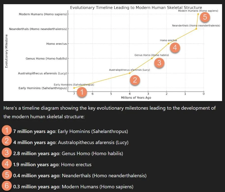
The evolutionary timeline leading to the development of the modern human skeletal structure is a fascinating journey that spans millions of years. This timeline, reveals the intricate changes that shaped our anatomy, enabling us to thrive in diverse environments.
The story begins with our early primate ancestors, who lived in trees and had skeletal structures adapted for arboreal life. These early primates had long, flexible limbs and grasping hands and feet, which allowed them to navigate the forest canopy efficiently. Around 7 to 5 million years ago, significant evolutionary shifts began to occur with the emergence of the earliest hominins.
One of the most pivotal changes in the hominin lineage was the adoption of bipedalism. Fossils of early hominins like Sahelanthropus tchadensis, Orrorin tugenensis, and Ardipithecus ramidus provide evidence of transitional adaptations for walking on two legs. These species exhibited changes in the pelvis, femur, and spine that supported bipedal locomotion, while still retaining some arboreal traits.
By around 4 million years ago, Australopithecus species, such as Australopithecus afarensis (exemplified by the famous “Lucy” fossil), showcased more advanced bipedal adaptations. Their pelvis and lower limb anatomy were more similar to modern humans, indicating a greater commitment to upright walking. However, they still possessed relatively long arms and curved fingers, suggesting they spent some time climbing trees.
The genus Homo, which emerged around 2.8 million years ago, marked a significant leap in the evolution of the human skeletal structure. Early Homo species, such as Homo habilis, had larger brains and more advanced tool-use capabilities. These changes were accompanied by further refinements in skeletal anatomy. The hands evolved for more precise manipulation, and the legs and feet became more adapted for long-distance walking and running.
Homo erectus, appearing around 1.9 million years ago, exhibited a skeletal structure even more suited for endurance running, a trait thought to be crucial for persistence hunting. Their long legs, narrow hips, and shorter arms reflected a body plan optimized for covering large distances on foot.
Neanderthals (Homo neanderthalensis), who lived from around 400,000 to 40,000 years ago, and modern Homo sapiens, who emerged around 300,000 years ago, further illustrate the diversity and specialization within the human lineage. Neanderthals had robust skeletal structures adapted to cold climates, with strong limbs and large nasal cavities for warming cold air. In contrast, modern humans evolved a more gracile frame, with a rounded skull, smaller brow ridges, and a pronounced chin.
Throughout this timeline, various environmental pressures, such as climate changes, dietary shifts, and the need for tool use, drove the anatomical adaptations observed in the fossil record. These changes not only enhanced our ancestors’ survival but also set the stage for the cultural and technological advancements that characterize human society today.
Cranial Morphology: Adaptations for Brain Size and Function
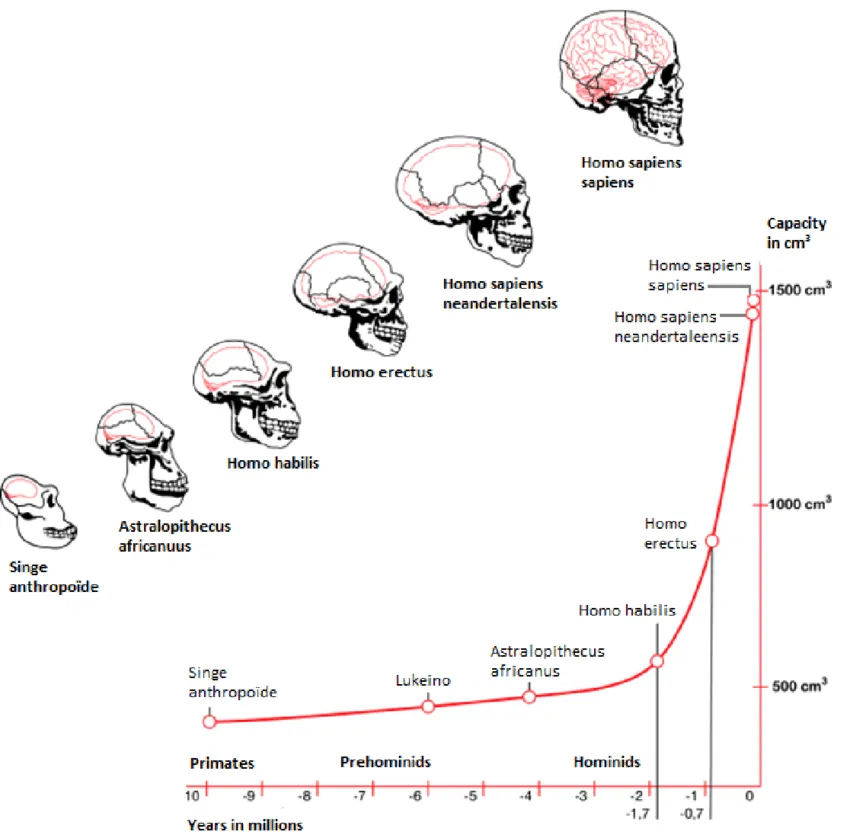
The evolution of cranial features in humans is a fascinating journey that highlights the interplay between structural adaptations and the development of advanced cognitive functions. Over millions of years, our ancestors’ cranial features underwent significant changes to accommodate larger brains, which are closely linked to enhanced cognitive abilities and complex behaviors.
One of the most notable changes in cranial features is the increase in brain size. Early hominins, such as Australopithecus afarensis (e.g., Lucy), had relatively small brains, averaging about 400-500 cubic centimeters (cc). In contrast, modern humans (Homo sapiens) have brain sizes averaging about 1350 cc. This tripling in brain volume over the course of human evolution required substantial modifications to the cranium.
The expansion of the brain led to a larger and more rounded cranial vault. Early hominins had low, elongated skulls, but as brain size increased, the cranial vault became higher and more globular. This change provided the necessary space for the growing brain. Additionally, the forehead became more vertical, a significant shift from the sloping foreheads of our ancestors. This vertical forehead is characteristic of modern humans and is associated with the frontal lobes, which are crucial for complex cognitive functions such as planning, decision-making, and social behavior.
Another significant change is the reduction in the size of the face and jaws. Early hominins had large faces with pronounced brow ridges and powerful jaws, necessary for processing a tough, fibrous diet. However, as tool use and cooking became more prevalent, the need for such robust chewing apparatus decreased. Consequently, the face became smaller and flatter, and the jaw muscles reduced in size. This reduction allowed for a more rounded cranium and provided additional space for the brain.
The evolution of the cranial base also played a crucial role. In early hominins, the foramen magnum, the hole through which the spinal cord passes, was positioned towards the back of the skull. As bipedalism evolved, the foramen magnum moved to a more central position under the skull, balancing the head on the spine and allowing for a larger braincase.
The development of a prominent chin in modern humans is another unique feature. While its exact purpose is still debated, it is believed to contribute to the structural integrity of the lower face as the jaws and teeth became smaller.
Bipedalism: Adaptations in the Pelvis and Lower Limbs
Bipedal locomotion, a defining characteristic of human evolution, required significant skeletal adaptations, particularly in the pelvis and lower limbs. These changes not only enabled efficient upright walking but also influenced other aspects of human physiology and behavior.
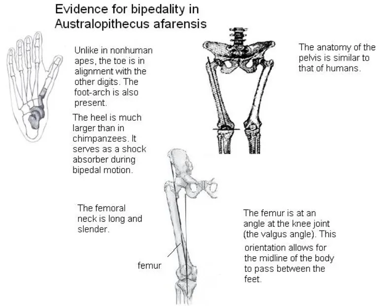
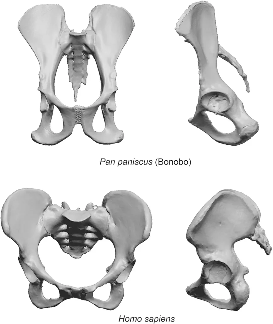
The pelvis underwent profound transformations to accommodate bipedalism. Early hominins, such as Australopithecus afarensis, exhibit the first major adaptations in pelvic structure. The pelvis became shorter and broader compared to that of quadrupedal primates. This change allowed for a more stable base of support for the upper body during upright walking. The iliac blades, which in quadrupeds are elongated and positioned more vertically, rotated to a more lateral position in bipedal hominins. This orientation provides better support for the organs and reduces stress on the lower back by distributing the weight more evenly across the pelvis.
Moreover, the sacrum, the triangular bone at the base of the spine, became wider and more robust. This adaptation further stabilized the pelvis, aiding in the efficient transfer of weight from the upper body to the lower limbs. The changes in the pelvic structure also affected the birth canal, which had to balance the demands of bipedal locomotion and the need to give birth to relatively large-brained infants.
The lower limbs also show significant adaptations for bipedalism. The femur, or thigh bone, developed a pronounced angle at the neck, bringing the knees closer together under the body’s center of gravity. This adaptation, known as the bicondylar angle, helps maintain balance during walking by minimizing lateral displacement of the body’s center of mass. In addition, the femoral head, which articulates with the pelvis, became larger and more spherical, allowing for a greater range of motion and distributing the mechanical load more effectively.
The knee joint also evolved to support bipedalism. The condyles at the lower end of the femur became more elliptical, providing better stability and shock absorption during walking. The tibia, or shin bone, developed a robust structure to support the increased weight load from the upper body.
The foot exhibits several adaptations for bipedalism as well. The arch of the foot, a unique feature among bipedal hominins, acts as a shock absorber and provides propulsion during walking. The big toe, or hallux, is aligned with the other toes, enhancing forward thrust and stability. In contrast, the big toe of quadrupedal primates is more divergent, aiding in grasping.
These skeletal adaptations for bipedalism are crucial in understanding the evolution of human locomotion. They not only facilitated efficient movement but also freed the hands for tool use and other functions, contributing to the development of complex behaviors and cultures. The transition to bipedalism represents a pivotal moment in human evolution, highlighting the intricate relationship between form and function in our lineage.
Vertebral Column: Adaptations for Upright Posture
The evolution of the vertebral column to support upright posture is a cornerstone in the development of bipedalism in humans. This transformation has profound implications for both mobility and spinal health, illustrating the intricate balance between evolutionary advantages and physiological challenges.
Early hominins, such as Australopithecus afarensis, exhibit the first signs of vertebral adaptations for bipedalism. The spine evolved to develop distinct curves—cervical, thoracic, and lumbar—that are crucial for maintaining balance and stability in an upright posture. The cervical curve in the neck and the lumbar curve in the lower back are particularly significant. These curves help distribute mechanical stress during walking and standing, acting as shock absorbers that mitigate the impact on the spine and lower limbs.
The lumbar region, in particular, shows pronounced evolutionary changes. The vertebrae in the lumbar spine became larger and more robust, supporting the increased weight of the upper body. The intervertebral discs, which act as cushions between the vertebrae, also adapted to handle greater compressive forces. Additionally, the lumbar vertebrae developed a wedged shape, with the front part being thicker than the back, which helps maintain the natural curve of the lower spine and supports upright posture.
Another critical adaptation is the change in the orientation and structure of the sacrum. In quadrupeds, the sacrum is positioned more horizontally, but in bipedal hominins, it became more vertical. This change helps in distributing the body’s weight through the pelvis to the lower limbs more effectively, enhancing stability while walking.
Despite these beneficial adaptations, the evolution of the vertebral column for bipedalism has also introduced challenges for spinal health. The S-shaped curvature, while essential for bipedal locomotion, predisposes humans to spinal issues such as herniated discs, scoliosis, and lower back pain. These conditions are less common in quadrupedal primates, whose spines are more linear and subjected to different mechanical stresses.
Lower back pain, one of the most common ailments in modern humans, is often attributed to the stresses and strains of maintaining an upright posture. The lumbar spine, bearing much of the body’s weight, is particularly vulnerable to degeneration and injury. Factors such as sedentary lifestyles, poor posture, and inadequate physical conditioning can exacerbate these issues, leading to chronic discomfort and disability.
Additionally, the cervical spine is prone to problems such as neck pain and cervical disc degeneration, which can result from the forward head posture often adopted during activities like reading, typing, or using mobile devices. This forward head posture increases the strain on the cervical vertebrae and associated muscles, leading to pain and dysfunction over time.
Limbs and Appendicular Skeleton: Tools of Mobility
The evolution of limb bones and joints has played a crucial role in facilitating human mobility and manipulation capabilities, reflecting our transition from arboreal life to bipedalism and complex tool use. This evolution showcases the intricate interplay between form and function, with significant changes in the structure of the limbs and joints that have enabled humans to adapt to diverse environments and tasks.
Upper Limbs
Shoulder Girdle and Arms: The evolution of the shoulder girdle has been instrumental in enhancing the range of motion of the arms. Early hominins, like Australopithecus afarensis, displayed shoulder joints that allowed for greater mobility, reflecting an arboreal lifestyle that required climbing and brachiation. Over time, as hominins adopted a more terrestrial lifestyle, the shoulder girdle evolved to support not just mobility but also stability, aiding in the manipulation of objects and tool use.
Hands and Fingers: One of the most significant evolutionary changes in human anatomy is the development of the hands and fingers. The increased dexterity and precision grip capabilities are attributed to the evolution of the opposable thumb. This thumb, positioned opposite the other fingers, allows for a powerful and precise grip, facilitating complex tasks such as tool making, writing, and delicate manipulations. The human hand’s flexibility and strength are crucial for survival and daily functioning.
Lower Limbs
Pelvis and Hip Joint: The pelvis underwent substantial changes to support bipedal locomotion. In early hominins, the pelvis was more bowl-shaped, providing a stable base for the upper body and supporting internal organs while walking upright. The hip joints evolved to be more robust and positioned differently, enabling efficient weight transfer and balance during bipedal movement. This evolution also helped in the stabilization required for prolonged walking and running.
Femur and Knee: The femur (thigh bone) evolved to be longer and stronger, angled inward from the hip to the knee. This inward angle, known as the valgus angle, aligns the knees under the body’s center of gravity, improving balance and walking efficiency. The knee joint itself became more specialized, with features that allow for a greater range of motion and stability, crucial for activities like running and jumping.
Feet and Ankles: The feet evolved from a grasping appendage in our primate ancestors to a structure optimized for bipedal locomotion. The development of a robust heel bone, arches, and aligned toes helped distribute body weight and absorb shock during walking and running. The big toe, in particular, evolved to be more aligned with the other toes, providing better support and propulsion during the push-off phase of walking.
Joint Adaptations
The evolution of joints has been essential in enhancing the functionality of limb bones. For instance, the ball-and-socket joint in the shoulders and hips provides a wide range of motion, while the hinge joints in the elbows and knees offer strength and stability for lifting, carrying, and mobility. The synovial joints, with their lubricating fluid, ensure smooth and pain-free movements, critical for everyday activities and complex manipulations.
Dental Evolution: Adaptations Reflecting Diet and Behavior
The evolution of dental morphology in humans reflects significant adaptations to dietary changes and behavioral shifts over millions of years. By examining the fossil record, scientists have uncovered how teeth evolved in response to varying dietary needs and environmental challenges, providing insights into the broader evolutionary story of hominins.
Early Hominins
Australopithecines: Early hominins, such as Australopithecines, exhibited dental features suited to a mixed diet. Their teeth were characterized by large, flat molars with thick enamel, ideal for grinding tough, fibrous plant material. The robust jaws and large chewing muscles supported the consumption of a varied diet, including fruits, leaves, and possibly meat. This dental morphology indicates that early hominins were generalists, capable of exploiting a wide range of food resources available in their environment.
Transition to Homo
Homo habilis and Homo erectus: As hominins transitioned to the genus Homo, significant changes in dental morphology occurred. Homo habilis, for example, had smaller molars and premolars compared to Australopithecines, with a reduction in enamel thickness. These changes suggest a shift toward a more diverse diet that included a greater proportion of meat. The development and use of tools for processing food, such as cutting and grinding, reduced the need for large teeth and powerful jaws. Homo erectus continued this trend, with further reductions in tooth size and jaw robustness. The advent of cooking and improved tool use likely played a role in these dental adaptations, as softer and more processed foods required less intensive chewing.
Anatomically Modern Humans
Homo sapiens: In anatomically modern humans, dental morphology has evolved to reflect further dietary and behavioral changes. Modern human teeth are generally smaller and less robust than those of earlier hominins. The reduction in tooth size and jaw strength corresponds with advances in food preparation techniques, such as cooking and grinding, which made food easier to chew and digest. Additionally, the advent of agriculture and the domestication of plants and animals introduced new dietary patterns, with a greater reliance on softer, processed foods like grains and cooked meats. These dietary changes reduced the selection pressure for large, powerful teeth and jaws.
Dental Adaptations and Behavior
Tool Use and Cultural Practices: The evolution of dental morphology is closely linked to the development of tool use and cultural practices. The use of tools for food processing and other activities reduced the mechanical demands placed on teeth. Cultural practices, such as cooking and the preparation of complex meals, further influenced dental evolution. These practices allowed for a greater variety of foods to be consumed, supporting dietary diversity and nutritional adequacy.
Diet and Health: Changes in dental morphology also reflect the impact of diet on overall health. The consumption of softer, processed foods reduced tooth wear and the incidence of dental diseases such as cavities and periodontal disease. However, the shift to agricultural diets high in carbohydrates and sugars increased the prevalence of dental caries in some populations. This illustrates the complex relationship between diet, dental morphology, and health outcomes in human evolution.
Social and Cultural Influences on Skeletal Evolution
The development of the human skeletal structure has been significantly influenced by social and cultural factors throughout history. These influences manifest in various ways, from dietary practices and technological advancements to lifestyle changes and cultural norms, shaping our bones and overall physical form.
Dietary Practices
Agricultural Revolution: The transition from a hunter-gatherer lifestyle to agriculture around 10,000 years ago brought significant changes to human skeletal structure. Early agricultural societies had diets heavily reliant on carbohydrates, such as wheat, rice, and maize, leading to a reduction in the robustness of jawbones and teeth. The softer, more processed agricultural foods required less intense chewing, which in turn influenced the development of smaller jaws and less pronounced dental arches.
Nutritional Deficiencies: With the advent of agriculture, there was also a marked increase in nutritional deficiencies due to a less varied diet. This often led to skeletal markers of malnutrition, such as enamel hypoplasia and signs of anemia in bones. In contrast, hunter-gatherer diets were more diverse and nutrient-rich, supporting more robust skeletal development.
Technological Advancements
Tool Use and Craftsmanship: The development and use of tools have had profound effects on human skeletal structure. Early hominins who developed stone tools had stronger hands and forearms due to the physical demands of tool-making and usage. As technology advanced, reducing the physical effort required for daily tasks, certain skeletal features, such as the robustness of the hands and arms, became less pronounced.
Sedentism and Urbanization: The move towards sedentary lifestyles with the rise of cities and civilizations also impacted skeletal development. The reduced physical activity associated with sedentism led to changes in bone density and muscle attachment sites. Urban living introduced new stresses, such as repetitive strain injuries, seen in skeletal remains from ancient civilizations.
Lifestyle and Physical Activity
Labor and Social Roles: Social roles and labor divisions have historically influenced skeletal structure. For instance, in societies where heavy physical labor was common, individuals typically had more robust bones with well-developed muscle attachment sites. Conversely, those in higher social strata with less physical labor often exhibited less pronounced skeletal robustness.
Sports and Physical Culture: In more recent history, the rise of organized sports and physical fitness culture has influenced skeletal development. Regular physical activity, especially during growth periods, contributes to stronger and denser bones. Conversely, a sedentary lifestyle, which has become more common in modern times, can lead to decreased bone density and increased susceptibility to osteoporosis.
Cultural Norms and Practices
Body Modification: Cultural practices such as cranial binding, foot binding, and corsetry have directly altered skeletal structures. Cranial binding, practiced in various cultures, reshaped the skulls of infants, leading to elongated or flattened skull shapes. Foot binding in China led to drastically altered foot bones, and corsetry in Europe caused changes to the ribcage and spine due to prolonged pressure.
Dietary Restrictions and Preferences: Cultural dietary restrictions, such as those seen in religious practices, have also shaped skeletal development. For example, vegetarian diets in certain religious groups have influenced bone health and development due to varying nutrient intakes.
Conclusion: A Legacy Written in Bone
The human body is a remarkable testament to our evolutionary journey. Every bone, every curve, tells a story of adaptation, resilience, and the relentless tinkering of natural selection. By studying evolutionary anatomy, we unlock the secrets inscribed in our very structure.
This knowledge transcends mere curiosity. It grants us a deeper understanding of ourselves, from the intricate mechanics of bipedalism to the powerful potential of our dexterous hands. It sheds light on the challenges faced by our ancestors and the ingenuity they displayed in overcoming them.
Furthermore, evolutionary anatomy informs diverse fields, and osteopathy is no exception. Osteopathic practitioners view the body as a holistic unit, where musculoskeletal imbalances can have cascading effects on overall health. Understanding how our skeletal structure evolved to function in certain ways allows osteopaths to identify these imbalances with greater precision.
For instance, knowledge of how bipedalism altered our spinal curvature helps osteopaths recognize and address misalignments that may contribute to back pain. Similarly, understanding the intricate relationship between cranial morphology and brain function can aid in treating headaches and neurological issues.
In essence, studying evolutionary anatomy is like deciphering an ancient manuscript written in bone. It’s a story of triumph over adversity, a testament to the enduring human spirit shaped by millions of years of adaptation. As we continue to unravel this narrative, we not only gain a deeper appreciation for our past but also illuminate the path towards a healthier and more fulfilling future. By incorporating this knowledge, osteopathy can offer a more targeted and effective approach to healthcare, promoting optimal well-being for all.
References
Books
- “The Body in Context: An Introduction to Evolutionary Anatomy” by David Bainbridge
- “Your Inner Beast: How Our Bodies Are Shaped by Evolution” by Robert W. Martin
- “Bones of Contention: Controversies in the Treatment of Musculoskeletal Pain” by Robert Shackleton (This book focuses on osteopathy but also touches on evolutionary anatomy)
Websites
- The Smithsonian National Museum of Natural History: https://humanorigins.si.edu/ (This website has a wealth of information on human evolution and anatomy)
- The American Museum of Natural History: https://www.amnh.org/explore/science-topics/human-origins (Another excellent resource for information on human evolution)
- The American Osteopathic Association: https://osteopathic.org/ (This website provides information on osteopathy and its principles)


