

Introduction
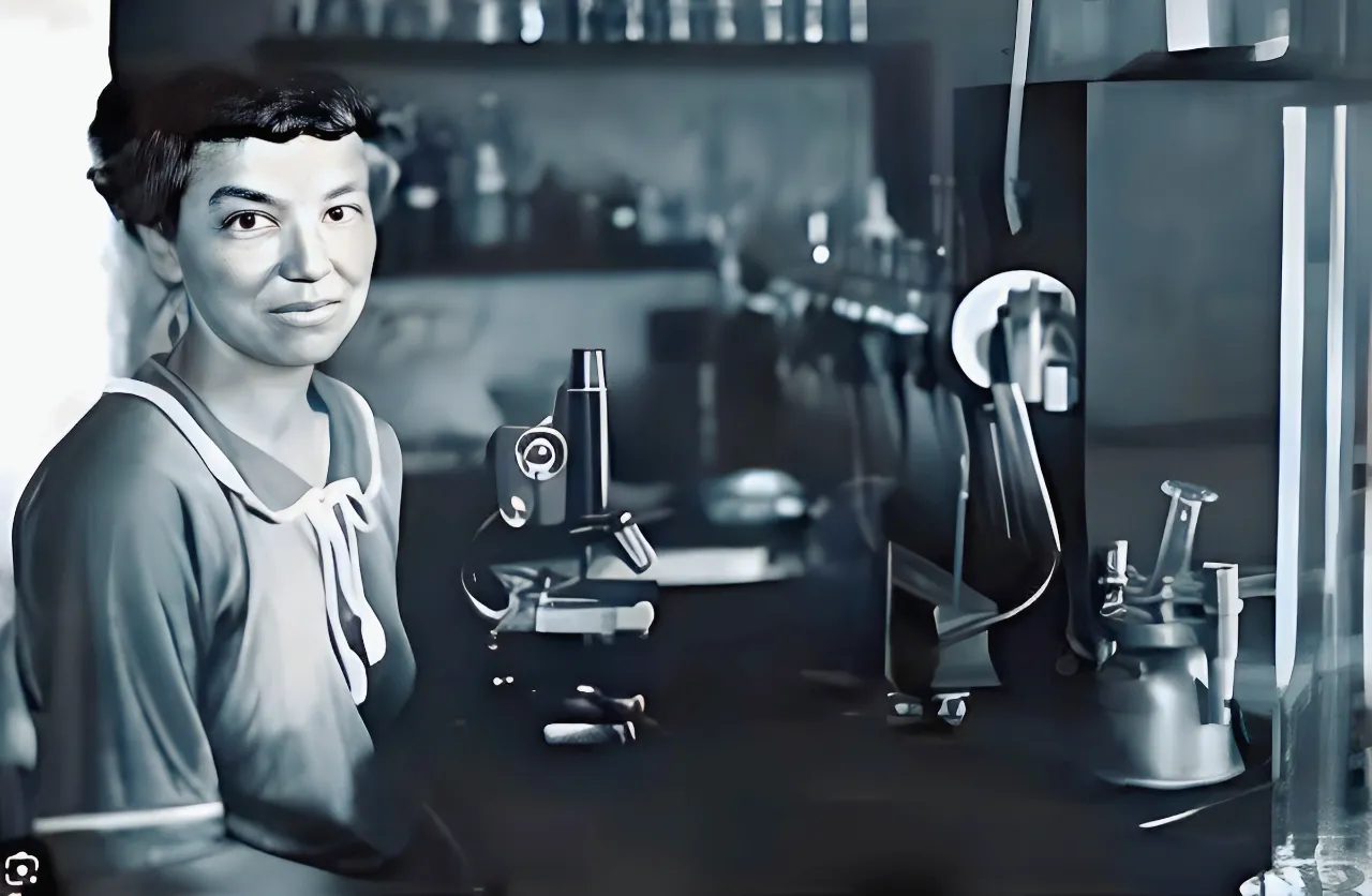
Louisa Burns (1869–1958) was a pioneering figure in the field of osteopathic medicine, making significant contributions to osteopathic research and practice in the early 20th century. A native of Ohio, United States, Burns was one of the first women to graduate from the American School of Osteopathy (now AT Still University) in 1901.
In 1907, researcher Louisa Burns began studying the mechanisms of reflex arcs in animal models to better understand the complex interactions between the viscera, spinal cord and soft tissues. His groundbreaking observations laid the foundation for understanding the interrelationship between the visceral and somatic systems, providing insights into the complex mechanisms governing the body’s response to various stimuli.
His notable research included the demonstration that electrical stimulation of the cervicx region caused muscle contractions near the lumbosacral joint. Similarly, stimulation of the cervix caused muscles near the second lumbar vertebra to contract. Additionally, electrical stimulation at the second lumbar vertebra caused regular and strong uterine contractions, accompanied by changes in uterine vessels and cervical rigidity.
Louisa Burns’ groundbreaking discoveries were not limited to reproductive physiology. She also explored the effects of electrical stimulation on tissue near the fourth dorsal vertebra, revealing an increase in pulse rate of up to 15 beats per minute. Additionally, stimulation of tissues near the fourth and fifth dorsal vertebrae caused vasoconstriction in the hands.
Louisa Burns’ work laid the foundation for understanding the interplay between the visceral and somatic systems, providing insights into the complex mechanisms governing the body’s response to various stimuli. His work has had considerable influence on the development of osteopathic principles and contributed to the evolving understanding of neuroanatomy and neurophysiology within the osteopathic profession.
As one of the first female osteopathic physicians, Louisa Burns left a lasting legacy, not only through her research, but also by breaking gender barriers in the field of osteopathic medicine. Her devotion to the advancement of osteopathy paved the way for future generations of women in the healthcare field. These observations paved the way for extensive osteopathic studies by future leaders in osteopathic education such as Wilbur Cole, DO, HV Halladay, DO, John Martin Littlejohn, MD, DO, William Smith, MD, DO, Irvin Korr , Ph.D., John Stedman Denslow, Ph.D., and William Johnston, DO, FAAO.
Key Contributions to Osteopathy
- Research on Viscero-Somatic Reflexes
Louisa Burns’ groundbreaking research established the connection between internal organs (viscera) and the musculoskeletal system, known as viscero-somatic reflexes. She demonstrated how disturbances in visceral function could manifest as musculoskeletal pain or dysfunction, laying the foundation for osteopathic principles emphasizing the body’s interconnected systems. - Impact on Reproductive Physiology
Burns conducted pioneering studies on the influence of spinal stimulation on reproductive organs. Her findings revealed that electrical stimulation of specific spinal segments could induce uterine contractions and alter cervical rigidity, highlighting the role of the nervous system in reproductive health. These discoveries contributed to the integration of osteopathy into maternal and obstetric care. - Cardiovascular Insights from Spinal Stimulation
Burns’ work extended to the cardiovascular system, where she explored the effects of spinal manipulation on heart rate and vascular tone. She observed that stimulation near the fourth dorsal vertebra increased pulse rate and caused vasoconstriction in the hands, providing early insights into the sympathetic nervous system’s role in regulating circulatory health. These findings underscored the broader systemic impact of osteopathic manipulative treatment.
The Medical Landscape of Louisa Burns’ Era: Challenges and Contributions
During the late 19th and early 20th centuries, the medical landscape was undergoing significant changes. Traditional allopathic medicine, with its focus on pharmaceutical treatments and surgical interventions, was the dominant form of healthcare. However, this period also saw the emergence of alternative medical practices, which sought to address the limitations and sometimes harmful effects of conventional medicine. Among these alternative practices, osteopathy, founded by Dr. Andrew Taylor Still in 1874, stood out as a holistic approach to healthcare that emphasized the body’s natural ability to heal itself.
Osteopathy was revolutionary in its approach, focusing on the musculoskeletal system’s role in health and disease. Dr. Still proposed that many diseases were the result of mechanical problems in the body and that manual manipulation of the musculoskeletal system could restore health. This approach was in stark contrast to the prevalent medical practices of the time, which often involved invasive procedures and the use of drugs that were not always well understood or safe.
During this era, the medical community was skeptical of osteopathy. The practice was often dismissed by mainstream physicians, who viewed it as unscientific and lacking in empirical evidence. Osteopaths were frequently marginalized, and their methods were criticized as unproven. Despite this, osteopathy began to gain a following, particularly among patients who were dissatisfied with conventional treatments.
It was within this challenging and often hostile environment that Louisa Burns emerged as a pioneering figure in osteopathy. Born in 1870, Burns became one of the first women to graduate from the American School of Osteopathy in Kirksville, Missouri, the institution founded by Dr. Still. Her entry into the field of osteopathy was itself a significant achievement, as women were largely excluded from professional medical training at the time.
Louisa Burns’ research was groundbreaking, particularly her work on viscero-somatic reflexes, which explored the connections between the internal organs (viscera) and the musculoskeletal system. Her studies provided empirical evidence that supported osteopathic principles, demonstrating that disturbances in one part of the body could manifest as problems in another, seemingly unrelated area. This concept was vital in legitimizing osteopathy as a scientific discipline.
Burns faced numerous challenges in her research. Funding was scarce, and she often had to work under difficult conditions with limited resources. Additionally, as a woman in a male-dominated field, she had to overcome significant biases and barriers to gain recognition for her work. Despite these obstacles, Burns persevered, driven by a deep commitment to advancing osteopathy and improving patient care.
Her contributions were not only scientific but also educational. Burns was a dedicated teacher who influenced many students and practitioners in the field. Her work laid the foundation for a more scientific approach to osteopathy, helping to bridge the gap between osteopathic principles and mainstream medical science.
Viscero-Somatic Reflexes
Viscerosomatic reflexes refer to reflex responses that occur at the musculoskeletal system in response to stimuli originating from internal organs (viscera). These reflexes result from the complex interaction between the autonomic nervous system, responsible for controlling internal organs, and the somatic system, responsible for musculoskeletal movement.
Precise definition of viscero-somatic reflexes
Viscero-somatic reflexes involve the transmission of sensory information from internal organs to the spinal cord, where it interacts with somatic motor neurons. This interaction results in muscular or sensory responses at the body level. These reflexes may play an important role in the regulation of visceral function and may also be involved in clinical manifestations when disrupted.
Importance in the pathological context
Viscerosomatic reflexes are significant in the pathological context, because abnormalities in these reflexes can be associated with functional or organic disorders. For example, irregularities in viscerosomatic reflexes may be seen in conditions such as gastrointestinal disorders, urological problems, or other visceral conditions.
Louisa Burns contributed to the development of concepts related to neurology. She emphasized the importance of viscero-somatic reflexes in the clinical evaluation of patients. According to Burns, abnormal viscero-somatic reflexes could be useful diagnostically by signaling neurological imbalances and providing indications of the functional health of internal organs.
Diagnostic utility of abnormal viscero-somatic reflexes according to Louisa Burns
“Somato-visceral reflexes are less circumscribed and less direct than viscero-somatic reflexes. Normal visceral activity depends in part on stimulation derived from somatosensory nerves…the possibility of recognizing abnormal viscero-somatic reflexes as an aid to diagnosis is inferred.”
Louisa Burns considered viscero-somatic reflexes to be valuable indicators in the assessment of patients. Abnormal responses in these reflexes could serve as alarm signals, indicating potential disturbances in the autonomic nervous system and visceral dysfunctions. Observation of these reflexes could point to specific areas requiring attention, allowing for more in-depth diagnosis and targeted intervention. It is important to note that interpreting these responses requires extensive clinical expertise, and professionals continue to evolve in their understanding and use of reflexes in practice.
Osteopathic manipulation and pregnancy
In 1907, Louisa Burns, DO, conducted a groundbreaking study that revolutionized our understanding of the relationship between pregnancy and musculoskeletal function. Her research delved into the intricate connections between electrical stimulation and muscular contractions in animals at various stages of pregnancy, offering valuable insights into the role of osteopathy in maternal health.
Through her meticulous experimentation, Burns discovered that electrical stimulation applied to the cervix elicited muscular contractions near the lumbosacral joint, a critical area of the spine. This finding underscored the profound interplay between neurological signals and musculoskeletal responses during pregnancy.
Furthermore, Burns observed that electrical stimulation targeted at the body of the uterus induced contractions in muscles adjacent to the second lumbar vertebra, highlighting the intricate network of nerves and muscles involved in the birthing process. This discovery shed light on the dynamic interactions between the reproductive organs and the lumbar spine, implicating osteopathic principles in maternal care.
Of particular significance was Burns’ observation that electrical stimulation applied to the second lumbar vertebra elicited regular and robust uterine contractions. This remarkable finding underscored the potential of osteopathic interventions to influence uterine activity through targeted spinal manipulation, opening new avenues for obstetric care.
Moreover, Burns documented that these uterine contractions induced by lumbar stimulation were accompanied by concurrent contraction of uterine vessels and rigidity of the cervix. Conversely, inhibition of tissues adjacent to the lumbosacral joint resulted in dilation of cervical vessels and relaxation of the cervix. These findings provided compelling evidence of the interconnectedness between spinal biomechanics and uterine physiology, highlighting the multifaceted role of osteopathy in promoting maternal well-being.
By elucidating the intricate mechanisms underlying pregnancy-related musculoskeletal changes, Burns’ research laid the foundation for integrating osteopathic principles into obstetric practice. Her pioneering work not only deepened our understanding of the physiological processes governing childbirth but also underscored the potential of osteopathic interventions to enhance maternal health outcomes.
Osteopathic manipulation and cardiovascular health
Louisa Burns’ pioneering research extended beyond the realm of pregnancy to explore the intricate connections between electrical stimulation and physiological responses in other areas of the body. Her investigations revealed fascinating insights into the effects of spinal manipulation on cardiovascular function, shedding light on the interplay between the sympathetic nervous system (SNS) and somatic dysfunction.
One notable finding from Burns’ studies was the impact of electrical stimulation near the fourth dorsal vertebra on pulse rate. She observed that such stimulation could elicit a significant increase in pulse rate, with measurements showing a rise of up to 15 beats per minute. This phenomenon highlighted the dynamic influence of spinal manipulation on cardiovascular dynamics, suggesting potential implications for managing cardiac health and function.
Furthermore, Burns documented the effects of stimulating tissues near the fourth and fifth dorsal vertebrae on the vascular system of the hands. She observed vasoconstriction in the hands following such stimulation, implicating the sympathetic nervous system in modulating peripheral vascular tone. This observation underscored the intricate relationship between spinal biomechanics and peripheral vascular regulation, offering new insights into the role of osteopathic interventions in circulatory health.
Somatic Dysfunction and Osteopathic Diagnosis
This understanding of the physiological effects of spinal manipulation led to the interpretation of a mechanism that would later be recognized as somatic dysfunction. Somatic dysfunction is defined as the disturbed or altered function of related components of the somatic system, encompassing skeletal, arthrodial, and myofascial structures, as well as associated vascular, lymphatic, and neural elements. Clinically, somatic dysfunction is identified through the TART criteria: Tissue texture changes, Asymmetry, Restriction of movement, and Tenderness (commonly assessed using counterstrain techniques).
By elucidating the physiological mechanisms underlying somatic dysfunction, Burns’ work provided clinicians with valuable diagnostic tools and therapeutic strategies for addressing musculoskeletal imbalances. The integration of her findings into osteopathic practice has enhanced our understanding of the complex interactions between spinal biomechanics, autonomic regulation, and overall health, paving the way for more effective patient care and management strategies.
Impact and Legacy: The Enduring Influence of Louisa Burns’ Research on Modern Osteopathy
Louisa Burns’ pioneering research on viscero-somatic reflexes has left a lasting legacy in the field of osteopathy, shaping modern practices and enhancing our understanding of the body’s interconnected systems. Her work provided critical scientific evidence that has influenced how osteopathic physicians diagnose and treat patients, emphasizing the importance of considering the entire body in the context of health and disease.
One of the most significant impacts of Burns’ research is the validation of the osteopathic principle that the body is an integrated whole. Her studies on viscero-somatic reflexes demonstrated that dysfunction in one part of the body, particularly the internal organs (viscera), could manifest as pain or dysfunction in the musculoskeletal system. This concept has become a cornerstone of osteopathic practice, guiding osteopaths to consider a broader range of potential causes when diagnosing and treating patients.
Application of Viscero-Somatic Reflexes in Modern Osteopathic Practice
Today, the understanding of viscero-somatic reflexes is integral to several aspects of osteopathic care. For instance, when a patient presents with chronic back pain, an osteopath might consider whether an underlying issue with an internal organ, such as the kidneys or the gastrointestinal system, could be contributing to the musculoskeletal pain. This holistic approach allows for more comprehensive care, addressing the root causes of symptoms rather than merely treating the symptoms themselves.
Another example of Burns’ influence is seen in the treatment of patients with conditions like irritable bowel syndrome (IBS). Osteopaths may employ techniques that target the musculoskeletal system, such as myofascial release or spinal manipulation, to alleviate discomfort and improve function in the digestive system. This approach is based on the understanding that the viscero-somatic reflexes can cause tension and pain in the body’s soft tissues, which, when addressed, can reduce symptoms related to the internal organs.
Burns’ research also plays a crucial role in osteopathic manipulative treatment (OMT). Techniques such as Chapman’s reflex points, which involve palpating specific areas of the body to relieve visceral dysfunction, are directly influenced by the concept of viscero-somatic reflexes. These methods are regularly used in clinical practice to treat conditions ranging from respiratory issues to gastrointestinal disorders, illustrating the enduring relevance of Burns’ work.
Educational Impact and Ongoing Research
Beyond clinical practice, Louisa Burns’ research has also had a profound impact on osteopathic education. Her findings are taught in osteopathic medical schools worldwide, ensuring that new generations of osteopaths are trained in the principles of holistic care and the importance of considering visceral contributions to musculoskeletal problems. Her work has inspired further research into the connections between the body’s systems, leading to new discoveries and innovations in osteopathic medicine.
In modern research, the principles of viscero-somatic reflexes continue to inform studies on the autonomic nervous system, chronic pain syndromes, and the role of the gut-brain axis in health and disease. Burns’ legacy is evident in the ongoing exploration of how systemic dysfunctions manifest as musculoskeletal pain and in the development of new therapeutic approaches that continue to push the boundaries of osteopathic medicine.
Historical Data vs Contemporary Validation
Louisa Burns’ work represented a major milestone in early 20th-century osteopathic research. However, as with all historical scientific contributions, her findings must be interpreted within the methodological limits of her time.
What Burns’ Historical Work Established
Based on experimental animal models and early neurophysiological techniques, Burns documented:
- Segmental relationships between visceral organs and somatic tissues.
- Reflex muscular responses following visceral or spinal stimulation.
- Cardiovascular changes associated with thoracic spinal stimulation.
- Evidence supporting bidirectional viscerosomatic and somatovisceral interactions.
Her findings provided one of the earliest experimental foundations for the concept of viscerosomatic reflexes within osteopathic medicine.
What Has Been Confirmed by Modern Research
Contemporary neurophysiology has validated several mechanisms consistent with Burns’ observations:
- Spinal afferent convergence between visceral and somatic pathways.
- Segmental facilitation, demonstrated in human subjects through electromyographic studies (Denslow).
- Spinal hyperexcitability as a recognized mechanism in central sensitization models.
- Autonomic modulation following spinal manipulation, suggested by studies measuring heart rate variability (HRV).
These findings strengthen the neurophysiological plausibility of viscerosomatic reflex mechanisms.
What Remains Partially Validated or Debated
Some aspects of early osteopathic reflex mapping remain under investigation:
- Precise organ-to-vertebral segment correlations.
- The exact magnitude and reproducibility of cardiovascular responses.
- Specific segmental control of uterine contractility.
- Quantifiable human replication of early experimental findings.
Modern research supports the general principle of neurosegmental interaction but does not always confirm highly specific one-to-one mappings described in early literature.
A Contemporary Integrative Perspective
Today, viscerosomatic reflexes are understood as part of a broader regulatory network involving:
- Spinal cord integration
- Autonomic nervous system balance
- Neural plasticity
- Central pain modulation
Modern osteopathy integrates these mechanisms within a systems-based model rather than a strictly mechanical or linear framework.
Balanced Epistemological Position
Burns’ contributions should be regarded as:
- Foundational experimental work
- Early empirical demonstration of viscerosomatic convergence
- A conceptual precursor to modern neurophysiological models
Her research opened a scientific pathway that continues to evolve, bridging manual medicine, autonomic regulation, and integrative physiology.
| Burns’ Claim | Modern Evidence | Level of Evidence |
|---|---|---|
| Viscerosomatic reflexes | Demonstrated spinal afferent convergence | High |
| Segmental facilitation | EMG findings (Denslow studies) | Moderate |
| Cardiac modulation via manipulation | Post-OMT heart rate variability (HRV) studies | Moderate |
| Segmental uterine control | Limited contemporary supporting data | Low to Moderate |
Conclusion: A Lasting Legacy and Ongoing Exploration
Louisa Burns’ pioneering research stands as a testament to the power of curiosity and exploration in the field of osteopathic medicine. Her groundbreaking work on viscero-somatic reflexes laid the foundation for a deeper understanding of the intricate connections between our internal organs and musculoskeletal system. Her investigations into the impact of spinal manipulation on cardiovascular health further broadened the scope of osteopathic practice.
Beyond her scientific contributions, Burns’ role as a trailblazing female osteopathic physician is equally significant. She challenged gender barriers and paved the way for future generations of women in healthcare.
While Burns’ discoveries provided a crucial stepping stone, the exploration of viscero-somatic reflexes and their role in osteopathic treatment continues. Modern research builds upon her work, delving further into the mechanisms of these reflexes and their potential for addressing various health conditions.
This ongoing journey of discovery underscores the dynamic nature of osteopathic medicine. As we gain a deeper understanding of the complex interactions within the body, osteopathic principles can continue to evolve, offering new avenues for promoting overall health and well-being.
References
Primary Historical Sources
Burns, L. (1907). Experimental Studies on the Physiology of the Spinal Cord. Journal of the American Osteopathic Association.
Burns, L. (1915). The Effect of Spinal Stimulation on Circulation. Journal of the American Osteopathic Association.
Burns, L. (1918). The Effect of Spinal Stimulation on Uterine Function. Journal of the American Osteopathic Association.
Development of the Concept of Segmental Facilitation
Denslow, J. S., Korr, I. M., & Krems, A. D. (1947). Quantitative Studies of Chronic Facilitation in Human Back Muscles. American Journal of Physiology.
Korr, I. M. (1975). Proprioceptors and Somatic Dysfunction: A Model of Facilitation. Journal of the American Osteopathic Association.
Modern Evidence on Autonomic Regulation
Bolton, P. S., & Budgell, B. (2006). Spinal Reflexes and Autonomic Regulation. Manual Therapy.
Cerritelli, F., et al. (2021). Osteopathic Manipulative Therapy and Autonomic Balance. Frontiers in Neuroscience.


