


Introduction
Patellofemoral syndrome brings together several conditions affecting the anterior region of the knee, including:
- Anterior Knee Pain Syndrome: A generic term encompassing pain felt in the front of the knee, often associated with activities such as running, jumping, or repetitive bending of the knee.
- Patellofemoral misalignment: A condition where the kneecap does not follow a normal path during knee movements, which can lead to abnormal rubbing and pain.
- Chondromalacial patella: A softening or deterioration of the cartilage of the kneecap, often accompanied by pain and cracking during knee movements.
- Iliotibial band syndrome: A condition in which the iliotibial band, a tendon extending from the pelvis to the shin, can rub against the outer part of the knee, causing pain.
The patella, as a sesamoid bone, sits at the front of the knee joint. It connects to the quadriceps tendon at the top and the patellar tendon at the bottom. Medial and lateral support of the patella is provided by the patellar reticulum.
During knee extension, the patella moves above the femoral condyles. In flexion, it slides under the condyles. This movement follows the longitudinal axis of the femur, reacting to the contraction of the quadriceps muscle. Traction of the patellar tendon follows the longitudinal axis of the tibia. This dynamic is influenced by the slight valgus angulation adopted by most knee joints in extension.
Anatomy of the Knee: Osteopathic Implications
The knee is a complex joint that plays a crucial role in the mobility and stability of the human body. Made up of several structures, including bones, ligaments, cartilage and muscles, the knee is subject to various conditions that can affect its function and cause pain. For osteopaths, understanding the anatomy of the knee is essential to effectively assess and treat associated musculoskeletal problems.
One of the main anatomical structures of the knee is the patellofemoral joint, where the patella articulates with the femur. Dysfunctions in this joint, such as misalignment or instability of the kneecap, can contribute to patellofemoral syndrome, a common condition characterized by pain in the front of the knee. Using osteopathic techniques such as joint manipulation and myofascial release, osteopaths can help restore alignment and mobility to the kneecap, thereby relieving associated symptoms.
In addition to the patellofemoral joint, the tibiofemoral joint between the femur and tibia is also important. Muscle imbalances, ligament strains or joint dysfunctions can lead to pain and movement limitations in this joint. Osteopaths can use joint mobilization techniques and rehabilitation exercises to restore muscle balance and improve knee mobility.
Ligaments also play a crucial role in knee stability. The medial and lateral collateral ligaments as well as the anterior and posterior cruciate ligaments stabilize the joint and limit excessive movement. Trauma, sprains or excessive strain can lead to ligament dysfunction and pain. Osteopathic techniques can help reduce ligament strain and promote healing of sprains by improving blood and lymphatic circulation around the joint.
In addition, the cartilaginous structures of the knee, such as the menisci, play an important role in shock absorption and joint load distribution. Meniscal injuries, such as tears, can cause pain and limitations in movement. Osteopaths may use myofascial release techniques and stretching to reduce tension around the menisci and promote healing.
Finally, muscles play a crucial role in the stability and movement of the knee. Muscle imbalances, tightness or weakness can contribute to knee disorders. Osteopaths often use myofascial release techniques, stretching and muscle strengthening exercises to restore muscle balance and improve knee function.
By integrating a thorough understanding of knee anatomy with specific osteopathic techniques, osteopaths can effectively evaluate and treat a variety of musculoskeletal knee disorders, seeking to restore optimal function and relieve pain.
Osteopathic Understanding of Patellofemoral Syndrome
Understanding patellofemoral syndrome from an osteopathic perspective involves an in-depth analysis of the underlying joint and muscular dysfunctions, as well as the patterns of compensation and dysfunction that manifest in this context.
At the joint level, patellofemoral syndrome is often associated with dysfunctions such as misalignments of the patella, reduced mobility of the patellofemoral joint and irregularities in the joint surfaces. These dysfunctions can lead to poor knee biomechanics, increasing the pressure placed on the patella during flexion and extension movements. Osteopathic techniques aim to restore joint alignment and mobility, which can reduce stress on the kneecap and relieve associated symptoms.
On the muscular level, patellofemoral syndrome is often linked to muscular imbalances between the different muscle groups around the knee, in particular the quadriceps, the hamstrings, the crow’s feet muscles and the stabilizing muscles of the knee. hip. These muscle imbalances can cause uneven traction on the kneecap, disrupting its normal movement and contributing to irritation and pain. Osteopathic techniques can help restore muscular balance by releasing excess tension and strengthening weakened muscles, which can reduce stress on the kneecap and promote better knee function.
Additionally, patellofemoral syndrome can lead to patterns of compensation and dysfunction throughout the body, as the individual attempts to relieve pain and maintain function. For example, changes in gait, posture, and movement patterns may develop to avoid causing increased pain in the affected knee. However, these adaptations can lead to muscle tension and imbalances in other parts of the body, which can worsen patellofemoral syndrome symptoms in the long term. Osteopaths consider these patterns of overall compensation and dysfunction when evaluating and treating patellofemoral syndrome, seeking to restore balance and coordination throughout the body to promote better function and relieve pain. .
Causes of patellofemoral syndrome
The causes of this syndrome are multifactorial and can include anatomical abnormalities, muscular imbalances, biomechanical factors and repetitive physical activities.
Anatomical abnormalities, such as a poorly positioned kneecap or irregularities in the shape of the femur, can contribute to the development of patellofemoral syndrome. For example, an overly tilted patella or shallow patellar cleft can increase pressure on the knee joint, leading to pain and associated symptoms.
Muscle imbalances also play a significant role in this condition. The quadriceps muscles, particularly the vastus medialis oblique (VMO), are essential for stabilizing the patella during movement. A weakening or imbalance in the quadriceps muscles can lead to misalignment of the kneecap, which can cause excessive friction and pain.
Biomechanical factors, such as foot and knee biomechanics, may also contribute to patellofemoral syndrome. For example, excessive pronation of the foot can cause excessive internal rotation of the tibia, which can alter the trajectory of the patella and increase forces on the knee joint.
Repetitive physical activities, especially those involving flexion and extension movements of the knee, can worsen the symptoms of patellofemoral syndrome. These activities may include running, jumping, walking uphill or downhill, and poorly performed strength training exercises.
Previous trauma to the knee, such as patella dislocations, ligament sprains, or fractures, can also increase the risk of developing patellofemoral syndrome. These injuries can lead to anatomical changes or alterations in the biomechanics of the knee, which can contribute to the appearance of symptoms.
List of causes
- Poor Patella Alignment: Abnormal alignment of the patella in relation to the femur can contribute to the syndrome.
- Abnormal Patella Tracking: Poor trajectory or tracking of the patella during knee movements.
- Weak Quadriceps: Weakness of the quadriceps muscles can lead to muscle imbalance.
- Periarticular Muscle Dysfunction: Muscle imbalances around the knee can disrupt stability.
- Ligament Hyperlaxity: Hyperlaxity of the ligaments can contribute to instability of the patella.
- Anatomical Anomalies of the Patella: Anatomical variations of the patella may be involved.
- Genetic Defects: Certain genetic predispositions can increase the risk of patellofemoral syndrome.
- Overuse of the Knee: Repetitive movements of the knee, especially with poor technique, can lead to the syndrome.
- Poor Posture: Poor body posture can influence knee mechanics.
- Biomechanical Defects: Abnormalities in the biomechanics of the knee during walking or running.
- Hormonal Changes: Hormonal changes, such as those occurring during growth, may influence the development of the syndrome in adolescents.
- Previous Trauma: Previous knee injuries, such as sprains or fractures, can increase the risk.
- Foot Defects: Foot problems, such as excessive pronation, can influence the biomechanics of the knee.
- Intense Physical Activities: Intense physical activities or repetitive movements can contribute to irritation of the kneecap.
- Ankle Instability: Ankle instability can affect the biomechanics of the knee.
- Obesity: Excess weight can increase pressure on the knees and contribute to the syndrome.
- Predisposing Genetic Factors: Predisposing genetic factors may play a role in susceptibility to the syndrome.
- Hip Strength Deficits: Deficits in hip muscle strength can affect knee stability.
- Improper Training Technique: Improper training technique can increase the risk of knee injuries.
- Repetitive Mechanical Stress: Repeated mechanical stress on the knee, especially without adequate recovery periods, can contribute to the syndrome.
Symptoms of patellofemoral syndrome
Here are some typical symptoms associated with this syndrome:
List of possible symptoms
- Pain in the front of the knee, often exacerbated by activities such as going up or down stairs, running, squatting or jumping.
- Feeling of pain or pressure under or around the kneecap, sometimes described as a dull ache or discomfort.
- Crepitus or grinding noise when moving the knee.
- Swelling of the knee in some cases.
- Feeling of weakness or instability in the knee, especially when going up or down stairs or when participating in sports activities.
- Pain in the front of the knee when sitting for long periods of time, such as when traveling by plane or car.
- Pain in the front of the knee after a prolonged period of inactivity, such as upon waking up in the morning.
- Feeling of pain or stiffness in the knee after sitting for a while with your knees bent, for example while watching a movie at the cinema.
- Sensation of pain or pressure when palpating the kneecap or its surroundings.
- Increased pain after running on a hard or inclined surface.
- Pain in the front of the knee when playing sports that involve repetitive flexion and extension movements, such as cycling or skiing.
- A feeling of “giving” or “triggering” the knee, where the knee seems to give or give way without warning.
- Pain in the front of the knee during or after going up or down stairs.
- Pain in the front of the knee when fully flexing the knee, such as when squatting.
- Difficulty bending or fully extending the knee due to pain or stiffness.
- Feeling of pain or stiffness in the knee in cold or wet weather.
- Pain in the front of the knee that is worse when sitting with your legs crossed.
- Feeling of pain or stiffness in the knee during or after a period of strenuous activity.
- Pain in the front of the knee that may radiate up the thigh or down the leg.
- Cracking or popping sensation in the knee during certain movements.
- Pain in the front of the knee when flexing or extending the leg, even without additional load.
- Difficulty getting up after sitting for a while due to knee pain or stiffness.
- Feeling of pain or tightness in the muscles around the knee, such as the quadriceps or thigh muscles.
- Pain in the front of the knee that may interfere with daily activities such as going up and down stairs or kneeling.
- Feeling of pain or discomfort in the knee that may disrupt sleep or overall quality of life.
Risk factors for having patellofemoral syndrome
List of risk factors
- Age and Growth: Patellofemoral syndrome is more common in adolescents during periods of rapid growth, when bone and muscle structures may be subject to significant changes.
- Gender: Women are generally more likely to develop patellofemoral syndrome than men. This may be related to anatomical differences, hormonal differences, and specific biomechanical patterns.
- Poor Patella Alignment: Abnormal alignment of the patella in relation to the femur, such as lateral tilt, can increase the risk of patellofemoral syndrome.
- Joint Instability: Joint instability, often due to anatomical or muscular factors, can contribute to the development of the syndrome.
- Muscle Weakness: Weakness of the muscles around the knee, particularly the quadriceps, can create muscular imbalances favoring the syndrome.
- Ligament Hyperlaxity: Hyperlaxity of the ligaments can increase excessive mobility of the patella and contribute to the syndrome.
- Intense Physical Activities: Participating in intensive physical activities or high-impact sports can increase risk, especially if the movements are repetitive.
- Biomechanical Problems: Abnormalities in the biomechanics of the knee, such as walking or running defects, can be risk factors.
- Hormonal Changes: Hormonal changes, such as those associated with pubertal growth in adolescents, may influence susceptibility.
- History of Knee Injuries: People with a history of previous knee injuries, such as sprains or fractures, may be more likely to develop the syndrome.
- Obesity: Excess weight can put increased pressure on the knees, increasing the risk of patellofemoral syndrome.
- Anatomical Abnormalities: Congenital or acquired anatomical anomalies, such as variations in the shape of the patella or femoral groove, may be risk factors.
- Family History: A family history of knee problems or patellofemoral syndrome may increase the risk.
Recommendation
- Knee Muscle in Good Condition: Strong muscles, quadriceps and hip abductors help keep the knee balanced during physical activity, but prevent deep squatting movements during weight training.
- Think alignment and technique: Flexibility and strength exercises to optimize your jumping, running and pivoting technique
- help the kneecap track properly : It is especially important that the outer hip muscles are exercised to prevent the knee from collapsing when you are squatting, landing from a jump, or dismounting. a step.
- Lose extra pounds: If you are overweight, losing weight takes the strain off your knees.
- Warm up: Before running or doing other exercises, warm up with about five minutes of light activity.
- Muscle Stretching: Promote flexibility with gentle stretching exercises.
- Increase intensity gradually: Avoid sudden changes in the intensity of your workouts.
- Use appropriate shoes: Make sure your shoes fit you well and absorb shock well. If you have flat feet, consider shoe inserts.
Osteopathic Evaluation
The assessment of patellofemoral syndrome by an osteopath involves a series of techniques aimed at identifying structural dysfunctions and soft tissue tensions that contribute to the patient’s symptoms. Here is an overview of the main evaluation techniques used:
1. Structural Assessment Techniques:
- Palpation: The osteopath uses their hands to palpate the relevant anatomical structures around the knee, such as the patella, femoral condyles, tibial plateau and ligaments. Palpation can detect any tenderness, swelling, warmth, or irregularity in these structures, which may indicate irritation or inflammation associated with patellofemoral syndrome.
- Mobility Tests: Specific tests are performed to assess knee mobility and overall joint function. This may include knee flexion, extension, rotation, and translation tests, as well as patella stability tests. These tests help identify movement restrictions and joint dysfunction that may contribute to patellofemoral syndrome symptoms.
2. Soft Tissue Assessment:
- Checking for Muscle Tension: The osteopath examines the surrounding muscles, such as the quadriceps, hamstrings, crow’s feet muscles and hip stabilizer muscles, looking for excessive muscle tension. Unbalanced muscle tension can contribute to poor knee biomechanics and increase pressure on the kneecap.
- Finding Restrictions in Fascia: Fascia is connective tissue that wraps and supports muscles, ligaments and organs. Restrictions in the thigh fascia, for example, can contribute to poor kneecap alignment and increased pressure on the patellofemoral joint.
- Evaluation of Other Joints: The osteopath may also evaluate adjacent joints, such as the hip and ankle, looking for blockages or dysfunctions that may affect the biomechanics of the knee.
Physical tests
- Watch the patient walk to see if the kneecaps are pointing toward each other (a sign of increased femoral anteversion or gluteus medius weakness).
- Check alignment.
- Look for a genu valgum (look at pronation of the foot)
- Check for atrophy of the vastus medialis oblique muscle.
- Observe the tracking of the patella as the knee moves in flexion and extension.
- Assess tissue restrictions to medial and lateral translation of the patella.
- Palpate the patella as the patient moves the knee through a range of motion to determine if crepitus is occurring and, if so, at what position.
- Hamstring muscle tension can be assessed by measuring resistance to passive knee extension with the hip flexed to 90° in the supine position.
Integration of Therapeutic Exercise
The integration of therapeutic exercise into the treatment of patellofemoral syndrome is an essential component to help restore optimal knee function and prevent relapses. Here is how therapeutic exercise can be incorporated into the osteopathic treatment plan:
- Muscle Strengthening: Exercises to strengthen the muscles around the knee, particularly the quadriceps, hamstrings, crow’s feet muscles, and hip stabilizer muscles, can help stabilize the patellofemoral joint and reduce the pressure exerted on the kneecap. Exercises such as knee extensions, hip flexions, lunges and squats may be prescribed based on the patient’s individual needs.
- Muscle Stretching: Stretching is important to maintain muscle flexibility and prevent excessive tension around the knee. Stretches targeting the quadriceps muscles, hamstrings, crow’s feet muscles, and hip muscles can help improve joint mobility and reduce pressure on the patellofemoral joint.
- Stabilization and Balance: Stabilization and balance exercises can help strengthen the stabilizing muscles of the knee joint, which is essential for maintaining good biomechanics and preventing injury. Exercises such as single leg work, side plank exercises, and balance exercises can be included in the exercise program to improve proprioception and knee stability.
- Correction of Posture and Body Mechanics: Patient education on correction of posture and body mechanics is also important. The osteopath can provide advice on how to avoid movements or positions that exacerbate the symptoms of patellofemoral syndrome, as well as strategies for improving daily posture.
- Gradual Progression: It is important that the therapeutic exercise is progressive and adapted to the patient’s individual abilities. The exercise program should be designed to allow for gradual progression in exercise intensity, duration, and complexity over time, based on the patient’s response and rehabilitation goals.
Case Studies and Treatment Examples
Here are two case studies illustrating examples of osteopathic treatment for patellofemoral syndrome, as well as the results obtained:
Case Study 1: Patient: Sarah, 28 years old, presented with chronic pain in the front of the right knee worsened by bending and climbing stairs. Assessment: Physical examination revealed tenderness to palpation of the patella, reduced mobility of the patellofemoral joint, and muscle tension in the quadriceps and crow’s feet muscles. Treatment: Osteopathic treatment included joint manipulation techniques to restore alignment and mobility of the kneecap, as well as myofascial release techniques to reduce muscle tension around the knee. Therapeutic exercises were also prescribed to strengthen the quadriceps and improve the stability of the knee joint. Results: After several treatment sessions, Sarah reported a significant reduction in pain and improvement in knee function. She was able to return to daily activities without significant limitations and maintained her progress with a home exercise program.
Case Study 2: Patient: Maxime, 35 years old, amateur football player, complaining of recurrent pain in the front of both knees during and after training. Assessment: Physical examination revealed bilateral patellar instability, hip stabilizing muscle weakness, and muscle tightness in the quadriceps and hamstrings. Treatment: Osteopathic treatment included joint mobilization techniques to improve patella stability, as well as exercises to strengthen the hip stabilizing muscles to correct muscle imbalances. Specific stretches have also been prescribed to reduce muscle tension around the knee. Results: After a few weeks of treatment and rehabilitation, Maxime noticed a significant reduction in pain and an improvement in the stability of his knees during physical activity. He was able to return to football training without experiencing significant pain and maintained his progress through a regular exercise program and continued attention to his body mechanics.
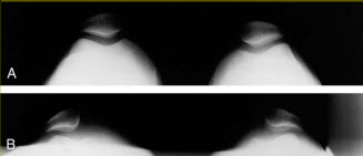
X-ray
- Tilt of the Patella: An x-ray may show an abnormal tilt of the patella relative to its normal axis.
- Abnormal Tracking: X-rays may reveal abnormal tracking of the patella during knee flexion and extension.
- Patellar Malalignment: X-rays may show malalignment of the patella in relation to the femur, which may be associated with lateral tilt or deviation.
- Erosion or Bone Changes: In some cases, bony changes, such as erosions or osteophytes, may be visible on x-rays, indicating joint wear and tear.
- Abnormal Joint Spacing: Changes in the spacing between the patella and femur may be observed.
- Lateral Displacement of the Patella: A lateral x-ray may show excessive lateral displacement of the patella.
- Joint Congruence: Assessment of joint congruence can show abnormalities in how the joint surfaces of the patella and femur align.
References
- Fulkerson JP. Diagnosis and treatment of patients with patellofemoral pain. Am J Sports Med. 2002 May-Jun;30(3):447-56. [PubMed]
- Thomeé R, Augustsson J, Karlsson J. Patellofemoral pain syndrome: a review of current issues. Sports Med. 1999 Oct;28(4):245-62. [PubMed]
- Hall R, Barber Foss K, Hewett TE, Myer GD. Sport specialization’s association with an increased risk of developing anterior knee pain in adolescent female athletes. J Sport Rehabil. 2015 Feb;24(1):31-5. [PMC free article] [PubMed]
- Witvrouw E, Callaghan MJ, Stefanik JJ, Noehren B, Bazett-Jones DM, Willson JD, Earl-Boehm JE, Davis IS, Powers CM, McConnell J, Crossley KM. Patellofemoral pain: consensus statement from the 3rd International Patellofemoral Pain Research Retreat held in Vancouver, September 2013. Br J Sports Med. 2014 Mar;48(6):411-4. [PubMed]
- Noehren B, Hamill J, Davis I. Prospective evidence for a hip etiology in patellofemoral pain. Med Sci Sports Exerc. 2013 Jun;45(6):1120-4. [PubMed]
- Herbst KA, Barber Foss KD, Fader L, Hewett TE, Witvrouw E, Stanfield D, Myer GD. Hip Strength Is Greater in Athletes Who Subsequently Develop Patellofemoral Pain. Am J Sports Med. 2015 Nov;43(11):2747-52. [PMC free article] [PubMed]
- Dye SF. The pathophysiology of patellofemoral pain: a tissue homeostasis perspective. Clin Orthop Relat Res. 2005 Jul;(436):100-10. [PubMed
- Macera CA. Lower extremity injuries in runners. Advances in prediction. Sports Med. 1992 Jan;13(1):50-7. [PubMed]
- Fairbank JC, Pynsent PB, van Poortvliet JA, Phillips H. Mechanical factors in the incidence of knee pain in adolescents and young adults. J Bone Joint Surg Br. 1984 Nov;66(5):685-93. [PubMed]
- Hart HF, Barton CJ, Khan KM, Riel H, Crossley KM. Is body mass index associated with patellofemoral pain and patellofemoral osteoarthritis? A systematic review and meta-regression and analysis. Br J Sports Med. 2017 May;51(10):781-790. [PubMed]
- DeHaven KE, Lintner DM. Athletic injuries: comparison by age, sport, and gender. Am J Sports Med. 1986 May-Jun;14(3):218-24. [PubMed]
- Taunton JE, Ryan MB, Clement DB, McKenzie DC, Lloyd-Smith DR, Zumbo BD. A retrospective case-control analysis of 2002 running injuries. Br J Sports Med. 2002 Apr;36(2):95-101. [PMC free article] [PubMed]
- Credit en partie: Jared M. Bump; Lindsay Lewis.
Summary: Symptoms include localized pain in the anterior knee, usually of in

 Weakness: Causes, Implications, and Solutions")


 injury of the knee")
 tear")