
Understanding inner knee pain and returning to manageable activity
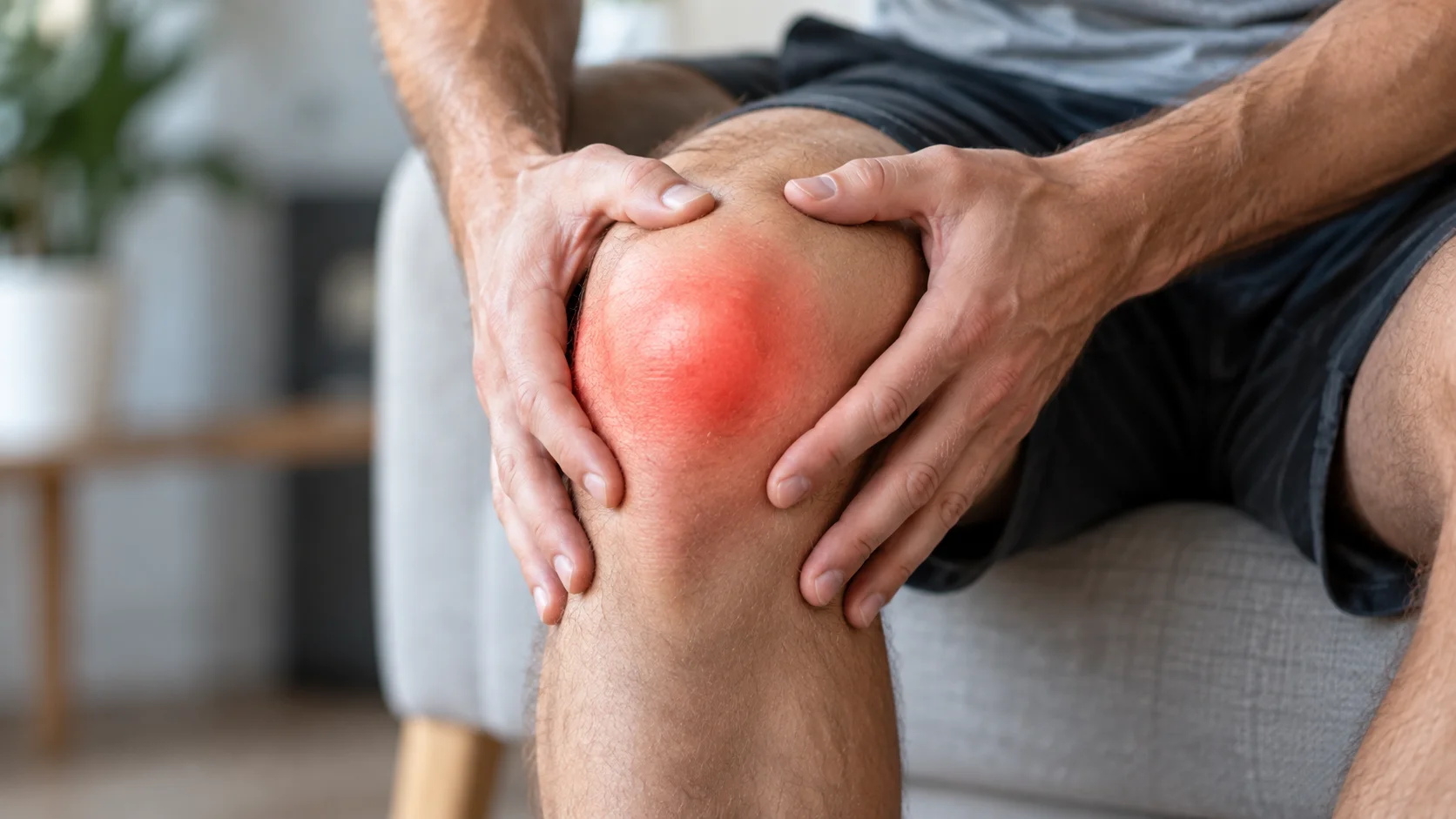
Pain on the inner side of the knee, a few centimetres below the joint, may suggest pes anserine pain syndrome. This name describes a painful region; it does not prove that a bursa is inflamed. “Pes anserine bursitis” remains a common term, but nearby tendons and tissues may also contribute to symptoms. Understanding what limits walking, stairs or sport matters more than immediately assigning a precise tissue diagnosis. [1]
Most presentations can be managed without invasive procedures. However, medial knee pain may have another origin, including the joint or a ligament. This article explains useful landmarks, situations requiring medical assessment and the principles of gradual rehabilitation. It provides information rather than a way to self-diagnose, and cannot replace an individual examination.
Anatomy: three tendons and a gliding region
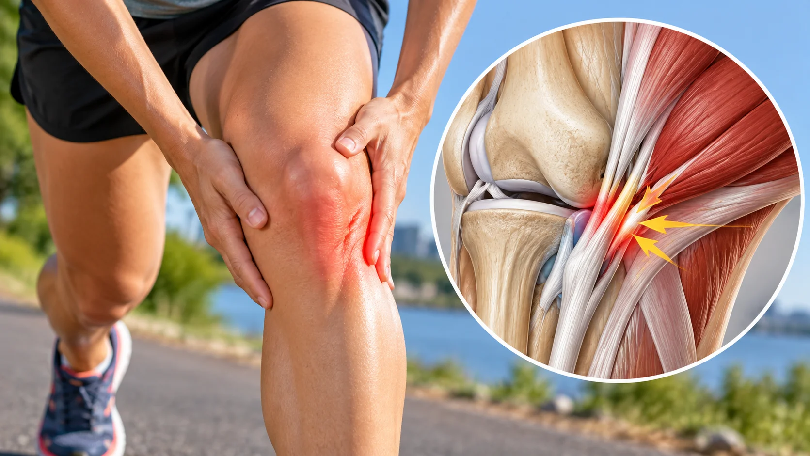
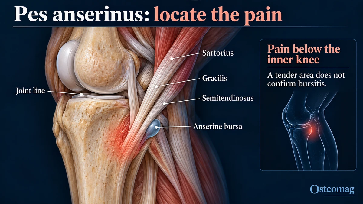
The pes anserinus consists of the sartorius, gracilis and semitendinosus tendons attaching to the anteromedial tibia. Their arrangement resembles a goose’s foot. These muscles help bend the knee and control the lower limb. The anserine bursa assists gliding relative to deeper structures, lying between the tendons and the medial collateral ligament. The tendon attachments and the boundaries of the bursa vary between individuals. [2]
The tender region is usually below the medial joint line rather than directly along it. This landmark helps guide examination, without identifying the painful tissue with certainty. Repeatedly pressing the same spot to “check” it can maintain local tenderness while adding little useful information. It is more helpful to describe where symptoms occur during ordinary movement.
Why pain may develop
A recent increase in stairs, hills, running or repetitive tasks may temporarily exceed the region’s tolerance. The whole pattern of activity deserves attention: walking at work, training, gardening and unusually demanding days all count. Overload does not necessarily imply a major injury. It mainly describes a possible mismatch between present demands and the capacity to manage them.
The syndrome may coexist with knee osteoarthritis. Ultrasound studies have found tendon or bursal changes in this population, but the findings do not always match symptoms. An association with osteoarthritis or body weight therefore cannot automatically explain an individual case. [3] Posture, foot shape or pelvic position should not be labelled the sole cause on the basis of a simple observation.
The useful question is practical: what changed before symptoms began, and which activities remain manageable? This approach avoids presenting the knee as fragile or “out of place.” It also helps identify pain that developed without a clear increase in activity and may require a broader assessment. A person’s description of the change is often more informative than a single static photograph.
Symptoms: recognising a pattern without jumping to conclusions
People often report pain below the inner knee, tenderness to touch, or discomfort on stairs and when rising from a chair. Longer walks and some bending movements may reproduce symptoms. Local swelling is possible, but its absence neither confirms nor excludes bursal involvement. The intensity can vary across the day and with different combinations of activity.
Location alone is not a sufficient test. In a 2000 study, visible bursitis was uncommon among women with a suggestive clinical presentation, most of whom had osteoarthritis. This finding highlights uncertainty; it does not mean that bursitis never occurs. [1] Another study found limited agreement between clinical classification and MRI. Imaging findings also need to be interpreted alongside symptoms. [4]
Clinical examination: assessing the knee and its function
The history covers symptom onset, possible injury, changes in activity, night symptoms and everyday difficulties. The clinician looks for swelling, unusual warmth, reduced movement or difficulty bearing weight. Tenderness below the joint is distinguished from joint-line pain, and neighbouring regions are examined when the history makes this relevant. The assessment should explain the person’s difficulties rather than simply reproduce pain.
Watching walking, a chair rise or a small step movement can help identify functional limitations. Strength assessment and selected ligament or meniscal tests depend on the history, without unnecessary repetition of painful movements. No isolated test turns a working hypothesis into certainty. [4]
In Osteomag’s Quebec context, the osteopath gathers information and recognises when referral is needed rather than making a medical diagnosis. Suspected joint injury, fracture or infection calls for assessment by an appropriately qualified healthcare professional. Palpated tension alone should not be used to explain findings that require medical investigation.
Inner knee pain: other possibilities to consider
| Possibility | Features to explore | Practical implication |
|---|---|---|
| Pes anserine pain syndrome | Tenderness below the medial joint line; load-related discomfort | Assess activities and neighbouring tissues |
| Knee osteoarthritis | Activity-related pain, stiffness and reduced movement | Address the knee as a whole |
| Meniscal involvement | Joint-line pain, twisting history or possible locking | Medical assessment for locking or persistent uncertainty |
| Medial collateral ligament injury | Trauma, pain along the ligament or instability | Assess stability and associated injuries |
| Bone involvement or referred pain | Atypical pain, difficulty bearing weight or symptoms from another region | Broaden assessment and consider targeted imaging |
| Infection or acute inflammation | A hot, red, swollen knee; possible fever | Urgent medical assessment |
These possibilities may overlap. A person with osteoarthritis may also have a tender anserine region. Care should reflect the functional problem and the consistency of findings rather than a single label. A review of knee bursae provides broader context for differentiating local pain from other conditions. [5]
Imaging: when it answers a useful question
Imaging is not automatically required for a compatible presentation without warning signs. Ultrasound can clarify fluid, tendon appearance or another superficial abnormality. It does not directly measure pain. A normal image does not make symptoms imaginary; an abnormality does not prove that it explains all of the discomfort. [3]
Radiographs are mainly useful for a bone or joint question. Typical osteoarthritis does not always require an X-ray for medical diagnosis. MRI may be considered if another lesion is suspected or the course remains unexplained. The clinician chooses the examination: the result should be capable of changing a decision, rather than merely producing another report. [6]
Warning signs and medical referral
When to seek help promptly
A very hot, red, swollen knee, especially with fever or feeling unwell, requires urgent assessment. Inability to bear weight after injury, a deformity or genuine locking that prevents movement also needs prompt evaluation. Significant acute pain should not automatically be attributed to the pes anserinus. [5]
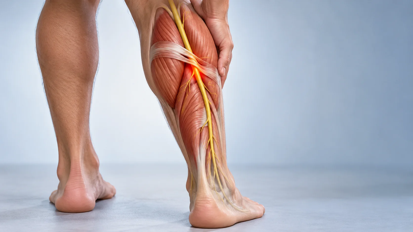
Sudden calf swelling, particularly on one side, also warrants urgent medical assessment. If it occurs with breathlessness or chest pain, call emergency services: this is outside the scope of a local knee pain problem. Do not delay assessment in order to try exercises or hands-on care. [11]
When to arrange a consultation
Pain that worsens despite activity adjustment, repeated instability, recurrent swelling or unusual night pain justifies consultation. Persistent symptoms without functional improvement should lead to a review of the working explanation and care plan. Continuing the same passive treatment indefinitely because a spot remains tender is not a useful way to assess progress.
Rehabilitation: finding a manageable load
The initial goal is to make daily activities manageable. Hills, long stair sequences or sessions causing a lasting flare can be reduced temporarily while tolerable movement continues. Prolonged complete rest is not a general strategy. A small study in people with osteoarthritis and anserine syndrome found improvement after physical treatment, without establishing a universal exercise programme. [7]
An individual starting point
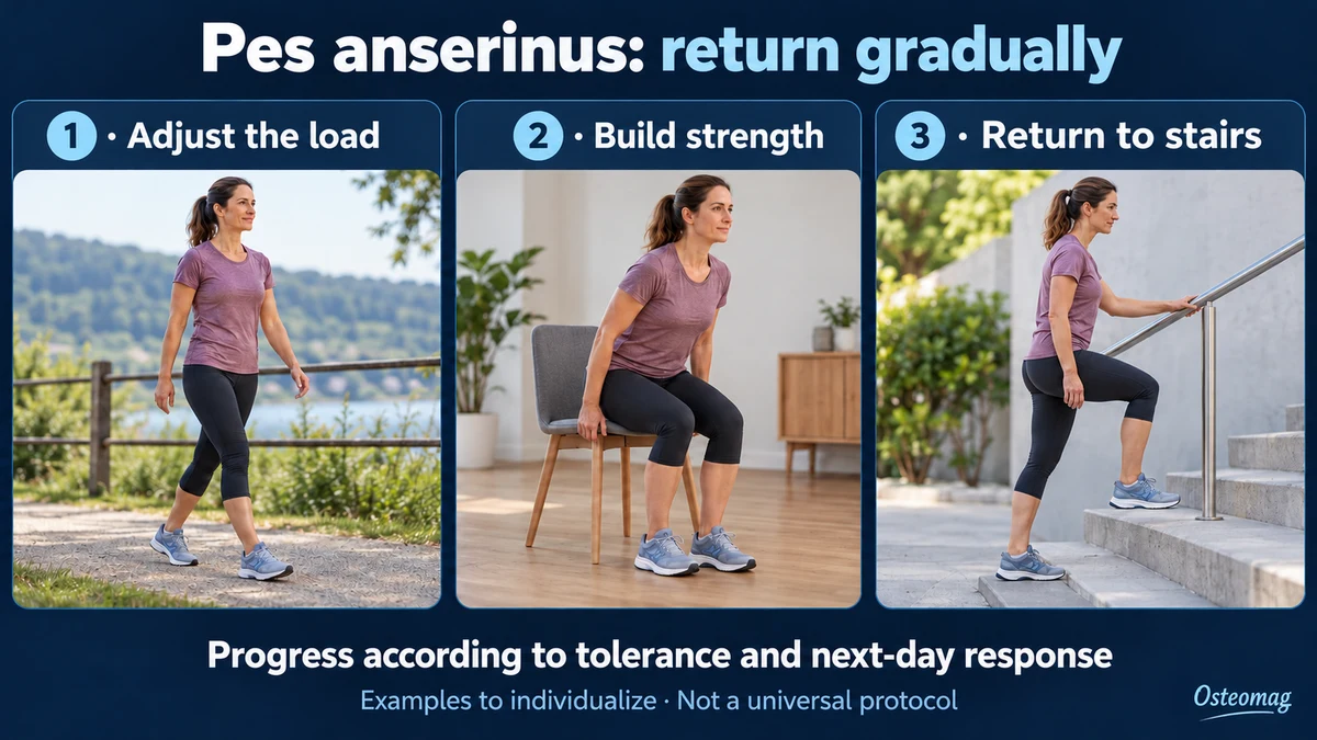
Depending on assessment, a programme may include an assisted chair rise and work for the quadriceps, hip muscles and hamstrings. Range, resistance and repetitions are chosen to allow controlled movement. An exercise causing sharp pain, locking or clear worsening is stopped and reassessed. These examples are not an identical prescription for everyone, and the starting level may need to be much easier than a demonstration suggests.
Stretching can be considered when an identifiable restriction interferes with an activity, without forcing directly through the painful region. Its value should be checked against function. A feeling of tightness does not automatically call for more intense stretching. The person should understand why an exercise is included, what it aims to improve and when to change it.
How to progress
Increase one variable at a time: walking duration, chair-rise repetitions, step height or resistance. The response during a session, later that day and the following morning helps adjust the dose. Lasting worsening or reduced walking capacity indicates that progression should be reduced and discussed. There is no numerical pain threshold validated for every anserine presentation.
A simple record can describe the activity and its consequence: “shorter walk, stairs easier afterwards” gives more useful information than constantly monitoring every sensation. The programme must fit real life. When work already places substantial demands on the knee, exercise is adjusted accordingly. Consistency is easier to maintain when the plan acknowledges those existing demands.
When osteoarthritis is also present
The strongest exercise evidence concerns knee osteoarthritis. A recent Cochrane review reports short-term improvements in pain and function, with varying certainty across comparisons. This supports active care when osteoarthritis coexists, without proving that a particular exercise directly treats the anserine bursa. [8] The programme and any support for weight management should be discussed without blame. [6]
A realistic role for osteopathic support
Osteopathic support can include discussing everyday demands, helping identify comfortable movement and encouraging adherence to active rehabilitation. Gentle soft-tissue work may be considered according to tolerance and consent. Its value should be judged against a concrete outcome: walking, standing from a chair or returning to an activity more easily. The person’s priorities guide that choice.
Trials of local treatments for anserine bursitis do not establish specific effectiveness for osteopathy. They involve selected populations and limited follow-up. [9] [10] Temporary relief does not demonstrate that the bursa has healed or that a mechanical cause has been corrected. Complementary care should encourage independence, respect warning signs and never delay necessary medical assessment.
Returning to activity and reducing recurrence
Return is guided by observable goals: usual walking without lasting aggravation, better stair tolerance and manageable strengthening exercises. For sport, duration, speed and terrain are reintroduced gradually. One good day does not immediately justify the previous training volume. Conversely, mild discomfort does not necessarily mean that another tissue injury has occurred. The overall pattern and recovery after activity matter.
There is no guaranteed recovery timeline. Symptoms, associated osteoarthritis, work demands and exercise tolerance influence the course. If progress stalls, reassess the problem rather than promise more sessions. The aim is to regain confidence in activity while retaining clear reasons to seek further assessment and a practical way to judge whether the plan is helping.
References
- [1] Uson J, et al. Pes anserinus tendino-bursitis: what are we talking about? Scand J Rheumatol. 2000;29:184–186. Source
- [2] Lee JH, et al. Pes anserinus and anserine bursa: anatomical study. Anat Cell Biol. 2014;47:127–131. Source
- [3] Toktas H, et al. Ultrasonographic assessment of pes anserinus tendon and pes anserinus tendinitis bursitis syndrome in patients with knee osteoarthritis. Mod Rheumatol. 2015;25:128–133. Source
- [4] Poor Accuracy of Clinical Diagnosis in Pes Anserine Tendinitis Bursitis Syndrome. Indian J Orthop. 2022. Source
- [5] Knee Bursae: A Comprehensive Review of Clinical Evaluation, Imaging Differentiation, and the Expanding Role of Biologic Therapies. 2025. Source
- [6] NICE. Osteoarthritis in over 16s: diagnosis and management. NG226. 2022. Source
- [7] Sarifakioglu B, et al. Comparison of the efficacy of physical therapy and corticosteroid injection in the treatment of pes anserine tendino-bursitis. J Phys Ther Sci. 2016;28:1993–1997. Source
- [8] Lawford BJ, et al. Exercise for osteoarthritis of the knee. Cochrane Database Syst Rev. 2024;12:CD004376. Source
- [9] Babaei-Ghazani A, et al. Ultrasound-Guided Pes Anserine Bursitis Injection Choices: Prolotherapy or Oxygen-Ozone or Corticosteroid: A Randomized Multicenter Clinical Trial. Am J Phys Med Rehabil. 2024;103:310–317. Source
- [10] Gouda W, et al. Comparing the Efficacy of Local Corticosteroid Injection, Platelet-Rich Plasma, and Extracorporeal Shockwave Therapy in the Treatment of Pes Anserine Bursitis: A Prospective, Randomized, Comparative Study. Adv Orthop. 2023;2023:5545520. Source
- [11] NHS. DVT (deep vein thrombosis). Reviewed 30 April 2026. Source