

Introduction
Paresthesia, often described as a “pricks and needles” sensation, is a widely known symptom although the name may escape many people. It usually occurs when prolonged pressure is placed on the nerves, such as when someone sits cross-legged for an extended period of time or falls asleep on one arm. Although paresthesia is usually harmless and resolves quickly without intervention, frequent or prolonged episodes can signal underlying problems that need medical attention.
Paresthesia is characterized by a burning, tingling, numbness, or even itching sensation, usually felt in the hands, arms, legs, or feet. However, it can also manifest itself in other parts of the body. This phenomenon is often caused by compression of the nerves, leading to a disruption in the normal transmission of sensory signals.
Most people have experienced temporary moments of paresthesia, often related to uncomfortable positions or pressure on the nerves. For example, sitting cross-legged for too long can compress nerves, causing a “pricks and needles” sensation. In these situations, paresthesia is usually painless and disappears once pressure on the nerves is released.
However, if paresthesia occurs recurrently, persists for long periods of time, or is associated with other symptoms, it is recommended to consult a healthcare professional. Indeed, paresthesia can sometimes be a sign of more serious conditions such as neurological disorders, nerve damage, circulatory problems, or underlying medical conditions such as diabetes.
Consulting an osteopath can be a beneficial step in determining the underlying cause of paresthesia. Osteopathy, as a discipline that focuses on overall health and movement of the body, can help evaluate the different influences on symptoms and offer tailored treatment approaches.
The information on this blog is for educational purposes only and not a substitute for professional medical advice. Do not attempt any maneuvers, exercises, or treatments described here without consulting a qualified healthcare professional. Improper application may lead to injury or complications. Always seek professional guidance for your specific health needs.
The Timeline of Paresthesia Discovery: A Historic Note
Ancient Foundations: Early Sensory Observations (Pre-17th Century)
The concept of abnormal sensations dates back to ancient medicine, though not specifically labeled as paresthesia. Early medical texts, such as those from Hippocrates (460–370 BCE), recognized sensory disturbances as signs of underlying imbalances in the body. Ancient Greek physicians attributed such phenomena to disruptions in the flow of humors, which were believed to regulate bodily functions. While their understanding lacked the anatomical specificity of modern science, these early observations laid the groundwork for future explorations.
Similarly, traditional Chinese medicine, as early as 1000 BCE, described abnormal sensations like tingling and numbness within the framework of qi (energy) blockages. Although these interpretations were metaphysical, they emphasized the importance of nerve-like systems in bodily health.
The Renaissance and Neurological Curiosity (17th–18th Century)
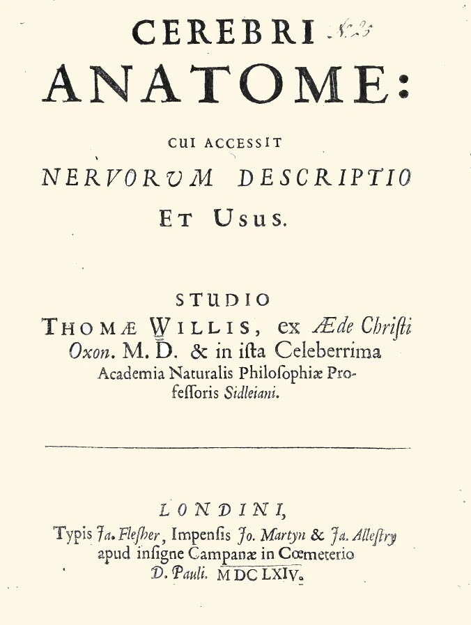
The Renaissance period marked a pivotal shift in the understanding of the human body, laying the groundwork for scientific inquiry into the nervous system. During the 1600s, Thomas Willis, widely regarded as the father of neuroscience, made significant strides in this area. In his landmark work, Cerebri Anatome (1664), Willis meticulously detailed the structure and function of the brain and nervous system, emphasizing their central role in sensation and movement. Although Willis did not explicitly discuss paresthesia, his groundbreaking descriptions of nerve function provided a conceptual framework that would shape centuries of research into sensory disturbances.
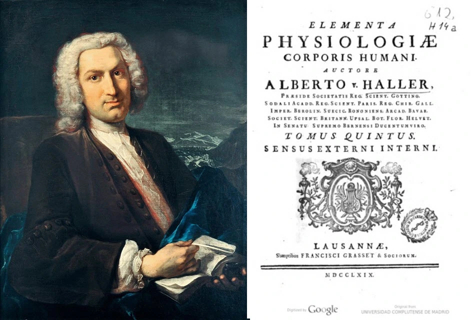
By the 18th century, the study of the nervous system advanced further through the work of pioneering anatomists such as Albrecht von Haller, whose contributions to physiology were transformative. Haller identified sensory nerves as the conduits for transmitting signals from peripheral receptors to the brain, establishing a foundational understanding of how sensations are perceived. His experiments demonstrated the link between nerve stimulation and sensory responses, offering insights into how disruptions in this delicate communication network could result in abnormal sensations like tingling or numbness—what we now recognize as paresthesia.
Through precise experiments, Haller demonstrated that nerves were not passive conduits but active participants in transmitting sensory and motor signals. His findings underscored the connection between the structural integrity of nerves and their functional output, offering early insights into how nerve injury, compression, or inflammation could give rise to sensory disturbances. By bridging the gap between anatomy and physiology, he laid the groundwork for the modern understanding of neurological disorders.
Haller’s legacy endures as one of the founding figures of experimental physiology. His contributions to understanding paresthesia and related conditions have influenced generations of neurologists and physiologists, making his work a cornerstone in the study of the human nervous system.
Caption: Albrecht von Haller’s Elementa Physiologiae revolutionized the understanding of nerve function, paving the way for the study of paresthesia.
These early investigations into the nervous system laid the scientific groundwork for understanding sensory disorders. The detailed anatomical studies of Willis and Haller inspired generations of researchers to explore the relationship between nerve function and sensory anomalies. This cumulative knowledge ultimately paved the way for 19th- and 20th-century neurologists, such as James Paget, Pierre Marie, and Charles Foix, to elucidate the mechanisms underlying paresthesia and other sensory conditions, advancing both diagnostics and treatment.
These contributions illustrate the profound impact of early scientific inquiry into the nervous system and its role in shaping modern neuroscience and clinical neurology.
19th Century: Clinical Recognition of Sensory Disturbances
1854: Sir James Paget and Nerve Compression
The first detailed clinical descriptions of paresthesia-like symptoms began to emerge in the mid-19th century, coinciding with advancements in medical science and the meticulous documentation of sensory disturbances. Among the pioneers was Sir James Paget, a distinguished British surgeon and pathologist, whose keen observations laid the groundwork for understanding the relationship between nerve compression and sensory abnormalities.
Paget documented cases of median nerve compression at the wrist, a condition that caused patients to experience numbness, tingling, and discomfort in their hands and fingers. These symptoms, now recognized as part of carpal tunnel syndrome, represented one of the earliest clinical connections between localized nerve compression and the abnormal sensations we now describe as paresthesia. Paget’s detailed accounts were instrumental in illustrating how structural and mechanical issues, such as repetitive strain or trauma to specific nerve pathways, could manifest as sensory disruptions.
His work not only advanced the understanding of neurological disorders but also underscored the importance of anatomical precision in diagnosing and treating conditions associated with paresthesia. By linking clinical observations with underlying anatomical causes, Paget set the stage for future discoveries in neurology and orthopedics.
Today, his contributions are celebrated as foundational in modern medicine, highlighting the importance of early recognition and intervention in conditions involving nerve compression. Paget’s legacy continues to influence how clinicians approach paresthesia, ensuring better outcomes for patients experiencing these often-debilitating symptoms. His work remains a testament to the value of detailed observation and its enduring impact on medical science.

Sir James Paget, one of the most influential figures in 19th-century medicine, is celebrated for his groundbreaking work in pathology and clinical observation. While best known for identifying Paget’s disease of bone and Paget’s disease of the breast, his keen observational skills also extended to neurological phenomena, including paresthesia.
Paresthesia, the abnormal sensation of tingling or “pins and needles,” often intrigued Paget as it could indicate underlying systemic or neurological disorders. He recognized that such sensations, while seemingly innocuous, were frequently early indicators of nerve compression, vascular dysfunction, or systemic illnesses. His writings emphasized the importance of linking clinical symptoms like paresthesia to their anatomical and physiological origins, which helped establish a framework for diagnosing neurological disorders.
Paget’s insistence on thorough clinical examination and the correlation of symptoms with pathological findings advanced the understanding of nerve-related sensory disruptions. His work laid the foundation for later neurologists, such as Pierre Marie and Charles Foix, who expanded on the interplay between anatomy and sensory disturbances.
James Paget’s holistic approach to medicine reminds us of the importance of careful observation in clinical practice. His contributions continue to inspire physicians and researchers in unraveling the complexities of conditions like paresthesia, ensuring his legacy endures in modern neurology.
Late 19th Century: Introduction of the Term “Paresthesia”
The term “paresthesia” The term “paresthesia” began gaining traction in medical literature during the late 19th century, marking a significant step forward in the field of neurology. Derived from the Greek words “para” (beside or abnormal) and “aisthesis” (sensation), the term was used to describe abnormal sensations, such as tingling, numbness, or a “pins and needles” feeling, often occurring without an identifiable physical stimulus. This nuanced terminology allowed physicians to categorize and discuss sensory disturbances with greater specificity, facilitating advances in clinical observation and diagnosis.
The Oxford English Dictionary cites early uses of “paresthesia” in medical texts from this period, underscoring the evolving need for a precise lexicon in the burgeoning field of neurology. As understanding of the nervous system expanded, terms like paresthesia became essential for describing phenomena that were previously misunderstood or dismissed. These abnormal sensations were often recognized as symptoms of underlying issues, such as nerve compression, vascular insufficiencies, or systemic neurological conditions.began appearing in medical literature during the late 19th century. Derived from the Greek words “para” (beside or abnormal) and “aisthesis” (sensation), it was used to describe abnormal sensations without clear physical stimuli. The Oxford English Dictionary cites early uses of the term in medical texts from this era, reflecting the growing need for precise language in neurology.
Early 20th Century: Advancements in Diagnosis and Anatomy
1913: Pierre Marie and Charles Foix’s Anatomical Discoveries
French neurologists Pierre Marie and Charles Foix made remarkable strides in advancing the understanding of paresthesia and its anatomical basis, significantly shaping modern neurology. Through their detailed and methodical postmortem examinations, they uncovered the critical role of the carpal ligament in compressing the median nerve, shedding light on a fundamental mechanism behind sensory disturbances. Their research highlighted how structural abnormalities, such as nerve compression, could manifest in symptoms like tingling, numbness, and pain, thereby providing a clearer picture of conditions like carpal tunnel syndrome.

Pierre Marie and Charles Foix, renowned pioneers in neurology, revolutionized the understanding of paresthesia—commonly experienced as tingling, numbness, or “pins and needles.” Their groundbreaking work bridged clinical observations and anatomical studies, providing insights that continue to shape modern medical practices.
In the early 20th century, paresthesia was often dismissed as an unexplained phenomenon. Marie and Foix, however, recognized it as a critical clue to underlying neurological dysfunctions. Their meticulous mapping of the nervous system and its vascular supply shed light on how disruptions in blood flow or nerve compression could result in sensory abnormalities. Through their work, they demonstrated how localized sensations, like paresthesia, often originated from broader structural or mechanical issues, such as nerve entrapments or vascular insufficiencies.
By correlating symptoms with specific anatomical lesions, they established a framework for diagnosing and managing paresthesia and similar neuropathic conditions. Their research also highlighted the importance of the spinal cord and peripheral nerves in sensory processing, paving the way for the development of diagnostic tools like electromyography and advanced imaging techniques.
Marie and Foix’s contributions extended beyond neurology, inspiring interdisciplinary approaches to healthcare. Their legacy underscores the importance of integrating clinical and anatomical insights to address complex conditions. Today, their work remains a cornerstone in understanding paresthesia, benefiting countless patients and advancing neurological research worldwide.
Through their visionary work, Marie and Foix transformed paresthesia from a medical mystery into a gateway for modern neurological exploration.re Marie and Charles Foix’s Anatomical Disco
Marie and Foix’s work was pioneering for its time, as it bridged the gap between clinical symptoms and their anatomical origins. By focusing on the interplay between the nervous system and surrounding tissues, they demonstrated how seemingly localized sensory disruptions often stemmed from broader mechanical or structural issues. This connection between anatomy and neurology helped lay the groundwork for understanding other neuropathic conditions and set the stage for future diagnostic techniques, including electromyography and imaging studies.
Moreover, their findings carried significant implications for both treatment and prevention. Recognizing the carpal ligament as a site of median nerve compression provided a basis for developing targeted interventions, such as surgical decompression and ergonomic adjustments, to alleviate symptoms. By addressing the root cause rather than just the symptoms, their work offered a blueprint for a more holistic approach to managing sensory disorders.
Marie and Foix’s contributions extend beyond their specific discoveries; they exemplify the value of integrating careful anatomical analysis with clinical inquiry. Their legacy continues to inspire neurologists, anatomists, and healthcare practitioners in their pursuit of understanding and treating the complex interplay between structure and function in the human body.
1930s–1950s: Progress in Neurology
The early 20th century marked a significant period of innovation and progress in the field of neurological diagnostics, as researchers sought to better understand the complexities of the nervous system. One of the most groundbreaking developments during this time was the introduction of electrodiagnostic studies in the 1930s. These techniques allowed scientists and clinicians to measure nerve conduction velocities, providing valuable insights into how nerves transmitted electrical signals and where disruptions in this process occurred.
Electrodiagnostic studies, such as nerve conduction studies (NCS) and electromyography (EMG), offered a direct method to evaluate the functional integrity of peripheral nerves and muscles. By stimulating nerves and recording their responses, these tools enabled the identification of abnormalities in nerve function with unprecedented precision. This advancement was particularly critical in diagnosing conditions that cause paresthesia, such as peripheral neuropathy, carpal tunnel syndrome, and radiculopathies.

This historical photograph features an early electromyograph (EMG) in use during the 1930s, marking a transformative era in the study and diagnosis of neurological conditions, including paresthesia. The electromyograph was a revolutionary invention, designed to measure electrical activity in muscles and nerves, providing invaluable insights into the functioning of the peripheral nervous system.
By recording the electrical signals generated during muscle contractions and nerve stimulations, the EMG enabled clinicians to detect abnormalities in nerve conduction and muscle responses. This technology was particularly instrumental in identifying the causes of sensory disturbances like paresthesia, which manifest as tingling, numbness, or other unusual sensations. For example, it allowed researchers to pinpoint nerve compression, such as in carpal tunnel syndrome, or detect systemic issues like diabetic neuropathy.
The device, though rudimentary compared to modern standards, was groundbreaking for its time. It allowed for objective and reproducible measurements, moving the field of neurology beyond subjective patient reports and basic clinical examinations. This innovation set the foundation for the advanced electrodiagnostic tools used today to assess nerve function and diagnose a wide range of neurological disorders.
This image captures the spirit of innovation and scientific curiosity that defined the early 20th century, highlighting how technological advancements like the electromyograph shaped our understanding of the nervous system and paved the way for modern diagnostic and therapeutic approaches in neurology.
For instance, in patients with carpal tunnel syndrome, electrodiagnostic tests could detect delayed signal transmission through the median nerve, confirming the presence and severity of nerve compression. Similarly, in cases of peripheral neuropathy, these studies could reveal patterns of nerve damage, such as slowed conduction or loss of signal, often associated with systemic conditions like diabetes or vitamin deficiencies.
The advent of these techniques significantly accelerated the understanding of nerve functions and dysfunctions. Clinicians were no longer limited to relying solely on physical examinations and patient-reported symptoms; they now had objective, quantifiable data to guide their diagnoses and treatment plans. These tools also contributed to the development of new treatment strategies, as a deeper understanding of nerve pathophysiology emerged.
By the mid-20th century, electrodiagnostic studies had become an essential component of neurological practice, paving the way for more sophisticated diagnostic and therapeutic approaches in the management of sensory disturbances like paresthesia. This period marked the beginning of an era in which technological innovation transformed the landscape of neurology and clinical care.
Mid-20th Century: Paresthesia in Clinical Focus
1950s–1960s: Dr. George S. Phalen’s Contributions
Dr. George S. Phalen of the Cleveland Clinic made groundbreaking contributions to the understanding of carpal tunnel syndrome. During the mid-20th century, Dr. Phalen conducted extensive research to explore the mechanisms behind this condition, which was becoming increasingly recognized as a prevalent issue among workers performing repetitive tasks. His meticulous studies revealed the critical role of repetitive stress and the anatomical narrowing of the carpal tunnel, a small passage in the wrist through which the median nerve travels.

This image features Dr. George S. Phalen, an esteemed physician and researcher whose groundbreaking work in the mid-20th century revolutionized the understanding and treatment of carpal tunnel syndrome, a leading cause of paresthesia in the hands. His clinical research at the Cleveland Clinic during the 1950s and 1960s was instrumental in linking repetitive stress and anatomical narrowing of the carpal tunnel to compression of the median nerve. These findings significantly advanced the medical community’s understanding of nerve compression syndromes and their associated symptoms, such as tingling, numbness, and hand weakness.
Dr. Phalen’s legacy is perhaps most closely tied to the development of the “Phalen’s Maneuver,” a diagnostic test that remains widely used today to identify carpal tunnel syndrome. This simple, yet effective, technique involves wrist flexion to reproduce characteristic symptoms, offering a quick and non-invasive method of diagnosis.
In addition to his contributions to diagnostic methodologies, Dr. Phalen’s research provided a strong foundation for modern surgical treatments, including carpal tunnel release surgery. These procedures, based on his pioneering insights, have helped millions of patients worldwide regain functionality and alleviate discomfort caused by nerve compression.
Beyond his specific contributions to carpal tunnel syndrome, Dr. Phalen’s broader research emphasized the importance of early diagnosis and comprehensive management of sensory and motor dysfunctions. His work continues to influence neurology, orthopedics, and rehabilitative medicine, cementing his place as a trailblazer in medical science. This portrait of Dr. Phalen captures the resolve and intellect of a visionary who left an indelible mark on the field of nerve compression research and treatment.
Phalen demonstrated that prolonged or repetitive pressure on the median nerve within this confined space could lead to a cascade of symptoms, including tingling, numbness, and weakness in the hands. These sensory disturbances, now understood as classic signs of carpal tunnel syndrome, were shown to arise from chronic nerve compression and inflammation, often exacerbated by occupational or lifestyle factors.
One of Dr. Phalen\u2019s most notable contributions was his development of the \u201cPhalen\u2019s Maneuver,\u201d a diagnostic test still widely used today. This simple but effective method involves flexing the wrist for a specified period to reproduce the symptoms of carpal tunnel syndrome, providing a practical tool for identifying the condition. By correlating clinical findings with his anatomical research, Phalen established a clear understanding of the condition\u2019s pathology, bridging the gap between symptoms and their underlying causes.
Phalen\u2019s work also laid the foundation for modern diagnostic imaging and electrodiagnostic studies, which have since refined the ability to pinpoint nerve compression and assess its severity. Moreover, his research informed the development of surgical interventions, such as carpal tunnel release surgery, designed to alleviate pressure on the median nerve by enlarging the carpal tunnel. These procedures, based on Phalen\u2019s pioneering insights, have become standard treatments for severe cases of the condition.
Beyond his direct contributions to carpal tunnel syndrome, Phalen\u2019s research emphasized the broader importance of understanding nerve compression syndromes and their systemic impact on sensory and motor function. His findings underscored the significance of early diagnosis and intervention, paving the way for more effective prevention and management strategies in clinical practice. Today, Dr. Phalen is remembered as a pivotal figure in neurology and orthopedics, whose work continues to influence the care of patients experiencing paresthesia and related conditions.
Modern Era: Comprehensive Understanding and Interdisciplinary Approaches (1970s–Present)
1970s: Advances in Neuroimaging
The advent of advanced imaging technologies, such as magnetic resonance imaging (MRI) and computed tomography (CT), marked a significant breakthrough in the diagnosis and understanding of paresthesia-related conditions. Prior to their development, clinicians often relied on clinical examinations and subjective reports from patients, which, while valuable, could not always pinpoint the exact cause of sensory disturbances. MRI and CT imaging changed this dynamic by offering detailed, non-invasive insights into the internal structures of the body.
MRI, with its ability to generate high-resolution images of soft tissues, allowed physicians to visualize nerve pathways, detect nerve compression, and identify issues such as herniated discs or spinal cord abnormalities that might contribute to paresthesia. This capability was particularly valuable in diagnosing conditions like sciatica, carpal tunnel syndrome, and cervical radiculopathy, where nerve impingement is a primary concern.
Similarly, CT scans provided a complementary perspective, excelling in imaging bones and detecting structural issues, such as fractures, that could indirectly affect nerve function. CT scans were also instrumental in identifying subtle anatomical variations or trauma that might contribute to chronic sensory disturbances.
These imaging tools not only enhanced diagnostic accuracy but also enabled earlier detection of conditions that might otherwise progress unnoticed. Furthermore, they provided critical information for planning treatments, such as surgery or targeted therapies, ensuring that interventions were precise and effective. The ability to correlate imaging findings with clinical symptoms represented a transformative step in understanding the mechanisms underlying paresthesia, ultimately improving patient outcomes and advancing neurological care.
1990s: Focus on Neuropathic Pain and Systemic Conditions
Research during the 1990s linked paresthesia to systemic conditions such as diabetes, autoimmune disorders, and vitamin deficiencies. These studies emphasized the importance of treating underlying causes rather than just managing symptoms.
21st Century: Integration of Holistic Care
In recent decades, there has been a shift toward interdisciplinary approaches to managing paresthesia. Neurologists, osteopaths, physical therapists, and alternative medicine practitioners now collaborate to address the multifaceted nature of sensory disturbances. Techniques like osteopathic manipulative treatment (OMT), acupuncture, and nerve gliding exercises are increasingly recognized for their therapeutic potential.
Common Types of Paresthesia
1. Carpal Tunnel Syndrome Paresthesia
Carpal tunnel syndrome (CTS) is a common condition caused by compression of the median nerve as it passes through the carpal tunnel, a narrow passageway in the wrist surrounded by bones and ligaments. The median nerve is responsible for providing sensation to the thumb, index, middle fingers, and part of the ring finger, as well as controlling some of the small muscles in the hand that allow for fine movements. When this nerve is compressed, it can result in paresthesia, which manifests as tingling, numbness, burning, or even a pins-and-needles sensation, particularly in the affected fingers.
One of the hallmark features of carpal tunnel syndrome is the worsening of symptoms at night, often disrupting sleep. This occurs because many people sleep with their wrists flexed, which increases pressure on the median nerve. Symptoms may also intensify after repetitive wrist movements or activities that involve gripping, typing, or prolonged use of tools. In more severe cases, individuals may experience hand weakness, difficulty gripping objects, and a tendency to drop items. Over time, untreated carpal tunnel syndrome can lead to atrophy of the thenar muscles at the base of the thumb, further impairing hand function.
Diagnosis of carpal tunnel syndrome involves a thorough clinical evaluation, including a detailed history of symptoms and a physical examination. Specific tests, such as Tinel’s sign (tapping on the median nerve to elicit tingling) or the Phalen’s maneuver (flexing the wrist to reproduce symptoms), are commonly used. In addition, nerve conduction studies and electromyography (EMG) are valuable tools to confirm the diagnosis and assess the severity of nerve compression by measuring how well electrical signals travel through the median nerve.
Treatment for carpal tunnel syndrome depends on the severity of symptoms and the underlying cause. Conservative approaches are often effective in mild to moderate cases. These include wearing a wrist splint, especially at night, to keep the wrist in a neutral position and reduce nerve compression. Avoiding repetitive activities that exacerbate symptoms, along with taking regular breaks during tasks, can also help. Over-the-counter pain relievers or anti-inflammatory medications may provide temporary relief, while corticosteroid injections can reduce inflammation and swelling around the median nerve.
For more severe or persistent cases, surgical intervention may be necessary. The most common procedure is carpal tunnel release surgery, which involves cutting the transverse carpal ligament to enlarge the tunnel and relieve pressure on the nerve. This surgery is often performed as an outpatient procedure and has a high success rate in alleviating symptoms and restoring hand function.
Early recognition and treatment are critical to prevent permanent nerve damage and muscle weakness. Lifestyle modifications, ergonomic adjustments, and prompt medical care can significantly improve outcomes for individuals with carpal tunnel syndrome.
2. Ulnar Nerve Paresthesia
Compression or irritation of the ulnar nerve, which runs from the neck down the arm and into the hand, often occurs at the elbow or wrist and can lead to a condition called cubital tunnel syndrome. This condition is characterized by paresthesia, presenting as tingling, numbness, or a “pins and needles” sensation in the ring and little fingers. The ulnar nerve is particularly vulnerable as it passes through the cubital tunnel at the elbow—a narrow space with minimal protective tissue—or through Guyon’s canal at the wrist.
Cubital tunnel syndrome is frequently caused by prolonged elbow flexion, such as when leaning on an armrest, holding a phone to the ear for extended periods, or sleeping with bent elbows. Repetitive strain, such as frequent bending of the elbow or direct pressure on the area, can also irritate the nerve. Athletes or workers performing repetitive elbow movements, such as those in manual labor or certain sports, are particularly at risk. In some cases, anatomical abnormalities, such as bone spurs or previous injuries, can further contribute to the compression of the ulnar nerve.
In addition to paresthesia, individuals may experience hand weakness, reduced grip strength, or difficulty with fine motor tasks. Over time, untreated cases may lead to muscle wasting in the hand, particularly in the small muscles that the ulnar nerve supplies.
Treatment typically begins with conservative approaches. Ergonomic adjustments, such as avoiding prolonged elbow flexion, using padded armrests, and wearing a splint to keep the arm straight during sleep, can significantly alleviate symptoms. Osteopathy therapy may help improve nerve mobility and reduce tension in the affected area. Anti-inflammatory medications or corticosteroid injections can be considered to reduce swelling around the nerve.
If symptoms persist or worsen despite these measures, surgical intervention may be necessary. Common procedures include ulnar nerve transposition, which moves the nerve to a less compressed position, or cubital tunnel release, which enlarges the space around the nerve to relieve pressure. Early diagnosis and management are crucial to prevent permanent nerve damage and ensure optimal recovery.
3. Sciatica Paresthesia
Sciatica is a condition that arises when the sciatic nerve, the largest nerve in the body, becomes compressed or irritated, most commonly due to a herniated disc in the lumbar spine. This compression results in a distinct form of paresthesia characterized by radiating numbness, tingling, or a “pins and needles” sensation that travels along the nerve’s pathway. These sensations typically start in the lower back and extend down the back of the thigh, calf, and sometimes even into the foot and toes.
In addition to paresthesia, sciatica may cause sharp, burning, or shooting pain, muscle weakness, or difficulty with leg movement. Symptoms often worsen during prolonged sitting, standing, or bending forward, as these positions can increase pressure on the sciatic nerve. Activities such as heavy lifting or sudden twisting motions may also exacerbate the condition.
Sciatica is usually diagnosed through clinical evaluation and imaging techniques like MRI or CT scans to identify the underlying cause of nerve compression. Treatment often begins with conservative measures, such as osteopathy to improve posture, strengthen core muscles, and reduce nerve pressure. Nonsteroidal anti-inflammatory drugs (NSAIDs) or corticosteroid injections can help manage pain and inflammation. In severe or persistent cases, surgical interventions, such as microdiscectomy or laminectomy, may be required to relieve nerve compression and restore normal function.
4. Diabetic Neuropathy
Diabetes, particularly when poorly managed, can lead to significant damage to the peripheral nervous system, resulting in a condition known as diabetic neuropathy. This type of nerve damage occurs when persistently high blood glucose levels cause chemical and structural changes in nerves and the blood vessels that supply them. Over time, this leads to impaired nerve signaling and the development of symptoms such as paresthesia, which often manifests as tingling, burning, numbness, or a pins-and-needles sensation.
Diabetic neuropathy frequently begins in the feet, as these are the farthest from the central nervous system and are particularly vulnerable to nerve damage. The condition may initially present with subtle symptoms, such as a mild tingling or burning sensation, but as it progresses, the symptoms can become more pronounced and may spread to the hands in what is referred to as a “stocking-and-glove” pattern. In addition to paresthesia, individuals with diabetic neuropathy may experience heightened sensitivity to touch (hyperesthesia), muscle weakness, and a loss of coordination or balance due to reduced sensory input from the affected areas.
The progression of diabetic neuropathy can lead to serious complications. For instance, the loss of sensation in the feet increases the risk of injuries, such as cuts, ulcers, or burns, which may go unnoticed and develop into infections or, in severe cases, lead to amputation. This underscores the importance of vigilant foot care for individuals with diabetes.
Management of diabetic neuropathy primarily focuses on controlling blood glucose levels, as sustained hyperglycemia is the root cause of nerve damage. Maintaining blood sugar within a target range through diet, exercise, and medication can slow the progression of neuropathy and prevent further damage. Early and consistent blood sugar management has been shown to significantly reduce the risk of developing neuropathy in diabetic patients.
Symptomatic treatments are also crucial in improving the quality of life for those experiencing paresthesia and other symptoms. Medications such as gabapentin, pregabalin, and duloxetine are commonly prescribed to manage nerve pain and reduce discomfort. Over-the-counter pain relievers may provide temporary relief, though they are typically less effective for neuropathic pain. In some cases, topical treatments like capsaicin cream or lidocaine patches can be applied directly to the affected areas to alleviate symptoms.
Additionally, lifestyle modifications play an important role in managing diabetic neuropathy. Regular exercise can improve circulation and nerve health, while a diet rich in essential nutrients, such as vitamin B12 and omega-3 fatty acids, supports nerve repair and maintenance. Smoking cessation and limiting alcohol intake are also recommended, as both habits can exacerbate nerve damage.
In more advanced cases, osteopathy and orthotic devices may help individuals cope with muscle weakness, balance issues, and mobility challenges. Preventive care, particularly regular foot exams and proper footwear, is essential to avoid injuries and complications.
Diabetic neuropathy is a progressive condition, but with early intervention, diligent blood glucose management, and a comprehensive care plan, its progression can be slowed, and symptoms can be effectively managed, improving the overall quality of life for those living with diabetes
5. Cervical Radiculopathy
Nerve root compression in the cervical spine, also known as cervical radiculopathy, is a condition that arises when the nerves branching out from the spinal cord in the neck become compressed or irritated. This compression is often caused by herniated discs, where the soft, gel-like center of an intervertebral disc pushes through its outer layer, or by arthritis, such as osteoarthritis, which can lead to bone spurs that narrow the spaces through which the nerves travel (foraminal stenosis). Other causes may include spinal injuries, degenerative disc disease, or tumors, all of which can reduce the space available for the nerve roots and lead to compression.
The condition results in paresthesia, characterized by abnormal sensations like tingling, numbness, or a “pins and needles” feeling. These symptoms commonly manifest in areas served by the affected nerve roots, including the neck, shoulders, arms, and hands. In more severe cases, individuals may also experience muscle weakness, difficulty with fine motor skills, or a loss of reflexes. For example, compression of the C6 or C7 nerve roots often leads to symptoms radiating down the arm and into the fingers, while C5 compression might result in shoulder weakness.
Cervical radiculopathy can significantly impact daily life, as the pain and sensory disturbances can make routine activities like writing, lifting objects, or even turning the head challenging. Symptoms often worsen with movements that increase pressure on the affected nerves, such as tilting the head backward or turning it to one side.
Diagnosis begins with a thorough medical history and physical examination, during which a healthcare provider will assess the range of motion in the neck, strength, and reflexes in the arms and hands. Specific tests, such as Spurling’s test (where the head is tilted and pressure is applied to reproduce symptoms), may help identify the affected nerve root. Imaging studies, including X-rays, MRI, or CT scans, are often used to visualize the cervical spine and confirm the presence of herniated discs, bone spurs, or other structural abnormalities. Additionally, nerve conduction studies and electromyography (EMG) may be performed to assess nerve function and pinpoint the site of compression.
Treatment options depend on the severity of symptoms and the underlying cause of the compression. Conservative management is often the first line of treatment. Physiotherapy plays a central role, with targeted exercises designed to strengthen the neck and shoulder muscles, improve posture, and relieve pressure on the nerve roots. Therapists may also use manual techniques, traction, or modalities such as ultrasound therapy to reduce pain and inflammation.
Pain relief can be achieved with nonsteroidal anti-inflammatory drugs (NSAIDs), corticosteroids, or muscle relaxants, while neuropathic pain medications like gabapentin or pregabalin may help manage tingling and numbness. In some cases, epidural steroid injections are used to reduce inflammation around the nerve root and provide more immediate relief.
If symptoms persist despite conservative measures or if there is significant nerve damage, weakness, or loss of function, surgical intervention may be necessary. Procedures such as anterior cervical discectomy and fusion (ACDF) or cervical disc replacement aim to remove the source of compression and stabilize the affected area. Advances in minimally invasive techniques have improved recovery times and outcomes for patients requiring surgery.
Early diagnosis and appropriate treatment are crucial to prevent long-term complications such as chronic pain, permanent nerve damage, or muscle atrophy. Patients are also encouraged to adopt lifestyle modifications, such as maintaining good posture, using ergonomic tools, and avoiding activities that strain the neck, to reduce the risk of recurrence. With a comprehensive approach to care, most individuals with cervical radiculopathy can achieve significant symptom relief and improved quality of life.
6. Multiple Sclerosis (MS) Paresthesia
Paresthesia is often one of the earliest and most common symptoms experienced by individuals with multiple sclerosis (MS), a chronic autoimmune disease that affects the central nervous system (CNS). In MS, the body’s immune system mistakenly attacks the myelin sheath, a protective covering that surrounds nerve fibers. This process, known as demyelination, disrupts the normal transmission of electrical signals along the nerves, leading to neurological symptoms such as paresthesia.
Paresthesia in MS can manifest as tingling, numbness, or a “pins and needles” sensation, often affecting the arms, legs, or face, although it may occur in other parts of the body as well. The sensations can range from mild and transient to more intense and persistent, significantly affecting daily life. For some individuals, paresthesia may be accompanied by other sensory disturbances, such as a burning sensation, a feeling of tightness (commonly referred to as the “MS hug”), or hypersensitivity to touch. These symptoms may occur suddenly and without warning, often coinciding with MS flare-ups or relapses, during which inflammation in the CNS is more active.
The nature of paresthesia in MS is unpredictable—it may come and go, persist for extended periods, or fluctuate in severity. These symptoms are frequently an early sign of nerve damage and may prompt further medical investigation, often leading to an MS diagnosis. Paresthesia can also be exacerbated by certain triggers, such as heat, stress, or fatigue, which are known to temporarily worsen MS symptoms.
Diagnosis of MS typically involves a combination of clinical evaluation, magnetic resonance imaging (MRI) to detect lesions in the CNS, and other diagnostic tests such as lumbar punctures or evoked potential studies. Identifying paresthesia as part of a broader pattern of neurological dysfunction is critical for timely diagnosis and treatment.
Management of paresthesia in MS focuses on both addressing the underlying inflammation and providing symptomatic relief. During active MS flare-ups, corticosteroids are commonly used to reduce inflammation and shorten the duration of symptoms. Disease-modifying therapies (DMTs), such as interferons, glatiramer acetate, or newer oral or infusion therapies, aim to slow the progression of MS, reduce the frequency of relapses, and prevent further nerve damage.
For symptom relief, neuropathic pain medications such as gabapentin, pregabalin, or amitriptyline may be prescribed to alleviate persistent or bothersome paresthesia. In some cases, osteopathy and occupational therapy can help individuals manage sensory disturbances and maintain functionality. Lifestyle changes, including regular exercise, stress management techniques, and avoiding heat exposure, may also help minimize symptoms.
Additionally, individuals with MS often benefit from a comprehensive care plan that includes support from neurologists, osteopathy, and counselors to address both the physical and emotional challenges of living with a chronic condition. While paresthesia itself may not always cause severe impairment, its presence can be distressing and may interfere with daily activities, making effective management crucial for maintaining quality of life.
In the long term, ongoing research into MS treatments offers hope for better symptom control and potentially curative therapies. For individuals with MS, early recognition and treatment of paresthesia and other symptoms play an essential role in managing the disease and preventing further complications.
7. Peripheral Neuropathy
Peripheral neuropathy is a condition that occurs when the peripheral nerves, which connect the brain and spinal cord to the rest of the body, become damaged. This nerve damage disrupts the normal transmission of signals between the CNS and peripheral parts of the body, leading to sensory, motor, or autonomic dysfunction. One of the hallmark symptoms of peripheral neuropathy is paresthesia, which manifests as tingling, burning sensations, numbness, or a “pins and needles” feeling, typically in the hands and feet. These abnormal sensations are often bilateral, symmetrical, and may progress over time, following what is commonly referred to as a “glove and stocking” distribution.
The causes of peripheral neuropathy are diverse and can include toxins, such as heavy metals or certain medications (e.g., chemotherapy agents), infections (e.g., shingles, Lyme disease, or HIV), autoimmune diseases (e.g., rheumatoid arthritis or lupus), diabetes, or vitamin deficiencies (notably B1, B6, and B12). Alcoholism and genetic disorders, such as Charcot-Marie-Tooth disease, can also lead to neuropathy. Identifying the root cause is crucial, as the treatment and prognosis depend heavily on addressing the underlying condition.
The symptoms of peripheral neuropathy often worsen at night, which can interfere with sleep and significantly affect quality of life. In addition to paresthesia, patients may experience burning pain, heightened sensitivity to touch (allodynia), muscle weakness, or loss of coordination and balance. Severe cases may result in profound numbness, increasing the risk of injuries due to an inability to feel pain or temperature changes. Motor neuropathy may cause difficulty with tasks requiring fine motor skills, while autonomic neuropathy can lead to symptoms like abnormal blood pressure, heart rate variability, or digestive issues.
Diagnosis involves a combination of clinical evaluation and diagnostic tests. A detailed medical history is essential to identify potential causes, such as exposure to toxins, recent infections, or chronic conditions like diabetes. Physical and neurological exams assess sensory, motor, and reflex functions. Further testing may include nerve conduction studies and electromyography (EMG) to evaluate nerve and muscle function. Blood tests can identify systemic causes, such as vitamin deficiencies, diabetes, or autoimmune markers. In some cases, a nerve biopsy or imaging studies like MRI may be required to pinpoint the source of nerve damage.
Management of peripheral neuropathy begins with treating the underlying cause. For example, in cases of vitamin deficiency, supplementation with the deficient vitamin (e.g., B12 injections) can reverse symptoms if addressed early. For diabetic neuropathy, strict blood sugar control is essential to slow progression. Patients exposed to toxins may benefit from discontinuing the harmful agent, while infections causing neuropathy are treated with antiviral or antibiotic medications.
Symptomatic treatment focuses on reducing discomfort and improving functionality. Neuropathic pain medications, such as gabapentin, pregabalin, or amitriptyline, are commonly prescribed to manage symptoms like burning and tingling. Topical treatments, such as capsaicin cream or lidocaine patches, may provide localized relief. For some patients, osteopathy is essential to maintain strength, coordination, and balance, particularly in cases of motor neuropathy.
Lifestyle modifications also play a crucial role in managing peripheral neuropathy. Regular exercise can enhance circulation and support nerve health, while a balanced diet rich in nutrients like omega-3 fatty acids and antioxidants can promote nerve repair. Avoiding alcohol and quitting smoking are also recommended, as these habits can exacerbate nerve damage.
Preventive care, particularly for patients with diabetes or other chronic conditions, is critical to avoiding complications. Routine foot care, including the use of protective footwear and regular inspections for injuries, is essential for patients with numbness to prevent infections or ulcers.
Peripheral neuropathy can be a progressive and debilitating condition, but early diagnosis and intervention can improve symptoms, prevent complications, and enhance the quality of life. With a comprehensive approach involving treatment of the root cause, symptom management, and lifestyle changes, many individuals can effectively manage this condition and maintain a better standard of living.
8. Stroke or Transient Ischemic Attack (TIA)
A stroke or a transient ischemic attack (TIA) can lead to the sudden onset of paresthesia, often experienced as tingling, numbness, or a “pins and needles” sensation on one side of the body. This occurs due to a disruption in blood flow to the brain, which affects the sensory nerve pathways responsible for processing sensations from various parts of the body. A stroke occurs when blood supply to a part of the brain is blocked or a blood vessel ruptures, causing brain cells to die due to lack of oxygen. In the case of a TIA, the blockage is temporary, and symptoms typically resolve within minutes to hours without causing permanent damage.
Paresthesia resulting from a stroke or TIA is often localized and may affect the face, arm, leg, or a combination of these, depending on the area of the brain involved. For example, a stroke affecting the sensory cortex or the thalamus can cause paresthesia in specific body regions. These abnormal sensations are frequently accompanied by other symptoms, such as muscle weakness, difficulty speaking (dysarthria), loss of coordination, or vision changes. In some cases, the paresthesia may present as the only symptom, which can lead to delayed recognition of the event.
The onset of symptoms in a stroke or TIA is typically sudden and may occur during periods of rest or activity. The FAST acronym is a widely used tool to recognize stroke symptoms:
- F (Face): Sudden drooping or numbness, often on one side of the face.
- A (Arms): Weakness or numbness in one arm or both, making it difficult to lift them.
- S (Speech): Slurred or difficult speech, or inability to speak.
- T (Time): Immediate action is crucial—calling emergency services without delay is essential.
A TIA, often referred to as a “mini-stroke,” serves as a warning sign for an impending stroke and should never be ignored, even if the symptoms resolve on their own. Research indicates that individuals who experience a TIA are at a significantly higher risk of having a full stroke in the near future.
Immediate medical intervention is critical to minimize brain damage and improve outcomes. In the case of an ischemic stroke (caused by a blood clot), thrombolytic therapy (e.g., tissue plasminogen activator or tPA) can be administered within a narrow therapeutic window (typically within 4.5 hours of symptom onset) to dissolve the clot and restore blood flow. Mechanical thrombectomy may be performed for larger clots. Hemorrhagic strokes (caused by a ruptured blood vessel) require surgical interventions or other measures to control bleeding and reduce pressure on the brain.
Diagnosis typically involves neuroimaging, such as a CT scan or MRI, to differentiate between ischemic and hemorrhagic strokes and identify the affected brain regions. Additional tests, such as carotid ultrasound, echocardiography, and blood work, may be conducted to determine the underlying cause and assess stroke risk factors.
Management and prevention focus on controlling modifiable risk factors. These include treating high blood pressure, managing diabetes, quitting smoking, reducing alcohol consumption, maintaining a healthy weight, and addressing high cholesterol levels through diet, exercise, or medications like statins. Antiplatelet drugs (e.g., aspirin) or anticoagulants may be prescribed to prevent further clot formation in at-risk individuals.
Rehabilitation plays a crucial role for stroke survivors, as lingering paresthesia, weakness, or other deficits may affect daily functioning. Physical therapy helps restore strength and mobility, while occupational therapy focuses on relearning skills for daily activities. Speech therapy may be needed for patients with language or swallowing difficulties.
The presence of sudden paresthesia, especially when accompanied by other stroke-related symptoms, is a medical emergency. Recognizing the signs and seeking immediate care can make the difference between recovery and severe, permanent disability. Public awareness and education about stroke symptoms are essential to ensure timely intervention and improve long-term outcomes.
9. Thoracic Outlet Syndrome
Thoracic outlet syndrome (TOS) is a condition caused by compression of the nerves, blood vessels, or both, in the thoracic outlet—the space between the collarbone (clavicle) and the first rib. This area serves as a passageway for critical structures, including the brachial plexus (a network of nerves that control movement and sensation in the arms and hands) and the subclavian artery and vein (which supply blood to and from the upper extremities). When these structures are compressed, it can lead to a range of symptoms that vary depending on whether the nerves, blood vessels, or both are affected.
One of the most common symptoms of TOS is paresthesia, characterized by tingling, numbness, or a “pins and needles” sensation in the arms, hands, and fingers. These sensations are typically caused by compression of the brachial plexus, a condition known as neurogenic thoracic outlet syndrome, which accounts for the majority of TOS cases. In addition to paresthesia, patients may experience muscle weakness, reduced grip strength, and difficulty performing fine motor tasks. Symptoms often worsen with overhead arm movements, such as reaching or lifting, as these actions further compress the already restricted space in the thoracic outlet.
In cases where blood vessels are compressed—a condition referred to as vascular thoracic outlet syndrome—symptoms may include discoloration of the affected arm or hand, swelling, a feeling of heaviness, and sensitivity to cold. Arterial compression may lead to reduced blood flow, resulting in pallor or cyanosis (a bluish discoloration), while venous compression can cause swelling and a purple or red tint due to poor venous return. In rare cases, vascular TOS can lead to the formation of blood clots, which may require urgent medical attention.
TOS is often associated with anatomical variations or abnormalities, such as an extra cervical rib, tight muscles, or fibrous bands that compress the thoracic outlet. Other contributing factors include poor posture, repetitive arm movements, carrying heavy loads, or trauma, such as whiplash or a clavicle fracture. Athletes who engage in repetitive overhead motions, such as swimmers or baseball pitchers, are particularly prone to developing TOS.
Diagnosis of TOS can be challenging due to the overlap of symptoms with other conditions, such as carpal tunnel syndrome or cervical radiculopathy. A thorough medical history and physical examination are critical, with specific tests like the Adson’s test, Roos test, or Wright’s test used to reproduce symptoms and assess for nerve or vascular compression. Imaging studies, including X-rays, MRI, or ultrasound, may be ordered to identify anatomical abnormalities or vascular involvement. Nerve conduction studies and electromyography (EMG) can help confirm neurogenic TOS.
Treatment for TOS typically begins with conservative approaches, especially in cases of neurogenic TOS. Osteopathy plays a central role in managing symptoms by improving posture, strengthening the shoulder girdle and neck muscles, and increasing flexibility. Specific exercises are designed to reduce pressure on the thoracic outlet by correcting muscle imbalances and enhancing the space available for the nerves and blood vessels. Postural correction, such as avoiding slouched positions and keeping the shoulders back and down, is essential to minimize compression. Patients may also benefit from ergonomic adjustments to their workspaces to reduce strain on the arms and shoulders.
In addition to osteopathy, pain management strategies may include nonsteroidal anti-inflammatory drugs (NSAIDs), muscle relaxants, or corticosteroid injections to reduce inflammation in the affected area. For vascular TOS, anticoagulants or thrombolytic therapy may be prescribed if blood clots are present.
If symptoms persist despite conservative treatment or if there is significant vascular involvement, surgical intervention may be necessary. Procedures such as first rib resection, scalenectomy (removal of scalene muscles), or decompression of the thoracic outlet are performed to relieve pressure on the nerves and blood vessels. These surgeries are typically reserved for severe or refractory cases and have high success rates when performed by experienced surgeons.
Lifestyle modifications are also crucial in managing TOS and preventing symptom recurrence. Avoiding repetitive overhead activities, maintaining good posture, and incorporating regular stretching and strengthening exercises can help reduce the risk of further compression. In cases related to work or sports, individuals may need to adjust their activities or use assistive devices to minimize strain on the thoracic outlet.
With early diagnosis and appropriate treatment, most individuals with thoracic outlet syndrome can achieve significant symptom relief and improve their quality of life. A comprehensive approach, combining conservative care, lifestyle changes, and, when necessary, surgical intervention, ensures the best outcomes for this complex and multifaceted condition.
10. Meralgia Paresthetica
Meralgia paresthetica is a condition caused by compression or irritation of the lateral femoral cutaneous nerve (LFCN), a sensory nerve that provides sensation to the outer thigh. Unlike other nerves, the LFCN has no motor function, so this condition does not lead to muscle weakness or motor deficits. Instead, the compression disrupts normal nerve signaling, resulting in symptoms such as tingling, burning, numbness, or a “pins and needles” sensation localized to the outer thigh (anterior and lateral regions). Some individuals may also experience heightened sensitivity (hyperesthesia) in the affected area or, conversely, reduced sensation (hypoesthesia).
The condition often arises due to external pressure or mechanical compression of the LFCN as it passes through the inguinal ligament near the front of the hip. Tight clothing, such as belts, corsets, or skinny jeans, is a common culprit, as it can restrict the nerve’s space and apply direct pressure. Obesity is another significant risk factor, as increased abdominal weight can place strain on the nerve pathway, particularly where it traverses tight anatomical structures. Prolonged standing, walking, or repetitive hip movements, such as those performed by athletes, may also lead to irritation or inflammation of the nerve. Additionally, conditions such as pregnancy, trauma to the pelvic region, or recent surgery involving the lower abdomen can predispose individuals to meralgia paresthetica by altering the nerve’s anatomy or increasing compression.
The symptoms of meralgia paresthetica can vary in intensity. Mild cases may present as an intermittent tingling or numb sensation, while more severe cases can cause persistent burning pain, discomfort with touch (allodynia), or even sharp, stabbing sensations. Activities that further compress the nerve, such as sitting for extended periods or wearing restrictive clothing, often exacerbate the symptoms. While the condition itself does not cause long-term nerve damage, the associated pain and discomfort can significantly affect an individual’s quality of life if left unaddressed.
Diagnosis is typically made through a detailed medical history and physical examination. A physician will evaluate the distribution of symptoms to determine if they align with the sensory territory of the LFCN. Specific tests, such as Tinel’s sign (tapping over the nerve to reproduce symptoms), may help confirm the diagnosis. Imaging studies like MRI or ultrasound may be used to rule out other conditions, such as herniated discs, hip joint abnormalities, or tumors, which can mimic the symptoms of meralgia paresthetica. In some cases, nerve conduction studies are performed to assess the function of the LFCN.
Treatment for meralgia paresthetica typically begins with conservative measures, which are highly effective in most cases. Weight loss is often recommended for individuals with obesity, as reducing abdominal weight can relieve pressure on the nerve. Avoiding tight clothing, belts, or other restrictive garments is crucial to prevent further compression. Modifying activities that aggravate symptoms, such as prolonged standing or walking, can also provide relief. Physical therapy exercises that focus on improving posture, core strength, and hip flexibility may help alleviate symptoms by reducing mechanical stress on the nerve.
In cases where symptoms persist despite conservative management, additional treatments may be required. Medications such as nonsteroidal anti-inflammatory drugs (NSAIDs) or neuropathic pain relievers like gabapentin or pregabalin can help manage pain and reduce nerve inflammation. Corticosteroid injections near the affected nerve may provide temporary relief by reducing localized swelling and irritation.
For severe or refractory cases, surgical intervention may be considered. Surgical options include nerve decompression, which involves relieving pressure on the LFCN, or neurectomy, which involves severing the nerve to eliminate pain. While effective, surgery is generally reserved for individuals whose symptoms significantly impact their daily lives and do not respond to other treatments.
Meralgia paresthetica is generally considered a benign condition, and with proper management, most individuals experience significant symptom relief. Adopting lifestyle modifications, such as wearing loose-fitting clothing, maintaining a healthy weight, and avoiding prolonged positions that compress the nerve, can prevent recurrence. Early recognition and intervention are key to ensuring that symptoms do not progress and quality of life is preserved.
Other
1. Tarsal Tunnel Syndrome
Tarsal tunnel syndrome occurs due to compression of the tibial nerve as it passes through the tarsal tunnel near the ankle. Symptoms include paresthesia, tingling, or burning in the sole of the foot and toes. It is often caused by repetitive use, trauma, or conditions like flat feet. Treatments include physical therapy, orthotics, anti-inflammatory medications, or, in severe cases, surgery to relieve nerve compression.
2. Brachial Plexus Injury
Injury to the brachial plexus, a network of nerves running from the neck to the arm, can cause paresthesia in the shoulder, arm, and hand. This often occurs due to trauma, such as in sports or car accidents, or as a result of nerve inflammation. Symptoms include numbness, tingling, and muscle weakness. Treatments focus on physical therapy and, in severe cases, surgical repair.
3. Spinal Stenosis
Spinal stenosis occurs when the spinal canal narrows, compressing the nerves or spinal cord. Paresthesia is a common symptom, often experienced as tingling or numbness in the arms, hands, legs, or feet, depending on the location of the compression. Symptoms are aggravated by standing or walking for prolonged periods. Treatment options include physical therapy, medications, epidural injections, or surgery4.
4. Radial Nerve Compression (Saturday Night Palsy)
Compression of the radial nerve, often caused by prolonged pressure on the arm (e.g., from falling asleep in an awkward position), results in paresthesia or numbness in the back of the hand and forearm. Symptoms are usually transient but can persist if the nerve damage is significant. Management includes rest, physical therapy, and splints
5. Intercostal Neuralgia
This condition involves irritation or compression of the intercostal nerves, which run along the ribs. It may cause paresthesia or tingling in the chest or upper abdominal area, often mimicking heart or lung issues. Causes include trauma, viral infections (e.g., shingles), or nerve entrapment. Treatment options include pain relievers, nerve blocks, or physiotherapy.
6. Cranial Nerve Paresthesia (Trigeminal Neuralgia)
Trigeminal neuralgia affects the trigeminal nerve, causing intense pain and paresthesia in the face, typically on one side. Triggers include talking, chewing, or touching the face. This condition can be caused by nerve compression or multiple sclerosis. Treatment includes anticonvulsants, nerve blocks, or surgery.
7. Restless Legs Syndrome (RLS)
RLS is a neurological condition characterized by uncomfortable sensations in the legs, such as tingling, crawling, or itching, which often occur at rest or during sleep. Although not always classified as paresthesia, the abnormal sensations are similar. Lifestyle changes, iron supplementation, and medications can help manage symptoms.
8. Hypocalcemia-Induced Paresthesia
Low calcium levels in the blood, known as hypocalcemia, can lead to paresthesia in the lips, tongue, fingers, and toes. It is often caused by conditions such as hypoparathyroidism, vitamin D deficiency, or kidney disease. Treatment focuses on restoring calcium and vitamin D levels.
9. Cervical Myelopathy
This condition arises from spinal cord compression in the cervical spine, often due to degenerative changes, trauma, or herniated discs. Paresthesia in the hands and feet, along with clumsiness, balance issues, and muscle weakness, are common symptoms. Treatment may involve surgery to decompress the spinal cord.
10. Guillain-Barré Syndrome (GBS)
GBS is an autoimmune disorder where the body attacks its peripheral nerves. Paresthesia often begins in the feet and hands, accompanied by weakness that can progress to paralysis. Early symptoms include tingling and numbness, and prompt treatment with immunoglobulin or plasma exchange is critical to halt progression.
Symptoms of paresthesia
Paresthesia, a phenomenon characterized by abnormal sensations like tingling, numbness, and a prickling or burning feeling, encompasses a spectrum of experiences that individuals may encounter in their daily lives. The tingling sensation, often likened to pins and needles, can range from mild to intense and may come and go unpredictably. Concurrently, there is a potential diminishment of tactile perception, where affected individuals might report a reduced ability to feel touch, pressure, or even changes in temperature. This numbness, a hallmark of paresthesia, contributes to the complex landscape of sensory irregularities.
A prevalent aspect of paresthesia is the subjective experience of a burning or prickling sensation. Individuals grappling with this condition might describe feelings akin to being pricked by numerous tiny needles or experiencing a mild burn. The discomfort associated with these sensations can vary, presenting itself as a mere nuisance in some instances and as a more significant challenge in others. Electric shock-like feelings can also occur suddenly and intensely, further adding to the diverse array of symptoms associated with paresthesia.
The manifestation of increased sensitivity in affected areas is another facet of paresthesia. Everyday activities, such as wearing tight clothing or placing pressure on specific body parts, can become discomforting due to heightened sensitivity. This heightened sensitivity is often intertwined with localized symptoms, wherein paresthesia affects specific areas such as the hands, feet, or face. Alternatively, the symptoms may radiate along nerves, impacting a larger anatomical region.
Paresthesia is not always a transient experience. It can present itself intermittently, occurring at specific times or under certain conditions, or it may persist chronically. Chronic paresthesia can be indicative of underlying medical conditions that require careful attention and management. Associated weakness in the affected areas, coupled with a sense of heaviness, can further complicate the experience for some individuals, influencing motor function and overall physical well-being.
Activities involving repetitive movements or prolonged positions may exacerbate paresthesia symptoms. For instance, typing for an extended period may worsen symptoms in the hands and fingers. It’s paramount to recognize that paresthesia, while a symptom in itself, is not a standalone medical condition. Rather, it serves as a signal, prompting the need for a comprehensive evaluation to identify underlying causes. Nerve compression, damage, metabolic imbalances, inflammatory processes, and systemic diseases are among the factors that contribute to these abnormal sensations. Seeking medical attention for a thorough diagnosis is crucial, as understanding and addressing the root cause are essential for effective management and treatment of paresthesia.
- Tingling Sensation: A tingling or tingling sensation is one of the most common symptoms of paresthesia. People often describe this feeling as “pins and needles.”
- Numbness: Numbness, a decrease in sensation, may accompany paresthesia. People may experience a lack of sensation or reduced sensation in the affected area.
- Burning or Stinging: Some individuals may experience burning or stinging sensations rather than “pricks and needles.” These sensations can be unpleasant but are usually not painful.
- Itching: Paresthesia may also be accompanied by itching, prompting the person to scratch the affected area.
- Abnormal Sensations: Abnormal sensations, such as heat waves, mild “electric shocks,” or cold sensations, may accompany paresthesia.
- Pain: In some cases, paresthesia may be associated with mild to moderate pain.
- Sensations of Tightness: Some individuals describe a feeling of tightness or constriction in the affected area.
Causes of paresthesia
Paresthesia, an intricate interplay of neurological signals and bodily responses, can find its roots in a myriad of underlying causes, contributing to the diverse tapestry of abnormal sensations experienced by individuals. One primary catalyst for paresthesia is nerve compression or irritation, where pressure on nerves, whether due to conditions like herniated discs or repetitive stress injuries, disrupts the normal transmission of signals, giving rise to sensations like tingling or numbness. Additionally, nerve damage, a consequence of conditions such as peripheral neuropathy linked to diabetes, autoimmune disorders, or certain medications, plays a significant role in the development of paresthesia, as damaged nerves send aberrant signals manifesting as tingling or numbness. Metabolic factors, encompassing imbalances in electrolytes like calcium, potassium, or sodium, can also influence nerve function, potentially triggering paresthesia, especially in cases of kidney disorders or nutritional deficiencies. Inflammatory processes within the body constitute another pathway to paresthesia, where conditions like rheumatoid arthritis or infections induce inflammation that disrupts the normal functioning of nerves, leading to abnormal sensations. Systemic diseases affecting the central nervous system, such as multiple sclerosis, contribute to paresthesia by damaging the protective myelin covering nerve fibers, impairing signal transmission and resulting in a spectrum of neurological symptoms. Psychological factors, while not direct causes, can exacerbate paresthesia symptoms, with stress and anxiety heightening the perception of abnormal sensations, underscoring the intricate connection between mind and body in this phenomenon. Furthermore, structural issues like spinal stenosis or tumors can exert pressure on nerves, prompting paresthesia. Trauma or injuries, particularly those impacting nerves directly, may elicit abnormal sensations as part of the body’s response to damage. Medications, with side effects that affect nerve function, can also contribute to paresthesia. Unraveling the cause of paresthesia demands a comprehensive examination, as its origins span a spectrum from localized nerve issues to systemic health conditions, showcasing the multifaceted nature of this sensory phenomenon. Addressing paresthesia effectively necessitates not only a keen understanding of these diverse causative factors but also a tailored approach to manage the underlying conditions, highlighting the intricate balance required in deciphering and mitigating the complexities of abnormal sensations within the human nervous system.
- Nerve Compression: Prolonged nerve compression can occur when a person remains in an uncomfortable position for an extended period of time. For example, sitting cross-legged or sleeping on one arm can compress nerves, leading to temporary paresthesia.
- Musculoskeletal Disorders: Musculoskeletal disorders, such as carpal tunnel syndrome, can lead to nerve compression, causing symptoms of paresthesia. These conditions often affect the hands and wrists.
- Circulation Problems: Circulatory problems, such as atherosclerosis or blood clots, can reduce blood flow to certain parts of the body, leading to paresthesia.
- Pinched Nerve: A pinched nerve, often caused by herniated discs or anatomical abnormalities, can cause paresthesia in the area where the nerve is affected.
- Diabetes: People with diabetes may develop peripheral neuropathies, which can cause symptoms of paresthesia in the hands and feet.
- Neurological Diseases: Neurological diseases such as multiple sclerosis or peripheral neuropathy can lead to feelings of paresthesia.
- Side Effects of Medications: Some medications, such as those used to treat cancer, HIV, or chemotherapy, can cause side effects including paresthesia.
- Stress and Anxiety: Stress and anxiety can contribute to feelings of paresthesia by affecting the nervous system.
- Nutritional Deficiencies: Certain nutritional deficiencies, particularly of B vitamins (such as vitamin B12), may be associated with symptoms of paresthesia.
Pathophysiology
One common cause is nerve compression or irritation. When nerves are compressed or pinched, their ability to transmit signals properly is compromised. This compression can result from conditions such as herniated discs, carpal tunnel syndrome, or repetitive stress injuries. In these cases, the pressure on nerves disrupts the normal flow of information, leading to paresthesia in the affected areas.
Similarly, nerve damage plays a significant role in the development of paresthesia. Conditions like peripheral neuropathy, often associated with diabetes, autoimmune disorders, or certain medications, can damage the nerves. As a result, the affected nerves may send abnormal signals, manifesting as tingling or numbness.
Metabolic factors also contribute to paresthesia. Imbalances in electrolytes, such as low levels of calcium, potassium, or sodium, can affect nerve function. These imbalances can arise from various conditions, including kidney disorders or nutritional deficiencies, leading to disturbances in the electrical signals conducted by nerves.
Inflammatory processes within the body can impact the nerves and cause paresthesia. Conditions like rheumatoid arthritis or infections can trigger inflammation, affecting the nerves’ ability to transmit signals properly. The release of inflammatory molecules can disrupt the normal functioning of nerve cells and contribute to abnormal sensations.
Moreover, systemic diseases affecting the central nervous system, such as multiple sclerosis, can result in paresthesia. In these cases, the protective covering of nerve fibers, known as myelin, is damaged, leading to impaired signal transmission and a range of neurological symptoms, including tingling or numbness.
Psychological factors, though not directly causing paresthesia, can exacerbate symptoms. Stress and anxiety can heighten the perception of abnormal sensations, making individuals more attuned to physical symptoms. The complex interplay between the mind and body highlights the importance of considering both physiological and psychological factors in understanding paresthesia.
- Nerve Compression: One of the common causes of paresthesia is prolonged compression of the nerves. When pressure is placed on a nerve for a prolonged period of time, it can lead to decreased blood flow and disruptions in the transmission of nerve signals. Uncomfortable positions, such as sitting cross-legged, can compress nerves and cause temporary paresthesia.
- Circulatory Impairments: Circulatory problems, such as atherosclerosis or blood clots, can reduce blood flow to certain parts of the body, depriving nerves of the oxygen and nutrients needed for them to function properly.
- Nerve Damage: Nerve damage, whether caused by trauma, injury, infection or other conditions, can disrupt the transmission of nerve signals, causing paresthesia.
- Neurological Disorders: Certain neurological disorders, such as multiple sclerosis, peripheral neuropathy or other diseases of the nervous system, can affect normal nerve function and lead to sensations of paresthesia.
- Diabetes: Diabetes can cause nerve damage, known as diabetic neuropathy, which can contribute to paresthesia, particularly in the hands and feet.
- Side Effects of Medications: Some medications, such as those used to treat cancer, HIV, or chemotherapy, can cause side effects including paresthesia.
- Stress and Anxiety: Stress and anxiety can affect the nervous system, leading to feelings of paresthesia.
- Nutritional Deficiencies: Certain nutritional deficiencies, particularly in B vitamins (such as vitamin B12), can contribute to paresthesia.
The Purpose of the Paresthesia System
The paresthesia system, commonly known as the sensory nervous system, plays a crucial role in our body. This intricate network of nerves and receptors serves as our body’s information highway, constantly relaying signals that allow us to perceive and interact with our environment. Paresthesia, often described as the sensation of tingling, prickling, or numbness, is a part of this system and serves various essential functions.
Sensory Nervous System as a Protective Mechanism
Firstly, the sensory nervous system acts as a sentinel, providing early warning signals to potential threats or harm. When we touch something hot, sharp, or potentially dangerous, the paresthesia system responds quickly, sending signals to the brain that trigger protective reflexes. This rapid communication allows us to withdraw our hand from a hot surface or react promptly to avoid injury.
This protective mechanism is not limited to external stimuli. Internally, the sensory nerves play a vital role in detecting changes within the body, such as inflammation, pressure, or tissue damage. For instance, paresthesia might occur as an early indicator of nerve compression or poor circulation, prompting us to address the issue before it escalates into a more serious condition. By serving as an alert system, the paresthesia system ensures that we remain vigilant and responsive to our body’s needs.
Role in Spatial Awareness
In addition to protection, the paresthesia system contributes significantly to our spatial awareness and coordination. It enables us to navigate our surroundings, perceive textures, and interact with objects. This sensory feedback loop is vital for activities that require precision and coordination, such as writing, typing, or even maintaining balance while walking.
The sensory nervous system provides constant input about the position and movement of our body parts, known as proprioception. This capability allows us to perform complex tasks without consciously thinking about them. For example, catching a ball or stepping over an obstacle relies on the seamless integration of sensory feedback and motor responses. Without the precise contributions of the paresthesia system, our ability to move efficiently and safely would be compromised.
Blood Flow Regulation and Adaptation
Beyond spatial awareness, the sensory nervous system plays a critical role in regulating blood flow and adapting to environmental changes. Specialized nerve receptors monitor temperature, pressure, and other physical stimuli, enabling the body to maintain homeostasis. For example, when exposed to cold temperatures, sensory nerves signal the brain to constrict blood vessels in the extremities, preserving core body heat. Conversely, during physical exertion, they help dilate blood vessels to increase oxygen delivery to muscles.
This system’s ability to adapt is also evident in its response to injury or repetitive stress. If a certain posture or activity places strain on a nerve, the paresthesia system may trigger sensations like tingling or numbness, encouraging us to adjust our behavior to prevent further harm. This adaptive feedback loop underscores the importance of the sensory nervous system in maintaining overall health and functionality.
Enhancing Emotional and Social Connections
Moreover, the paresthesia system is integral to our sense of touch and plays a fundamental role in our social interactions. The gentle touch of a loved one, a handshake, or a pat on the back all contribute to our emotional well-being. Sensory nerves are finely tuned to detect not only temperature and pressure but also the subtle nuances of texture and vibration, allowing us to experience the richness of the world around us.
Touch has a profound impact on our emotional state and mental health. Positive sensory experiences can reduce stress, foster a sense of security, and even promote healing. Research highlights the therapeutic effects of touch, with studies showing that it can lower cortisol levels, enhance mood, and improve the immune response. This underscores the role of the paresthesia system in nurturing not just physical health but also emotional resilience.
Integration with Other Systems
Beyond the immediate physical functions, the paresthesia system is deeply interconnected with other bodily systems. It influences hormonal balance, stress responses, and even immune function. For example, the sensory nerves communicate with the autonomic nervous system, which regulates vital functions like heart rate, digestion, and respiration. This interplay ensures that the body can adapt to varying demands and maintain equilibrium.
The therapeutic potential of this system extends into practices like osteopathy, massage therapy, and acupuncture, which harness the sensory nervous system to promote relaxation, reduce pain, and enhance overall well-being. By engaging the paresthesia system through targeted interventions, these therapies tap into its ability to regulate and restore balance.
- Nerve Protection:
- Paresthesia may be a response to nerve compression or sustained pressure on the nerves. This sensation serves as a warning that a nerve is compressed or compromised.
- When pressure is placed on a nerve, it can disrupt its normal function. Paresthesia prompts individuals to adjust their position or relieve pressure, thereby preventing possible nerve damage.
- Regulation of Blood Flow:
- Paresthesia can also be related to blood circulation. Decreased blood flow to certain parts of the body, whether due to compression or circulatory problems, can lead to feelings of tingling or numbness.
- By experiencing paresthesia, individuals may become aware of compromised blood flow, prompting them to treat the underlying problem and restore adequate circulation.
- Danger Report:
- The sensation of paresthesia can act as an alarm system, signaling potential danger or harm to the body.
- For example, when a person adopts a position that compresses the nerves for a prolonged period of time, the resulting paresthesia encourages them to change posture to avoid nerve damage.
- Neurological Feedback:
- Paresthesia provides valuable information about the state of the nervous system. Changes in sensation may indicate neurological conditions or disorders.
- Neurological disorders, such as neuropathies or myelin diseases, can manifest as symptoms of paresthesia, alerting individuals and healthcare professionals to possible problems.
- Adaptation and Learning:
- The sensation of paresthesia helps individuals adapt and learn from their experiences. For example, someone who frequently feels tingling in certain positions may learn to avoid those positions to avoid discomfort.
Osteopathic Solutions for Paresthesia: Restoring Balance to the Musculoskeletal System
Paresthesia, characterized by tingling, numbness, or a “pins and needles” sensation, can range from mildly inconvenient to severely debilitating. Often associated with conditions affecting the nervous system, musculoskeletal alignment, or circulatory health, paresthesia can significantly impact an individual’s quality of life. Osteopathy, with its holistic approach to health, offers promising solutions by addressing the root causes rather than merely alleviating symptoms.
Restoring Balance to the Musculoskeletal System
At the core of osteopathic practice is the understanding that the body functions as an interconnected whole. Misalignments, or somatic dysfunctions, in the musculoskeletal system can place undue pressure on nerves, leading to paresthesia. For example, spinal misalignment may impinge on nerve roots, while poor posture or muscle tension might exacerbate nerve compression in areas such as the wrists, shoulders, or lower back.
Osteopathic manipulative treatment (OMT) seeks to restore optimal alignment and mobility. Techniques such as soft tissue manipulation, myofascial release, and gentle mobilizations can alleviate tension and release pressure on affected nerves. For individuals with carpal tunnel syndrome or sciatica, common causes of paresthesia, OMT can help by improving the structural balance around the median or sciatic nerve.
Furthermore, osteopaths assess the broader context of a patient’s condition. For instance, biomechanical imbalances in the pelvis may contribute to lower limb paresthesia, while issues in the cervical spine can cause tingling in the hands or arms. By identifying and addressing these underlying dysfunctions, osteopathic care not only relieves symptoms but also reduces the risk of recurrence.
Enhancing Circulation and Nerve Health
Healthy circulation is critical for nerve function. Blood vessels deliver oxygen and nutrients essential for nerve repair and maintenance. Poor circulation, as seen in conditions like diabetes or peripheral artery disease, often leads to nerve damage and paresthesia.
Osteopathic techniques aim to enhance blood flow and lymphatic drainage. Rhythmic stretching, joint articulation, and cranial osteopathy can stimulate vascular and lymphatic systems, promoting healing in affected areas. Improved circulation reduces inflammation around nerves, facilitating their recovery and function.
Additionally, osteopaths focus on optimizing the health of adjacent tissues. For example, releasing fascial restrictions can decrease localized ischemia, while improving joint mobility can ensure adequate blood supply to surrounding nerves. Such interventions are particularly beneficial for individuals recovering from injuries or surgeries that might impair nerve function.
Osteopaths also guide patients in incorporating lifestyle changes to support circulation and nerve health. Recommendations might include regular physical activity, avoiding prolonged static positions, and maintaining a balanced diet rich in essential vitamins and minerals like B-complex vitamins and magnesium, which are vital for nerve health.
Managing Stress and Improving Lifestyle
Chronic stress is a known contributor to nerve dysfunction. Prolonged stress triggers the release of cortisol, a hormone that, when elevated over time, can damage nerve tissue and impair its ability to regenerate. Stress-related muscle tension may also lead to nerve compression, further exacerbating paresthesia.
Osteopathy addresses stress holistically. Techniques such as craniosacral therapy and gentle myofascial release not only improve physical symptoms but also induce a state of relaxation, reducing stress levels. By activating the parasympathetic nervous system, osteopathic care can help modulate the body’s stress response and support overall nerve health.
Lifestyle advice is integral to osteopathic treatment. Patients may be encouraged to adopt stress management practices such as mindfulness, meditation, or yoga. Incorporating regular physical activity can help reduce stress hormones, while promoting endorphin release to enhance mood and nerve function.
Sleep is another critical factor. Poor sleep quality often accompanies stress and can hinder the body’s natural repair processes. Osteopaths may provide guidance on sleep hygiene, recommending practices like establishing a regular sleep schedule, creating a relaxing bedtime routine, and optimizing the sleep environment.
A Holistic Path to Recovery
Paresthesia can have diverse causes, including trauma, chronic conditions, and lifestyle factors. Osteopathy’s holistic approach enables practitioners to tailor treatments to the unique needs of each patient. By addressing musculoskeletal alignment, enhancing circulation, and promoting a balanced lifestyle, osteopaths can effectively alleviate symptoms and support long-term nerve health.
Importantly, osteopathic care is often complementary to other medical treatments. Patients with underlying conditions like diabetes, multiple sclerosis, or neuropathy may benefit from an integrated care plan that includes osteopathy alongside conventional medical interventions.
Prevention and Self-Care for Paresthesia
Taking proactive steps to prevent paresthesia and manage its symptoms can significantly enhance overall well-being. By incorporating ergonomic practices, maintaining a balanced diet, and adopting stress reduction techniques, individuals can support their nervous system health and minimize the occurrence of paresthesia.
Ergonomic Practices and Postural Adjustments
Improper posture and repetitive movements are common culprits of nerve compression, which can lead to paresthesia. Adopting ergonomic practices and postural adjustments can reduce pressure on nerves and improve overall comfort.
- Workspace Ergonomics: Ensure your workstation promotes good posture. Adjust your chair so your feet rest flat on the floor, and keep your computer monitor at eye level to avoid straining your neck and shoulders. Use ergonomic keyboards and mouse pads to support wrist alignment and minimize the risk of carpal tunnel syndrome.
- Frequent Breaks: Avoid prolonged periods in one position. Stand up, stretch, and move around every 30-60 minutes to relieve pressure on nerves and improve circulation.
- Postural Awareness: Maintain a neutral spine position when sitting or standing. Avoid slouching or crossing your legs for extended periods, as these habits can compress nerves and restrict blood flow.
- Supportive Accessories: Use lumbar cushions, wrist braces, or supportive footwear to align the body properly during daily activities and reduce strain on vulnerable areas.
Dietary Recommendations for Nervous System Health
Nutrition plays a critical role in maintaining healthy nerve function. A well-balanced diet rich in essential nutrients can protect nerves, promote repair, and reduce inflammation.
- Vitamin B Complex: B vitamins, particularly B12, B6, and B1, are crucial for nerve health. They support nerve repair and reduce the risk of neuropathies. Foods like fish, eggs, dairy products, fortified cereals, and leafy greens are excellent sources of these vitamins.
- Antioxidants: Consuming foods rich in antioxidants, such as berries, nuts, seeds, and dark chocolate, helps combat oxidative stress that can damage nerves.
- Omega-3 Fatty Acids: These healthy fats, found in fatty fish, flaxseeds, and walnuts, are essential for maintaining the myelin sheath that protects nerves.
- Magnesium and Potassium: These minerals support nerve function and muscle relaxation. Include bananas, avocados, spinach, and nuts in your diet.
- Hydration: Staying well-hydrated improves circulation and supports nerve health. Aim for at least 8-10 glasses of water daily.
- Limit Harmful Substances: Reduce alcohol consumption and avoid smoking, as both can damage nerves and impair circulation.
Stress Reduction Techniques
Stress and anxiety can exacerbate paresthesia by increasing nerve sensitivity and disrupting normal bodily functions. Incorporating stress management techniques into your routine can help alleviate symptoms.
- Mindfulness Practices: Meditation and deep-breathing exercises can calm the nervous system and improve blood flow, reducing the perception of abnormal sensations.
- Regular Exercise: Physical activity, such as yoga, tai chi, or walking, promotes circulation, releases endorphins, and helps manage stress.
- Adequate Sleep: Prioritize restful sleep, as it is crucial for nerve repair and overall health. Create a consistent sleep schedule and a relaxing bedtime routine.
- Time for Relaxation: Engage in hobbies, spend time with loved ones, or practice gratitude to lower stress levels and enhance emotional well-being.
By integrating these prevention and self-care strategies into daily life, individuals can reduce the frequency and severity of paresthesia, ensuring a healthier and more comfortable existence.
When to Seek Professional Help: Recognizing Chronic or Severe Symptoms
Taking charge of your health means knowing when it’s time to seek professional care. While many aches, tingling sensations, or discomforts resolve on their own, persistent or severe symptoms may indicate underlying issues requiring medical attention. Osteopathy, with its holistic approach, can be invaluable in addressing these concerns by identifying root causes and promoting natural healing.
Recognizing Chronic or Severe Symptoms
It’s essential to understand the difference between minor, transient discomfort and symptoms that warrant professional evaluation. Chronic symptoms—those lasting for weeks or months—often signal deeper problems. Similarly, severe symptoms that significantly disrupt daily activities or worsen over time should never be ignored.
Some key indicators that it’s time to seek help include:
- Persistent Paresthesia: Tingling, numbness, or a “pins and needles” sensation that doesn’t subside may point to nerve compression, neuropathy, or systemic conditions like diabetes.
- Severe Pain: Intense or sharp pain, especially in the back, neck, or joints, may indicate structural misalignment, disc herniation, or inflammation.
- Loss of Function: Difficulty in movement, muscle weakness, or coordination problems can be signs of nerve or musculoskeletal issues requiring immediate attention.
- Radiating Symptoms: Pain or numbness radiating to other areas, such as sciatica (pain from the lower back to the leg), often suggests nerve involvement.
- Worsening Symptoms: Any condition that progressively deteriorates, such as increasing pain or spreading numbness, needs prompt evaluation.
- Other Alarming Signs: Symptoms accompanied by fever, weight loss, or sudden onset of paralysis require urgent medical care, as they may indicate serious health conditions.
Diagnostic Processes and Osteopathic Evaluation
When you visit a healthcare professional, including an osteopath, the diagnostic process begins with a comprehensive evaluation. Osteopaths aim to understand the whole picture by assessing your symptoms, medical history, and lifestyle factors. Here’s what you can expect:
Initial Consultation
An osteopath will begin with an in-depth discussion about your symptoms. Questions might include:
- How long have you experienced the discomfort?
- Are there specific triggers or patterns?
- Have you noticed any other symptoms, such as swelling, stiffness, or fatigue?
This step helps identify potential causes and informs the next stages of assessment.
Physical Examination
A hands-on examination is central to osteopathic care. The practitioner will:
- Assess Posture and Alignment: Checking for imbalances or misalignments in the spine, pelvis, and joints.
- Evaluate Range of Motion: Testing the flexibility and mobility of affected areas to pinpoint restrictions or dysfunctions.
- Palpate for Tension or Tenderness: Using their hands to detect areas of tightness, inflammation, or muscle strain.
Specialized Tests
In some cases, additional diagnostic tools may be necessary. An osteopath might recommend:
- Imaging Studies: X-rays, MRIs, or CT scans to examine structural issues or rule out fractures and disc problems.
- Neurological Tests: Evaluations to check nerve function, reflexes, and sensory responses.
- Blood Work: To identify systemic issues like vitamin deficiencies, diabetes, or autoimmune conditions.
The Osteopathic Approach
Once a diagnosis is established, osteopaths employ a range of treatments tailored to your needs. These may include osteopathic manipulative treatment (OMT), advice on ergonomic adjustments, or guidance on exercises to support recovery. Importantly, osteopaths collaborate with other healthcare providers when necessary, ensuring comprehensive care.
Seeking professional help early not only alleviates discomfort but also prevents minor issues from escalating into more complex conditions. If you’re experiencing chronic or severe symptoms, don’t hesitate to reach out to a qualified osteopath or healthcare provider for an accurate diagnosis and effective treatment plan.
Scientific Insights into Osteopathy’s Role in Managing Paresthesia
“An Osteopathic Approach to Notalgia Paresthetica” (2021)
This case study and literature review discuss osteopathic manipulative treatment (OMT) for notalgia paresthetica, a condition marked by chronic itch or paresthesia. The study proposes a novel autonomic-neurologic-lymphatic model and highlights the efficacy of OMT in improving symptoms by addressing underlying somatic dysfunctions.
Link to full text
“The Study of Pain” (2022)
This research focuses on osteopathy’s role in treating chronic pain and associated symptoms like paresthesia. The authors detail clinical improvements through targeted OMT sessions that address pain sources and sensory disturbances.
Link to full text
“Osteopathy Modulates Brain–Heart Interaction in Chronic Pain Patients” (2021)
The study employs advanced imaging techniques to show that OMT improves neurophysiological pathways, alleviating symptoms such as tingling or numbness in chronic pain conditions.
Link to article
“Osteopathic Manipulative Treatment for Facial Numbness After Whiplash Injury” (2013)
This case study examines the application of OMT for a patient with facial numbness due to a whiplash injury. The findings demonstrate significant symptom relief and improved mobility.
Link to article
“Effectiveness of OMT for Carpal Tunnel Syndrome” (2015)
The pilot project evaluates OMT’s effectiveness in reducing paresthesia and numbness in carpal tunnel syndrome patients. Results indicate significant improvements in nerve function and symptom reduction.
Link to article
“Integrated Osteopathic-Neurologic Examinations with Musculoskeletal Treatment” (2022)
This comprehensive approach integrates osteopathy with neurology, highlighting the benefits of musculoskeletal treatments for acute and chronic dysfunctions, including paresthesia.
Link to article
“Managing Neuropathic Pain” (2007)
This article addresses how osteopathy’s holistic focus aids in managing neuropathic pain, including numbness and tingling, through individualized diagnostic and therapeutic techniques.
Link to article
“Sensations Experienced During Osteopathy in Cranial Field Treatment” (2014)
This study explores patient-reported outcomes during cranial osteopathy, documenting improved sensory function and reduced paresthesia in treated individuals.
Link to article
“Treatment Modalities for Notalgia Paresthetica Including OMT” (2020)
This article explores various treatment options for notalgia paresthetica, with OMT showing substantial promise in relieving symptoms and improving quality of life.
Link to article
“Nociception, Pain, Neuroplasticity, and OMT” (2018)
This research connects neuroplastic changes in chronic conditions to OMT, showing significant reductions in symptoms like numbness and tingling through neurophysiological adjustments.
Link to article
Frequently Asked Questions About Paresthesia
1. What is paresthesia?
Paresthesia refers to abnormal sensations, such as tingling, numbness, burning, or “pins and needles,” typically felt in the hands, feet, arms, legs, or other parts of the body. It can be temporary or chronic, depending on the underlying cause.
2. What causes paresthesia?
Paresthesia can be caused by nerve compression (e.g., from sitting awkwardly or repetitive movements), nerve damage (as in diabetes or peripheral neuropathy), circulatory problems, vitamin deficiencies, infections, or systemic conditions like multiple sclerosis or stroke.
3. Is paresthesia a serious condition?
Temporary paresthesia, such as that caused by sitting in an awkward position, is usually harmless and resolves on its own. However, chronic or recurring paresthesia may indicate an underlying medical condition that requires evaluation and treatment.
4. What are the most common symptoms of paresthesia?
The most common symptoms include:
- Tingling or “pins and needles.”
- Burning or prickling sensations.
- Numbness or reduced sensation.
- A feeling of tightness or electrical shock-like sensations. These may occur intermittently or persist, depending on the cause.
5. How is paresthesia diagnosed?
Diagnosis involves a thorough medical history, physical examination, and often additional tests like nerve conduction studies, electromyography (EMG), blood tests (to check for deficiencies or systemic diseases), and imaging (e.g., X-ray, MRI) to identify the cause
6. Can stress or anxiety cause paresthesia?
Yes, stress and anxiety can trigger paresthesia. Hyperventilation and heightened nerve sensitivity during anxiety episodes can lead to tingling or numbness, especially in the hands, feet, or face.
7. What are the treatment options for paresthesia?
Treatment depends on the cause. Options include:
- Lifestyle changes (e.g., improving posture, avoiding repetitive movements).
- Medications (e.g., pain relievers, nerve pain medications like gabapentin).
- Physical therapy for nerve compression or injury.
- Supplements for deficiencies (e.g., vitamin B12).
- Surgical interventions for severe cases of nerve compression.
8. Can paresthesia be prevented?
In many cases, yes. Preventive measures include:
- Maintaining proper posture.
- Avoiding repetitive motions.
- Wearing ergonomic clothing and footwear.
- Managing underlying health conditions, like diabetes or high blood pressure.
- Staying active and maintaining a healthy weight.
9. When should I see a doctor for paresthesia?
You should consult a doctor if:
- Paresthesia is persistent or recurring.
- It is accompanied by pain, muscle weakness, or loss of coordination.
- It affects one side of the body (could indicate a stroke or TIA).
- It is associated with other concerning symptoms, such as vision changes or speech difficulties.
10. What are the potential complications of untreated paresthesia?
Untreated paresthesia can lead to complications depending on the cause. For example:
- Nerve damage may progress, leading to permanent sensory or motor loss.
- Underlying conditions like diabetes or stroke may worsen.
- Severe cases may impact quality of life, making daily tasks difficult.
Conclusion: Addressing Paresthesia Holistically
Paresthesia, with its often perplexing sensations of tingling or numbness, can significantly impact daily life. Addressing this condition requires more than symptom management—it calls for a comprehensive, holistic approach that considers the interconnectedness of the body and mind. This is where osteopathy shines, offering a framework for not just alleviating discomfort but also improving overall health and well-being.
The Importance of a Holistic Approach
Treating paresthesia holistically involves understanding the underlying causes rather than focusing solely on temporary relief. Nerve health is closely tied to the musculoskeletal, circulatory, and nervous systems, as well as to lifestyle factors like stress, nutrition, and physical activity. By addressing these elements together, a more lasting solution can be achieved.
Osteopathy provides a unique perspective in this regard. It seeks to restore balance within the body, alleviating nerve compression, improving circulation, and promoting relaxation. Techniques such as osteopathic manipulative treatment (OMT) can resolve structural imbalances, while guidance on posture, exercise, and stress management further enhances recovery.
This integrated approach is particularly valuable for individuals dealing with chronic or recurring paresthesia. It empowers patients to understand their condition, make informed choices, and adopt preventive measures that support long-term health.
Encouragement to Seek Tailored Solutions
Every individual’s experience with paresthesia is unique, shaped by factors like medical history, lifestyle, and the specific underlying cause. As such, a one-size-fits-all approach rarely provides effective results. Tailored solutions, crafted through a detailed evaluation by a qualified professional, are essential for meaningful improvement.
Osteopaths specialize in personalized care, developing treatment plans that address each patient’s specific needs. Whether your paresthesia is linked to a musculoskeletal issue, circulatory problem, or systemic condition, an osteopath’s holistic approach ensures that no contributing factor is overlooked. Moreover, they work collaboratively with other healthcare providers when necessary, ensuring you receive comprehensive care.
If you’ve been living with persistent or severe paresthesia, now is the time to take proactive steps toward relief. By seeking professional help, you can gain a deeper understanding of your condition and access solutions designed to restore comfort and functionality to your daily life.
Moving Forward
Paresthesia may be a complex condition, but it is one that can often be managed effectively with the right approach. Holistic care, as exemplified by osteopathy, offers not only symptom relief but also a path to greater overall health. Take the first step by consulting a trusted healthcare provider and exploring personalized solutions that prioritize your unique needs and long-term well-being.
References
General Background on Paresthesia
- Mayo Clinic: General information about causes, symptoms, and medical evaluation of paresthesia.
Mayo Clinic Staff (2022). “Paresthesia Overview.” Mayo Clinic. Available here. - National Institute of Neurological Disorders and Stroke (NINDS): Detailed explanation of neurological conditions, including paresthesia.
NINDS (2022). “Nervous System Disorders.” National Institute of Health. Available here. - Cleveland Clinic: Description of nerve-related conditions and their treatments, including sensory disturbances like paresthesia.
Cleveland Clinic (2022). “Peripheral Neuropathy.” Cleveland Clinic. Available here.
Osteopathy in Treating Paresthesia
- An Osteopathic Approach to Notalgia Paresthetica: Case study and review of osteopathic solutions for chronic nerve-related symptoms.
Fuller, D.B. (2021). “An Osteopathic Approach to Notalgia Paresthetica.” ResearchGate. PDF Link. - Osteopathic Manipulative Treatment for Chronic Pain Conditions: Demonstrates the role of OMT in reducing symptoms of chronic nerve compression.
Cerritelli, F., Chiacchiaretta, P., et al. (2021). “Osteopathy and Chronic Pain Management.” Scientific Reports. Full Text.
Causes and Mechanisms of Paresthesia
- Neurological Foundations: Overview of conditions like multiple sclerosis and peripheral neuropathy that lead to paresthesia.
Campbell, S.M., Winkelmann, R.R. (2012). “Neurological Foundations of Paresthesia.” Journal of Clinical Medicine. NCBI Link. - Pathophysiology of Paresthesia: Explains the biochemical and structural disruptions leading to sensory disturbances.
Pelletier, R., Bourbonnais, D. (2018). “Nerve Damage and Neuropathic Symptoms.” Journal of Osteopathic Medicine. ScienceDirect Link.
Stress and Lifestyle Influences
- The Role of Stress in Neuropathies: Discusses how psychological stress can exacerbate sensory disturbances like paresthesia.
Galluzzi, K.E. (2007). “Stress and Neuropathic Pain Management.” Journal of Osteopathic Medicine. De Gruyter Link. - Integrative Approaches to Stress and Nerve Health: Holistic care strategies for managing paresthesia through lifestyle changes.
Mulcahy, J., Vaughan, B. (2014). “Stress Management in Neurological Conditions.” Evidence-Based Complementary Medicine. SAGE Link.
Circulatory and Musculoskeletal Considerations
- Improving Circulation for Nerve Health: Focuses on the link between circulation and nerve function, highlighting osteopathy’s role.
Burnham, T., Higgins, D.C., et al. (2015). “Circulatory Health in Nerve Disorders.” Journal of Osteopathic Medicine. De Gruyter Link.


")


