

Introduction
The patella, or kneecap, is a small bone situated in front of the knee joint, vital for proper knee function and movement. However, when the patella is positioned higher than normal within the femoral groove, a condition known as Patella Alta, it can have significant implications for knee health and function. Patella Alta alters the biomechanics of the knee joint, leading to various issues such as patellar instability, anterior knee pain, and increased risk of cartilage damage and osteoarthritis. Understanding the complexities of Patella Alta is paramount for healthcare professionals, athletes, and individuals, as it can affect daily activities, sports performance, and long-term joint health. This condition can arise from multiple factors, including congenital abnormalities, skeletal growth patterns, muscular imbalances, or traumatic injuries. Moreover, certain anatomical variations, such as a shallow trochlear groove or a high-riding patella tendon, can contribute to the development of Patella Alta.
The impact of Patella Alta on knee health extends beyond mere discomfort or inconvenience; it can significantly impair mobility, stability, and quality of life. Individuals with Patella Alta may experience recurrent episodes of knee pain, swelling, and giving-way sensations, particularly during activities that involve repetitive knee movements, such as running, jumping, or climbing stairs. Furthermore, patellar misalignment associated with Patella Alta can lead to abnormal stress distribution within the knee joint, predisposing the articular cartilage to degenerative changes and early-onset osteoarthritis. Left unaddressed, Patella Alta may progress over time, exacerbating symptoms and increasing the likelihood of debilitating knee conditions requiring surgical intervention.
Given the multifaceted nature of Patella Alta and its potential repercussions, early detection and comprehensive management are essential for optimizing knee health and minimizing long-term complications. Healthcare professionals must conduct thorough clinical assessments, including physical examinations, imaging studies, and functional evaluations, to accurately diagnose Patella Alta and determine its underlying causes and severity. Treatment strategies may encompass a combination of conservative measures, such as activity modification, physical therapy, bracing, and non-steroidal anti-inflammatory medications, aimed at reducing symptoms, improving knee alignment, and enhancing muscular support around the joint.
Anatomy of the knee
The knee joint stands as a marvel of anatomical engineering, facilitating a wide range of movements crucial for daily life. Its complex structure and biomechanics make it a pivotal point in the human musculoskeletal system. Understanding the intricacies of knee anatomy is essential for healthcare professionals, athletes, and anyone interested in maintaining optimal joint health. At the heart of this intricate structure lies the patella, commonly known as the kneecap. The patella’s role in knee function is profound, contributing significantly to mechanical efficiency, stability, and load distribution within the joint. By exploring the anatomy of the knee and delving into the unique functions of the patella, we gain valuable insights into the mechanisms of knee injuries, pathologies, and rehabilitation strategies, thus empowering us to preserve and enhance knee health and function.
The knee joint comprises several key components, including bones, ligaments, tendons, and cartilage, all working synergistically to facilitate movement while maintaining stability. The femur, the longest bone in the body, forms the upper portion of the knee joint, articulating with the tibia, the larger of the two lower leg bones, and the smaller fibula. These bones are connected by a network of ligaments, such as the anterior cruciate ligament (ACL) and the posterior cruciate ligament (PCL), which provide stability and prevent excessive movement. The menisci, C-shaped cartilage structures located between the femur and tibia, act as shock absorbers, cushioning the joint and enhancing its congruence during weight-bearing activities.
Amidst this intricate framework, the patella assumes a central role, nestled within the tendon of the quadriceps muscle at the front of the knee. Its unique positioning allows it to glide smoothly over the femoral groove during knee flexion and extension, thus increasing the leverage of the quadriceps muscle and reducing friction within the joint. Additionally, the patella serves to protect the underlying structures of the knee, such as the articular cartilage and synovial membrane, from external trauma and excessive forces.
Furthermore, the patella plays a vital role in load distribution across the knee joint, particularly during activities that involve weight-bearing or dynamic movements. By increasing the moment arm of the quadriceps tendon, the patella enhances the efficiency of muscle contraction, allowing for greater force production while minimizing the risk of tendon overload or injury. Its presence also contributes to joint stability by helping to maintain proper tracking of the patellofemoral joint and preventing lateral displacement of the patella during movements.
In essence, the patella serves as a crucial anatomical landmark and functional component of the knee joint, influencing its stability, mobility, and overall performance. By understanding its unique contributions to knee function, we can better appreciate the complexities of knee anatomy and pathology, thereby guiding clinical practice, injury prevention strategies, and rehabilitation efforts aimed at optimizing knee health and function.
Understanding Patella Alta
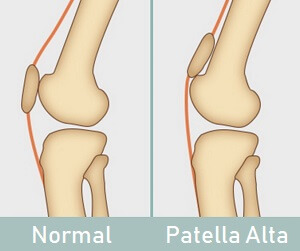
Patella Alta, also known as high-riding patella or high patella, is a condition characterized by the abnormal positioning of the patella, or kneecap, higher than its typical anatomical location within the femoral groove. This deviation from the norm can have significant implications for knee health and function, potentially leading to various symptoms and complications. Patella Alta is typically diagnosed through a combination of clinical assessment, imaging studies, and functional evaluations, allowing healthcare professionals to identify its underlying causes and determine appropriate management strategies tailored to individual needs.
Several factors can contribute to the development of Patella Alta, including congenital abnormalities, skeletal growth patterns, muscular imbalances, and traumatic injuries. Congenital factors, such as genetic predisposition or developmental anomalies in patellar morphology, may result in abnormal patellar positioning from birth. Skeletal growth patterns, such as rapid longitudinal growth of the femur relative to the patella tendon, can also influence the alignment of the patella within the knee joint, leading to Patella Alta over time. Additionally, muscular imbalances or weaknesses in the quadriceps and hamstring muscles may disrupt the dynamic stability of the patellofemoral joint, contributing to patellar malalignment and subsequent Patella Alta. Traumatic injuries, such as patellar dislocations or fractures, can also alter the normal biomechanics of the knee joint, potentially causing or exacerbating Patella Alta.
Diagnosing Patella Alta involves a comprehensive evaluation of the patient’s medical history, symptoms, and physical examination findings, supplemented by imaging modalities such as X-rays, magnetic resonance imaging (MRI), or computed tomography (CT) scans. During the physical examination, healthcare professionals may assess patellar height using various clinical tests, such as the Insall-Salvati ratio, Caton-Deschamps index, or Blackburne-Peel index, which compare the length of the patellar tendon to the patellar length or patellar height relative to the femur. Imaging studies provide valuable insights into the anatomical relationship between the patella, femur, and tibia, helping to confirm the diagnosis of Patella Alta and identify associated structural abnormalities or pathologies.
Furthermore, functional assessments, such as gait analysis, dynamic knee stability tests, and muscle strength evaluations, may aid in understanding the biomechanical factors contributing to Patella Alta and guiding appropriate treatment interventions.
Causes of Patella Alta
List of causes
- Congenital Abnormalities: Some individuals may be predisposed to Patella Alta due to genetic factors or developmental anomalies in patellar morphology. These congenital abnormalities can lead to abnormal patellar positioning from birth, contributing to the development of Patella Alta over time.
- Skeletal Growth Patterns: Rapid longitudinal growth of the femur relative to the patellar tendon can influence the alignment of the patella within the knee joint. As the femur grows, it may outpace the growth of the patellar tendon, causing the patella to ride higher within the femoral groove. Skeletal growth patterns play a significant role in the development of Patella Alta, particularly during periods of rapid growth and development, such as adolescence.
- Muscular Imbalances: Weaknesses or imbalances in the quadriceps and hamstring muscles can disrupt the dynamic stability of the patellofemoral joint, leading to patellar malalignment and subsequent Patella Alta. The quadriceps muscles, in particular, play a crucial role in controlling the movement and positioning of the patella during knee flexion and extension. Imbalances in muscle strength or activation patterns can result in abnormal forces acting on the patella, contributing to its elevated position within the knee joint.
- Traumatic Injuries: Acute or chronic traumatic injuries to the knee, such as patellar dislocations, fractures, or ligamentous tears, can disrupt the normal biomechanics of the joint and alter the positioning of the patella. Traumatic injuries may damage the supportive structures surrounding the patella, including the patellar tendon, quadriceps muscles, or ligaments, leading to instability and malalignment of the patellofemoral joint. In some cases, traumatic injuries may directly cause or exacerbate Patella Alta, necessitating appropriate management and rehabilitation to restore normal knee function.
- Joint laxity: Individuals with increased joint laxity, or hypermobility, may be more prone to Patella Alta due to instability within the patellofemoral joint. Excessive joint laxity can allow for greater patellar movement and malalignment, predisposing the patella to ride higher within the femoral groove.
- Patellar tendon length: Variations in patellar tendon length can influence the positioning of the patella within the knee joint. A longer-than-normal patellar tendon may result in higher patellar placement, while a shorter tendon may lead to a lower patellar position. These anatomical variations can contribute to the development of Patella Alta.
- Quadriceps muscle tightness: Tightness or contracture of the quadriceps muscles can exert abnormal forces on the patella, pulling it upwards and contributing to its elevated position within the knee joint. Quadriceps muscle tightness may result from factors such as prolonged sitting, inadequate stretching, or muscle imbalances.
- Patellar instability: Individuals with a history of recurrent patellar dislocations or subluxations may develop Patella Alta as a secondary consequence of patellar instability. Chronic patellar instability can lead to structural changes within the patellofemoral joint, including patellar malalignment and elevation.
- Overuse injuries: Repetitive stress or overuse of the knee joint, particularly in activities that involve high-impact movements or excessive loading, can contribute to the development of Patella Alta. Overuse injuries may result in inflammation, degenerative changes, or structural abnormalities within the patellofemoral joint, ultimately affecting patellar alignment.
- Neuromuscular conditions: Certain neuromuscular conditions, such as cerebral palsy or muscular dystrophy, can affect muscle tone, coordination, and control, potentially leading to abnormalities in patellar positioning and alignment. Individuals with neuromuscular conditions may be at increased risk of developing Patella Alta due to underlying musculoskeletal impairments.
- Obesity: Excess body weight can exert increased pressure on the knee joint, leading to biomechanical alterations and patellar malalignment. Obesity is associated with an increased risk of developing Patella Alta and other knee-related conditions due to the additional mechanical stress placed on the joint during weight-bearing activities.
- Anatomical variations in the femoral trochlea: Irregularities or abnormalities in the shape or depth of the femoral trochlea, the groove in which the patella articulates, can affect patellar tracking and alignment, potentially leading to Patella Alta.
- Leg length discrepancy: Significant differences in leg length can disrupt the normal biomechanics of the lower extremities, altering forces acting on the patella and contributing to patellar malalignment and elevation.
- Joint hypermobility syndrome: Individuals with joint hypermobility syndrome may exhibit excessive range of motion in multiple joints, including the knee, predisposing them to instability and malalignment of the patella.
- Ligamentous laxity: Excessive laxity or looseness in the ligaments surrounding the knee joint, such as the medial patellofemoral ligament (MPFL) or lateral collateral ligament (LCL), can compromise joint stability and increase the risk of Patella Alta.
- Patellofemoral dysplasia: Structural abnormalities or dysplastic changes in the patella or femur can disrupt the normal alignment of the patellofemoral joint, leading to maltracking of the patella and subsequent Patella Alta.
- Connective tissue disorders: Certain connective tissue disorders, such as Ehlers-Danlos syndrome or Marfan syndrome, can affect the integrity of ligaments, tendons, and cartilage, predisposing individuals to joint instability and malalignment, including Patella Alta.
- Joint inflammation: Chronic inflammation within the knee joint, as seen in conditions such as rheumatoid arthritis or inflammatory synovitis, can disrupt normal joint mechanics and contribute to patellar malalignment and elevation.
- Post-surgical complications: Surgical procedures involving the knee joint, such as knee arthroscopy or ligament reconstruction, may result in post-operative complications such as scar tissue formation, joint stiffness, or altered biomechanics, potentially leading to Patella Alta.
- Degenerative changes: Age-related degenerative changes, such as osteoarthritis or chondromalacia patellae, can affect the structural integrity of the knee joint and alter patellar alignment, increasing the risk of developing Patella Alta.
- Overtraining or improper training techniques: Athletes who engage in repetitive or high-impact activities without adequate rest or proper training techniques may experience overuse injuries or muscular imbalances, predisposing them to Patella Alta and other knee-related conditions.
- Patellar tendinopathy: Chronic overuse or repetitive stress on the patellar tendon, as seen in activities like running or jumping, can lead to degenerative changes within the tendon, potentially affecting its length and contributing to Patella Alta.
- Hormonal influences: Hormonal fluctuations, such as those occurring during puberty or pregnancy, can affect musculoskeletal growth and development, potentially influencing patellar alignment and contributing to Patella Alta.
- Leg alignment abnormalities: Varus or valgus alignment of the lower extremities, characterized by inward or outward angulation of the knees, respectively, can affect patellar tracking and alignment, predisposing individuals to Patella Alta.
- Joint hyperextension: Excessive hyperextension of the knee joint, particularly during activities like gymnastics or ballet, can place increased stress on the patellofemoral joint and alter patellar positioning, leading to Patella Alta.
- Foot pronation or supination: Abnormal foot mechanics, such as excessive pronation (inward rolling) or supination (outward rolling), can affect lower limb alignment and biomechanics, potentially influencing patellar tracking and contributing to Patella Alta.
- Patellar maltracking: Dysfunctional movement patterns or maltracking of the patella within the femoral groove, often associated with factors like muscle weakness or tightness, can lead to abnormal patellar positioning and elevation, resulting in Patella Alta.
- Joint effusion: Accumulation of fluid within the knee joint, as seen in conditions like synovitis or joint inflammation, can alter joint mechanics and contribute to patellar malalignment and elevation.
- Previous knee surgeries: Individuals with a history of knee surgeries, such as meniscal repair or cartilage restoration procedures, may experience alterations in joint biomechanics or structural integrity, potentially predisposing them to Patella Alta.
- Overuse of high-heeled shoes: Prolonged wearing of high-heeled shoes can alter lower limb alignment and increase patellofemoral joint pressure, potentially contributing to patellar malalignment and elevation.
- Aging-related changes: Age-related changes in muscle strength, flexibility, and joint mechanics can affect patellar tracking and alignment, increasing the risk of developing Patella Alta, particularly in older adults.
Diagnosis and Assessment Methods of patella alta
Diagnosing Patella Alta involves a comprehensive evaluation that combines clinical assessment, imaging studies, and functional evaluations. Here are some common diagnosis and assessment methods:
List
- Clinical assessment: A thorough physical examination is essential for evaluating patellar alignment and identifying signs of Patella Alta. Healthcare professionals may assess patellar height using various clinical tests, such as the Insall-Salvati ratio, Caton-Deschamps index, or Blackburne-Peel index. These measurements compare the length of the patellar tendon to the length of the patella or its height relative to the femur.
- Range of motion assessment: Evaluating the range of motion of the knee joint can provide valuable insights into patellar mobility and alignment. Healthcare professionals may assess knee flexion and extension, as well as any limitations or abnormalities in movement patterns that may indicate Patella Alta.
- Imaging studies: Radiographic imaging, such as X-rays, magnetic resonance imaging (MRI), or computed tomography (CT) scans, is commonly used to visualize the anatomical relationship between the patella, femur, and tibia. X-rays are particularly useful for assessing patellar height and alignment, while MRI or CT scans can provide detailed images of soft tissue structures and detect any associated abnormalities or pathologies.
- Patellar tilt assessment: Assessing the tilt of the patella, or its inclination relative to the femoral condyles, can help identify abnormalities in patellar tracking and alignment. Healthcare professionals may use imaging modalities such as MRI or CT scans to evaluate patellar tilt and assess its contribution to Patella Alta.
- Gait analysis: Observing the patient’s gait and dynamic knee function can provide valuable information about patellar stability, tracking, and alignment during weight-bearing activities. Gait analysis may involve visual observation, motion analysis systems, or pressure-sensitive platforms to assess knee biomechanics and detect any abnormalities associated with Patella Alta.
- Functional evaluations: Functional assessments, such as single-leg squats, step-down tests, or hop tests, can help identify biomechanical deficits or weaknesses in the lower extremities that may contribute to Patella Alta. These tests evaluate dynamic knee stability, muscle strength, and neuromuscular control, providing valuable insights into the underlying causes of the condition.
- Provocative maneuvers: Performing specific provocative maneuvers, such as the apprehension test or J-sign test, can elicit symptoms or signs indicative of patellar instability or malalignment associated with Patella Alta. These maneuvers involve applying stress to the patellofemoral joint or altering knee position to assess patellar mobility and stability.
- Physical examination for muscle imbalances: Healthcare professionals may conduct a physical examination to assess for muscle imbalances around the knee joint, particularly focusing on the quadriceps and hamstring muscles. Weakness or tightness in these muscles can contribute to patellar malalignment and elevation, thus exacerbating Patella Alta.
- Palpation of bony landmarks: Palpation of bony landmarks around the knee joint, such as the superior pole of the patella, tibial tuberosity, and femoral condyles, can help healthcare professionals identify any abnormalities in patellar position and alignment. Palpation may also reveal signs of inflammation, tenderness, or crepitus associated with Patella Alta.
- Functional knee tests: Functional knee tests, including the Thessaly test, Clarke’s sign, or Noble compression test, can help assess patellofemoral joint stability and detect any abnormalities in patellar tracking or alignment. These tests involve applying stress to the knee joint and observing for pain, clicking, or apprehension, which may indicate Patella Alta or patellar instability.
- Electromyography (EMG): Electromyography can be used to assess muscle activity and coordination around the knee joint, providing valuable information about muscle function and neuromuscular control. EMG recordings can help identify abnormalities in muscle activation patterns or timing that may contribute to Patella Alta and guide targeted rehabilitation strategies.
- Three-dimensional (3D) motion analysis: Three-dimensional motion analysis systems can capture and analyze dynamic knee movements during functional tasks, providing detailed insights into patellar tracking, alignment, and joint biomechanics. 3D motion analysis allows for precise measurement of patellar motion and can help identify abnormal movement patterns associated with Patella Alta, guiding treatment planning and rehabilitation.
Effects of Patella Alta on Knee Health
Instability and Dislocation Risks
Patella Alta is associated with an increased risk of patellar instability and dislocation, posing significant challenges to knee health and function. Here’s how Patella Alta contributes to instability and the risk of dislocation:
List of effects
- Altered patellar tracking: Patella Alta disrupts the normal biomechanics of the patellofemoral joint, causing the patella to ride higher within the femoral groove. This altered positioning can lead to abnormal patellar tracking, where the patella may shift laterally or tilt out of alignment during knee movement. As a result, the patella may become more susceptible to instability, increasing the risk of dislocation.
- Reduced patellar stability: The patella plays a crucial role in stabilizing the knee joint, particularly during activities that involve dynamic movements such as running, jumping, or changing direction. In individuals with Patella Alta, the elevated position of the patella may compromise its stability, making it more prone to subluxation or complete dislocation, especially under conditions of increased stress or sudden changes in direction.
- Impaired quadriceps function: Patella Alta can affect the function of the quadriceps muscles, which play a vital role in controlling patellar movement and stability during knee flexion and extension. Muscle imbalances or weaknesses associated with Patella Alta may result in inadequate quadriceps support, further predisposing the patella to instability and increasing the risk of dislocation.
- Structural abnormalities: Patella Alta is often associated with underlying structural abnormalities within the patellofemoral joint, such as shallow trochlear grooves, patellar dysplasia, or ligamentous laxity. These structural variations can exacerbate patellar instability and increase the likelihood of dislocation, particularly in cases where the patella fails to engage properly within the femoral groove during knee movement.
- Previous injury or trauma: Individuals with a history of previous patellar dislocations or traumatic knee injuries may be at increased risk of recurrent instability, particularly if underlying anatomical abnormalities such as Patella Alta are present. Previous injury or trauma can weaken the supportive structures around the knee joint, making it more susceptible to recurrent episodes of instability and dislocation.
- Ligamentous laxity: Excessive laxity or looseness in the ligaments supporting the knee joint, such as the medial patellofemoral ligament (MPFL) or lateral collateral ligament (LCL), can contribute to patellar instability and predispose individuals to dislocation events.
- Hypermobile patella: Patellar hypermobility, characterized by excessive movement or subluxation of the patella within the femoral groove, is commonly observed in individuals with Patella Alta and increases the risk of recurrent instability and dislocation.
- Malalignment of lower limb: Abnormal lower limb alignment, such as genu valgum (knock-knees) or genu varum (bow-legs), can affect patellar tracking and alignment, predisposing individuals to patellar instability and dislocation.
- Traumatic events: Acute traumatic events, such as a direct blow to the knee or a sudden twisting motion, can cause patellar dislocation in individuals with Patella Alta, particularly if underlying structural abnormalities or muscle weaknesses are present.
- Repetitive stress or overuse: Repetitive stress or overuse of the knee joint, particularly in activities that involve high-impact movements or excessive loading, can contribute to patellar instability and increase the risk of dislocation events over time.
- Abnormal patellar shape or morphology: Structural abnormalities in the shape or morphology of the patella, such as patellar dysplasia or trochlear dysplasia, can disrupt normal patellar tracking and stability, predisposing individuals to instability and dislocation.
- Joint inflammation or synovitis: Chronic inflammation within the knee joint, as seen in conditions such as rheumatoid arthritis or synovial plica syndrome, can weaken the supportive structures around the patella and increase the risk of instability and dislocation.
- Neuromuscular conditions: Certain neuromuscular conditions, such as cerebral palsy or muscular dystrophy, can affect muscle tone, coordination, and control, increasing the risk of patellar instability and dislocation in affected individuals.
- Improper rehabilitation or return to activity: Inadequate rehabilitation following a previous patellar dislocation or injury can lead to residual muscle weakness, joint instability, and altered movement patterns, predisposing individuals to recurrent instability and dislocation events.
- Overpronation or oversupination of the foot: Abnormal foot mechanics, such as overpronation (excessive inward rolling) or oversupination (excessive outward rolling), can affect lower limb alignment and biomechanics, potentially contributing to patellar instability and dislocation.
- Joint hyperextension: Excessive hyperextension of the knee joint, particularly during activities like dancing or gymnastics, can place increased stress on the patellofemoral joint and increase the risk of patellar instability and dislocation.
- Environmental factors: Environmental factors such as uneven terrain or inappropriate footwear can increase the risk of falls or traumatic events that may result in patellar dislocation, particularly in individuals with underlying Patella Alta.
- Participation in high-risk sports: Participation in high-risk sports or activities that involve frequent changes in direction, jumping, or pivoting movements, such as basketball, soccer, or volleyball, can increase the risk of patellar instability and dislocation in individuals with Patella Alta.
- Joint effusion or swelling: Accumulation of fluid within the knee joint, as seen in conditions such as knee effusion or synovitis, can compromise joint stability and increase the risk of patellar instability and dislocation events.
- Excessive weight: Excess body weight can increase the mechanical stress placed on the knee joint during weight-bearing activities, potentially compromising joint stability and increasing the risk of patellar instability and dislocation.
- Inadequate warm-up or stretching: Inadequate warm-up or stretching before physical activity can lead to muscle tightness, reduced joint mobility, and altered movement patterns, increasing the risk of patellar instability and dislocation events.
- Psychological factors: Psychological factors such as fear of reinjury or lack of confidence in knee stability can affect an individual’s movement patterns and predispose them to patellar instability and dislocation events.
- Hormonal changes: Hormonal fluctuations, such as those occurring during the menstrual cycle or pregnancy, can affect ligament laxity and joint stability, potentially increasing the risk of patellar instability and dislocation in susceptible individuals.
- Delayed or untreated injuries: Delayed or untreated injuries to the knee joint, such as patellar tendon tears or osteochondral defects, can weaken the supportive structures around the patella and increase the risk of patellar instability and dislocation over time.
Impact on Cartilage Health
Patella Alta can have a significant impact on cartilage health within the knee joint, potentially leading to degenerative changes, cartilage damage, and early-onset osteoarthritis. Here’s how Patella Alta affects cartilage health:
List of impacts
- Altered biomechanics: Patella Alta disrupts the normal biomechanics of the knee joint, leading to abnormal patellar tracking, increased contact pressures, and altered load distribution within the patellofemoral joint. These biomechanical alterations can result in excessive stress on the articular cartilage, particularly on the posterior surface of the patella and the femoral trochlea, predisposing the cartilage to degeneration and wear.
- Increased contact pressures: The elevated position of the patella within the femoral groove in individuals with Patella Alta can lead to increased contact pressures between the patella and the femur during weight-bearing activities. These elevated pressures can exceed the capacity of the articular cartilage to withstand load, resulting in cartilage compression, deformation, and ultimately, degenerative changes.
- Patellar maltracking: Patella Alta is often associated with patellar maltracking, where the patella fails to glide smoothly within the femoral groove during knee movement. This maltracking can cause uneven distribution of forces across the articular surfaces of the patella and femur, leading to focal areas of cartilage wear, erosion, and degeneration over time.
- Chondral defects: Prolonged patellar malalignment and instability associated with Patella Alta can lead to the development of chondral defects or cartilage lesions within the patellofemoral joint. These defects may range from superficial fissures or softening of the cartilage to full-thickness defects involving the underlying subchondral bone, compromising joint integrity and function.
- Synovial inflammation: Chronic patellar malalignment and instability can result in synovial inflammation and joint effusion, further exacerbating cartilage damage and degeneration. Inflammatory mediators released within the joint space can contribute to cartilage degradation, matrix breakdown, and the progression of osteoarthritis in individuals with Patella Alta.
- Early-onset osteoarthritis: The cumulative effects of altered biomechanics, increased contact pressures, patellar maltracking, and cartilage damage associated with Patella Alta can accelerate the onset and progression of osteoarthritis within the patellofemoral joint. Individuals with Patella Alta may experience symptoms of osteoarthritis, such as joint pain, stiffness, and functional limitations, at an earlier age than those without the condition.
- Subchondral bone changes: Patella Alta can lead to abnormal loading patterns on the subchondral bone beneath the cartilage, potentially causing remodeling, sclerosis, or cyst formation. These changes in the subchondral bone can further exacerbate cartilage damage and contribute to the progression of osteoarthritis.
- Meniscal injury: Patella Alta may increase the risk of meniscal injury due to altered joint mechanics and increased contact pressures within the knee joint. Meniscal tears or degeneration can accelerate cartilage wear and compromise joint stability, further worsening the impact on cartilage health.
- Posterior patellar impingement: Individuals with Patella Alta may experience posterior patellar impingement, where the elevated patella contacts the femoral condyles during knee flexion. This impingement can lead to cartilage damage on the posterior aspect of the patella and the femoral trochlea, contributing to degenerative changes and osteoarthritis.
- Cartilage fissures and delamination: Prolonged patellar malalignment and instability associated with Patella Alta can result in the development of cartilage fissures, cracks, or delamination within the patellofemoral joint. These structural abnormalities compromise cartilage integrity and increase the risk of progressive cartilage loss over time.
- Cartilage softening and thinning: Chronic patellar maltracking and instability can lead to cartilage softening, thinning, and loss of proteoglycan content within the patellofemoral joint. These changes make the cartilage more susceptible to mechanical damage and accelerate the progression of osteoarthritis.
- Focal cartilage lesions: Patella Alta may result in the formation of focal cartilage lesions or defects within the patellofemoral joint, particularly in areas of increased contact pressures or abnormal loading. These lesions can compromise joint function, increase pain, and contribute to the development of osteoarthritis.
- Cartilage inflammation: Altered joint mechanics and increased synovial inflammation associated with Patella Alta can trigger an inflammatory response within the articular cartilage, leading to cartilage degradation, matrix breakdown, and the release of catabolic mediators that further exacerbate cartilage damage.
- Cartilage wear patterns: Patella Alta can lead to distinctive wear patterns on the articular surfaces of the patella and femur, characterized by areas of focal abrasion, fibrillation, or denudation. These wear patterns reflect abnormal joint mechanics and increased contact pressures, indicating cartilage degeneration and osteoarthritic changes.
- Secondary osteochondral lesions: Individuals with Patella Alta may develop secondary osteochondral lesions, involving both cartilage and underlying bone, as a result of chronic patellar malalignment and instability. These lesions can compromise joint integrity, impair function, and accelerate the progression of osteoarthritis.
- Compromised joint lubrication: Altered joint mechanics associated with Patella Alta can disrupt the normal distribution of synovial fluid within the knee joint, compromising joint lubrication and reducing the ability of the cartilage to withstand friction and mechanical stress. This can further exacerbate cartilage wear and degeneration over time.
- Joint stiffness: Patella Alta can lead to joint stiffness and decreased range of motion in the knee joint, which may contribute to abnormal loading and stress on the articular cartilage during movement, potentially accelerating cartilage degeneration and osteoarthritic changes.
- Cartilage erosion: Prolonged patellar malalignment and instability associated with Patella Alta can lead to cartilage erosion, characterized by gradual loss of cartilage thickness and integrity over time. Cartilage erosion can compromise joint function and exacerbate symptoms of pain and stiffness.
- Cartilage fibrillation: Chronic patellar maltracking and instability may result in cartilage fibrillation, characterized by the development of small, irregular surface irregularities or fraying within the articular cartilage. These fibrillations can progress to deeper cartilage damage and contribute to the development of osteoarthritis.
- Increased susceptibility to mechanical stress: Patella Alta renders the articular cartilage more susceptible to mechanical stress and shear forces, particularly during weight-bearing activities or movements that involve knee flexion and extension. This increased susceptibility can lead to accelerated wear and tear on the cartilage, hastening the onset of osteoarthritic changes.
- Impaired shock absorption: Altered joint mechanics associated with Patella Alta can impair the ability of the cartilage to absorb shock and dissipate mechanical forces during weight-bearing activities. This can result in increased stress concentration on localized areas of the cartilage, promoting cartilage degeneration and contributing to the progression of osteoarthritis.
Symptoms and Clinical Presentation
Patella Alta can manifest with a variety of symptoms and clinical presentations, which can vary in severity depending on the underlying cause, degree of patellar malalignment, and individual factors. Here are some common symptoms and clinical features associated with Patella Alta:
Likst of symptoms
- Anterior knee pain: One of the hallmark symptoms of Patella Alta is anterior knee pain, which may be localized to the front of the knee or felt more diffusely around the patellofemoral joint. The pain is often exacerbated by activities that involve knee flexion, such as climbing stairs, squatting, or running.
- Patellar instability: Individuals with Patella Alta may experience episodes of patellar instability, characterized by feelings of the patella slipping, shifting, or giving way during movement. This instability may be accompanied by audible or palpable sensations of clicking, popping, or grinding within the knee joint.
- Swelling and inflammation: Chronic patellar malalignment and instability associated with Patella Alta can lead to synovial inflammation and joint effusion, resulting in swelling, warmth, and tenderness around the patellofemoral joint. Swelling may exacerbate pain and limit knee mobility, particularly during weight-bearing activities.
- Crepitus: Patella Alta may be accompanied by crepitus, or a sensation of grinding or crunching within the knee joint, especially during movements that involve patellar tracking, such as knee flexion or extension. Crepitus may be audible or palpable and is often indicative of cartilage wear and degeneration.
- Reduced range of motion: Patella Alta can restrict knee mobility and range of motion, particularly during activities that require full knee flexion or extension. Individuals may experience limitations in their ability to fully straighten or bend the knee, contributing to functional impairments and activity limitations.
- Weakness or instability: Muscle weakness or imbalance around the knee joint, particularly in the quadriceps and hip abductor muscles, may accompany Patella Alta and contribute to feelings of knee instability, reduced joint control, and altered movement patterns.
- Difficulty with stairs or slopes: Individuals with Patella Alta may experience difficulty ascending or descending stairs, ramps, or inclines, due to increased stress on the patellofemoral joint and exacerbation of symptoms such as pain, instability, and weakness.
- Functional limitations: Patella Alta can impact daily activities and functional tasks that require knee stability and mobility, such as standing up from a seated position, walking, or participating in sports or recreational activities. Functional limitations may vary depending on the severity of symptoms and the individual’s activity level.
- Visible patellar prominence: In severe cases of Patella Alta, the elevated position of the patella within the knee joint may be visually apparent, with the patella appearing more prominent or elevated compared to its normal anatomical position.
- Recurrent patellar dislocations: Individuals with severe or untreated Patella Alta may be at increased risk of recurrent patellar dislocations or subluxations, where the patella partially or completely moves out of its normal position within the femoral groove.
- Pain with prolonged sitting: Individuals with Patella Alta may experience discomfort or pain with prolonged sitting, particularly with the knees flexed, such as during car rides, movie watching, or desk work. This pain may be exacerbated by the compressive forces on the patellofemoral joint.
- Difficulty with squatting: Patella Alta can make squatting movements challenging due to increased stress on the patellofemoral joint and altered biomechanics. Individuals may experience pain, instability, or a feeling of “giving way” in the knee during squatting activities.
- Instability during single-leg activities: Activities that require single-leg support, such as standing on one leg or performing single-leg exercises, may be particularly challenging for individuals with Patella Alta due to reduced knee stability and control. They may experience feelings of wobbliness or unsteadiness in the knee.
- Increased pain with activity: Pain associated with Patella Alta often worsens with physical activity, especially activities that involve repetitive knee movements, such as running, jumping, or cycling. The increased stress on the patellofemoral joint during these activities can exacerbate symptoms.
- Altered biomechanics during gait: Individuals with Patella Alta may exhibit altered biomechanics during walking or running, characterized by changes in stride length, foot placement, or knee alignment. These biomechanical alterations may be compensatory mechanisms to reduce pain or instability in the knee.
- Feeling of “giving way”: Patella Alta can result in a sensation of the knee “giving way” or feeling unstable during weight-bearing activities, such as walking on uneven surfaces or navigating stairs. This feeling may be due to patellar maltracking or instability within the patellofemoral joint.
- Difficulty with leg extension: Straightening the knee joint fully (leg extension) may be challenging for individuals with Patella Alta due to increased tension on the patellar tendon and limited mobility in the knee. They may experience pain or discomfort at the end range of motion.
- Anterior knee tenderness: Palpation of the patella or the surrounding soft tissues may elicit tenderness or discomfort in individuals with Patella Alta, particularly at the superior pole of the patella or along the patellar tendon insertion.
- Audible clicking or popping: Patella Alta may be associated with audible clicking, popping, or snapping sounds within the knee joint during movement, particularly with activities that involve patellar tracking, such as squatting or stair climbing.
- Pain with passive patellar mobilization: Passive mobilization of the patella by a healthcare professional during a physical examination may reproduce symptoms of pain or discomfort in individuals with Patella Alta, indicating sensitivity or irritation of the patellofemoral joint.
Preventive Measures and Lifestyle Considerations
Implementing preventive measures and lifestyle considerations can help manage and reduce the risk of complications associated with Patella Alta. Here are some strategies to consider:
List of preventive measures
- Strengthening exercises: Engaging in a structured exercise program focused on strengthening the muscles around the knee, particularly the quadriceps, hamstrings, and hip abductors, can help improve knee stability and reduce the risk of patellar instability and dislocation.
- Neuromuscular training: Incorporating neuromuscular training exercises, such as balance and proprioception drills, can enhance joint proprioception and control, improving dynamic knee stability and reducing the risk of falls or traumatic injuries.
- Flexibility and mobility exercises: Performing regular stretching and mobility exercises for the lower extremities can help maintain optimal joint range of motion and prevent muscle tightness or imbalance that may contribute to patellar malalignment and instability.
- Proper footwear: Wearing appropriate footwear with adequate support and cushioning can help reduce stress on the knee joint during weight-bearing activities and promote optimal lower limb alignment, reducing the risk of patellar maltracking and instability.
- Activity modification: Avoiding high-impact activities or sports that involve repetitive knee movements, sudden changes in direction, or excessive jumping can help reduce the risk of exacerbating symptoms associated with Patella Alta and prevent further joint damage.
- Gradual progression of activities: Gradually increasing the intensity, duration, and frequency of physical activities can help minimize the risk of overuse injuries and gradual onset of symptoms associated with Patella Alta. Incorporating adequate rest periods and cross-training activities can also promote overall joint health and recovery.
- Proper biomechanics: Maintaining proper body mechanics and movement patterns during daily activities and exercise can help reduce excessive stress on the knee joint and promote optimal patellar tracking and alignment. Avoiding activities that place undue stress on the knees or promote poor posture can help prevent aggravation of symptoms.
- Weight management: Maintaining a healthy body weight through a balanced diet and regular exercise can help reduce the mechanical stress placed on the knee joint and decrease the risk of developing or exacerbating symptoms associated with Patella Alta.
- Protective bracing or taping: Using patellar braces or kinesiology tape during physical activities can provide additional support and stability to the knee joint, reducing the risk of patellar instability and improving functional performance, particularly during high-risk activities or sports.
- Regular monitoring and follow-up: Individuals with Patella Alta should undergo regular monitoring and follow-up with a healthcare professional, such as an orthopedic surgeon or physical therapist, to assess symptom progression, monitor joint health, and adjust treatment strategies as needed.
- Proper warm-up and cool-down: Implementing a comprehensive warm-up routine before engaging in physical activity and incorporating a cooldown period afterward can help prepare the muscles and joints for exercise, reduce the risk of injury, and promote optimal recovery.
- Cross-training: Diversifying exercise routines by incorporating a variety of low-impact activities, such as swimming, cycling, or elliptical training, can help reduce repetitive stress on the knee joint and provide alternative cardiovascular and strength-training benefits.
- Postural awareness: Maintaining good posture during daily activities and exercise, including sitting, standing, and walking, can help distribute weight evenly across the joints and minimize excessive stress on the knees, reducing the risk of aggravating symptoms associated with Patella Alta.
- Injury prevention strategies: Implementing injury prevention strategies, such as using proper lifting techniques, avoiding sudden changes in activity level, and gradually increasing exercise intensity, can help minimize the risk of acute injuries and exacerbation of symptoms related to Patella Alta.
- Footwear modification: Choosing footwear with appropriate arch support, cushioning, and stability features can help improve lower limb alignment and reduce the risk of biomechanical abnormalities that may contribute to patellar malalignment and instability.
- Orthotic support: Using custom orthotic inserts or shoe inserts prescribed by a healthcare professional can provide additional support and alignment for the feet and lower limbs, helping to reduce excessive pronation or supination and promote optimal patellar tracking.
- Pain management strategies: Incorporating pain management techniques, such as ice therapy, heat therapy, or over-the-counter pain medications, can help alleviate symptoms of pain and inflammation associated with Patella Alta, allowing for better participation in physical activity and rehabilitation.
- Balance and proprioception training: Including balance and proprioception exercises, such as single-leg stance, stability ball exercises, or balance board drills, can help improve joint proprioception, neuromuscular control, and dynamic stability, reducing the risk of falls or injuries.
- Education and self-management: Educating individuals with Patella Alta about their condition, including potential triggers, exacerbating factors, and self-management strategies, empowers them to take an active role in their care and make informed decisions about lifestyle choices and treatment options.
- Regular monitoring and assessment: Regularly monitoring symptoms, functional status, and joint health through self-assessment or periodic evaluations by a healthcare professional can help track progress, identify any changes or concerns, and adjust treatment strategies accordingly to optimize outcomes.
Conclusion
Patella Alta is a complex orthopedic condition characterized by elevated positioning of the patella within the femoral groove, often leading to patellar malalignment, instability, and potential complications such as cartilage damage and osteoarthritis. Understanding the anatomy, biomechanics, and clinical implications of Patella Alta is crucial for accurate diagnosis and effective management.
Throughout this discussion, we’ve explored various aspects of Patella Alta, including its definition, anatomy, causes, diagnosis, symptoms, and impact on knee health. We’ve also discussed preventive measures, lifestyle considerations, and ongoing research directions aimed at improving our understanding and management of this condition.
Moving forward, continued research efforts focused on biomechanics, imaging modalities, genetic factors, non-surgical interventions, and surgical techniques will be essential for advancing our knowledge and enhancing clinical care for individuals with Patella Alta. By implementing preventive strategies, promoting patient education, and exploring innovative treatment approaches, we can strive to optimize outcomes, reduce complications, and improve the quality of life for individuals affected by Patella Alta.
Ultimately, a multidisciplinary approach involving collaboration between clinicians, researchers, and patients is essential for addressing the challenges posed by Patella Alta and ensuring comprehensive, evidence-based care tailored to the individual needs of each patient. Through ongoing dedication to research, education, and clinical practice, we can continue to make strides in the management of Patella Alta and improve outcomes for patients worldwide


 Weakness: Causes, Implications, and Solutions")


