

Clinical evidence indicates that 50% of all patients seen in hospital with an initial diagnosis of angina are indeed from a source other than cardiac. Pectoralis minor myofascial pain syndrome resembles that of an angina attack (1).
Introduction: When chest pain deceives everyone
Sudden chest pain, oppressive or radiating to the left arm… In the collective imagination as in emergency rooms, these symptoms immediately evoke a cardiac emergency. And for good reason: angina pectoris and myocardial infarction represent vital issues where every minute counts. However, in a significant number of cases, the tests come back normal, the electrocardiogram is reassuring, and the cardiac markers in the blood do not indicate any myocardial distress. The patient then leaves with a vague diagnosis or simple “stress”, without a concrete answer to their pain. What if it was in fact a myofascial syndrome of the pectoralis major muscle ?
This entity, still little known to the general public – and sometimes underestimated even in the medical community – can dramatically mimic cardiac pain. This phenomenon, called pseudoangina pectoris , creates diagnostic confusion that can generate anxiety, lead to repeated examinations and delay actual management. The pectoralis major muscle, particularly when infiltrated by myofascial trigger points, can refer pain to the anterior thorax, shoulder, left arm, or even jaw, in a picture that is confusing for the clinician and worrying for the patient.
Myofascial pain syndrome is a functional musculoskeletal disorder in which a muscle has one or more active trigger points. These hypersensitive areas cause local and referred pain, often at a distance from the affected muscle site. In the case of the pectoralis major, referred pain can perfectly mimic an angina attack, with oppression, chest tension, and sometimes shortness of breath or anxiety. This is due to the very specific topography of the muscle, its sternocostal attachments, its nervous connections, and the influence of stress on its tone.
Emergency medicine rightly favors an approach of excluding serious pathologies. But once these have been ruled out, it is essential to refer the patient for a rigorous musculoskeletal assessment. This is where osteopathy can play a key role. Thanks to its fine palpation, its comprehensive approach, and its knowledge of the myofascial chains, the osteopath is able to identify the myofascial origin of certain so-called unexplained pains. The case of the pectoralis major is a prime example.
It is not uncommon for a patient who has consulted urgently for “left chest pain,” after undergoing reassuring tests, to feel helpless in the face of persistent symptoms. This diagnostic wandering can increase their anxiety and maintain a vicious cycle of pain. The osteopath, by identifying localized tension in the pectoralis major muscle, by locating a painful trigger point on palpation, and by tracing the map of the referred pain, can offer this patient a coherent, concrete explanation, and above all a therapeutic solution.
The challenge here goes beyond simply identifying a pain point. It’s about reconciling the patient’s subjective experience—their oppressive sensation, their fear of a heart condition—with a very real, but often ignored, bodily cause. This bodily and integrative approach helps restore inner security, along with physical relief. From this perspective, the pectoralis major muscle becomes both the vector of pain and the key to its resolution.
In this article, we will explore in detail the anatomical features of the pectoralis major muscle, the concept of myofascial syndrome, the mechanisms of referred pain mimicking angina, and specific osteopathic assessment and treatment methods. We will also discuss how to distinguish this functional pathology from a true cardiac emergency, and why it is crucial for healthcare professionals, as well as patients, to be informed of this diagnostic possibility.
Understanding that some chest pains do not come from the heart, but from a tense or overworked muscle, is not a minimization. On the contrary, it is a way to broaden our understanding of the body and move beyond the binary logic of “serious or psychological.” Between the two lies a subtle bodily reality, which osteopathy is particularly well placed to listen to and treat.
Functional anatomy of the pectoralis major
The pectoralis major muscle is often reduced, in the collective imagination, to a symbol of strength or masculine aesthetics. However, behind its apparent simplicity lies a complex structure, at the intersection of emotional, biomechanical, and postural tensions. To understand how it can cause pain mimicking angina, it is essential to explore its functional anatomy in detail.
A fan-shaped muscle, at the crossroads of several territories
The pectoralis major is a broad, flat, triangular, fan-shaped muscle. It extends from the rib cage to the humerus, occupying the anterior portion of the chest wall. It is made up of three distinct portions:
- The clavicular portion , which originates on the medial half of the clavicle.
- The sternal portion , which inserts onto the manubrium and the body of the sternum.
- The costal portion , which attaches to the cartilages of the first six ribs and sometimes to the sheath of the rectus abdominis muscle.
These three bundles converge laterally to insert into a common tendon on the crest of the greater tubercle of the humerus, at the level of the anterior lip of the intertubercular groove.
This particular fan-shaped arrangement allows the muscle to participate in several complex movements, but also makes it vulnerable to differential tensions between its bundles, which can cause areas of overload and local or referred pain.
Innervation and vascularization: a sensitive map
The pectoralis major is innervated by the lateral and medial pectoral nerves , originating from the brachial plexus (roots C5 to T1). These nerves carry not only motor information, but also a proprioceptive and nociceptive component.
This innervation crosses territories also involved in cardiac chest pain, which contributes to the phenomenon of viscero-somatic convergence : the brain can confuse the real origin of the pain signal, wrongly attributing muscular pain to a more serious visceral origin, such as that of the heart.
Vascularization is provided by branches of the thoracoacromial and lateral thoracic arteries , which run close to the surface of the muscle and can be influenced by persistent myofascial tensions.
Biomechanical role: more than a strength muscle
Functionally, the pectoralis major is an adductor, flexor, and medial rotator of the humerus . It plays a fundamental role in pushing movements (such as push-ups or the bench press), but also in gripping activities when the arm is brought forward.
However, its function goes far beyond simple motor skills: it is involved in shoulder stabilization , particularly in prolonged postures with the arms forward (working on a screen, driving, caring for children, etc.). In this context, repeated stress or muscular imbalance (particularly with antagonist muscles such as the rhomboids or the scapula fixators) can lead to chronic hypertonicity of the pectoralis major, favoring the emergence of active trigger points.
Interactions with breathing and emotions
Due to its costal and sternal attachments, the pectoralis major also participates in accessory breathing . In situations of stress, hyperventilation, or diaphragmatic blockage, this muscle can contract excessively to help lift the rib cage. This respiratory hyperactivity can develop over time, particularly in anxious individuals, and further increase myofascial tension.
On the somato-emotional level, many patients describe a feeling of chest tightness, oppression or anxiety , linked to this anterior area of the thorax. The pectoralis major is often involved in this type of picture, as if it “enclosed” the sternum, reinforcing a posture of withdrawal and protection. This clinical observation is particularly relevant in the osteopathic approach, where body reading integrates physical and psycho-emotional dimensions.
The breeding ground for referred pain
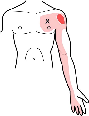
When trigger points occur in the pectoralis major – often located near the sternum or in its middle portion – the pain can radiate to the anterior chest wall , the inner side of the left arm , or even the breast region or the axilla . This referred pain is particularly fearsome because it closely mimics that of angina pectoris.
The patient then describes a dull, heavy, sometimes burning pain , which increases with exertion or certain postures, and which does not respond to conventional treatments. In these cases, detailed knowledge of the functional anatomy of the pectoralis major allows the therapist to guide the palpatory examination and to formulate a relevant hypothesis.
Understanding Myofascial Syndrome: Triggers, Physiology, and Referred Pain
Myofascial syndrome is one of the most common musculoskeletal disorders, yet one of the least understood in the differential diagnosis of chronic pain. At the root of this syndrome are myofascial trigger points , which are real knots of tension in the muscular structure. In a careful osteopathic approach, they take on their full importance, particularly when they are responsible for referred pain mimicking much more serious pathologies, such as pseudo-anginal chest pain.
What is a trigger point?
A trigger point is a hypersensitive area located in a taut band of skeletal muscle. On palpation, it is often felt as a small ball or cord. When activated, it can produce:
- Spontaneous or provoked local pain ,
- A characteristic and reproducible referred pain ,
- A decrease in joint range of motion or functional weakness of the affected muscle,
- Sometimes local autonomic signs (redness, sweating, paresthesia).
These points can be active (produce spontaneous pain) or latent (asymptomatic but painful when pressed, can be activated under stress or overwork).

Origin and pathophysiological mechanisms
Trigger point activation depends on several factors:
- Repeated microtrauma or prolonged contractions,
- Postures maintained over time, without muscle relaxation,
- Emotional shocks or chronic stress, which induce muscular hypertonia,
- Direct trauma , surgeries, or prolonged immobilization.
Physiologically, the most widely accepted theory is that of local energy crisis : a vicious cycle of ischemia, hypoxia, and accumulation of metabolic waste leads to the release of pain-producing substances (substance P, bradykinin, serotonin) that maintain pain. In parallel, a spinal reflex loop is established, reinforcing contraction and pain perception.
Pain that travels: the phenomenon of referred pain
Trigger points are known for their ability to project pain far from their location. For example, a point in the upper trapezius can generate temporal pain. It is this specificity that makes myofascial pain syndrome difficult to diagnose and often confused with other pathologies.
Referred pain is neither random nor psychosomatic: it follows reproducible maps described by authors such as Travell & Simons. For each muscle, one or more projection territories are well known. In the case of the pectoralis major, pain can project to the chest, shoulder, left arm, and even the mammary region , creating a clinical illusion of cardiac pathology.
This phenomenon is reinforced by neuronal convergence at the level of the spinal cord: sensory input from muscles, skin, and viscera use similar pathways, which can disrupt the central interpretation of the pain signal. The brain, in the absence of a “precise GPS,” can then attribute a visceral origin to pain of myofascial origin.
Clinical consequences of myofascial syndrome
Patients with myofascial pain syndrome often have no visible lesions on imaging. However, they suffer from constant pain, which can be disabling. This pain can worsen with stress, fatigue, or certain postures, and respond poorly to conventional treatments (anti-inflammatories, analgesics, reassuring but ineffective imaging).
In addition, the existence of an active trigger point can modify motor patterns , lead to postural compensations, and fuel distant joint dysfunctions. Hence the importance of a detailed manual assessment and a comprehensive therapeutic approach.
The role of osteopathy in myofascial treatment
The osteopath, thanks to his targeted palpation , is able to identify trigger points, even when they are latent or hidden in complex patterns. Once located, several approaches are possible:
- Manual inhibition of the painful point by prolonged compression,
- Specific or post-isometric stretches ,
- Myofascial techniques to release local and global tension,
- Overall postural rebalancing to avoid recurrences.
The osteopathic approach is not limited to the muscle: it takes into account the fascia, the muscle chains, the neurovegetative system, and the emotional experience of the patient, which can often be involved in the chronicity of the syndrome.
Chest pain without cardiac injury – pseudoangina pectoralis major
When a patient experiences deep, anterior chest pain radiating into the left arm and sometimes accompanied by oppression or a feeling of discomfort, the first hypothesis raised is that of a cardiac problem . This caution is justified, because a myocardial infarction or unstable angina are life-threatening emergencies. However, there is a common but often neglected situation: pseudoangina of myofascial origin , and more precisely linked to the pectoralis major muscle .
Pain that perfectly mimics angina pectoris
Pseudoangina is a form of non-cardiac chest pain of musculoskeletal origin, but whose clinical characteristics can closely mimic those of true angina pectoris. The patient describes a dull or burning pain, poorly localized, increased by certain movements or postures , sometimes triggered at rest or under stress, with radiation to the shoulder, the inner side of the left arm, or even the jaw.
In the case of the pectoralis major , the location is often parasternal , sometimes slightly lateralized, and the pain may increase on deep palpation of the muscle or during isometric contraction. This muscle has a referred pain zone that largely overlaps with the territory of anginal pain, which makes confusion possible, even for experienced clinicians.
Differential features: what distinguishes pseudo-angina from true angina
Although the two pictures may look similar, several elements can guide the clinician towards a myofascial origin:
| Pseudoangina (muscular) | Angina pectoris (cardiac) |
|---|---|
| Pain reproducible on palpation | Pain not influenced by palpation |
| Triggered by certain postures | Triggered by exertion or stress |
| Can last several hours | Often lasts a few minutes |
| Not relieved by trinitrin | Typical relief from trinitrin |
| Normal cardiac imaging and tests | Possible abnormalities on ECG or ultrasound |
| Irregular, non-systematic irradiation | Classic irradiation (left arm, jaw) |
It is crucial to clarify that only a thorough medical examination can definitively rule out a cardiac cause. The goal is not to deny the importance of cardiac examinations, but to consider a relevant differential diagnosis when examinations are normal and pain persists.
The role of stress and breathing
An often overlooked element is the link between stress, breathing, and muscle tension . In chronic stress, the body often adopts high thoracic breathing , with increased involvement of accessory muscles such as the pectoralis major. This muscular hyperactivity, against a background of postural fatigue or physical overload, can generate or maintain an active myofascial trigger point in this muscle.
Furthermore, the anxiety generated by chest pain – especially when a cardiac cause has been suspected – increases bodily alertness and can amplify the perception of pain, creating a vicious circle between tension, pain and anxiety.
Emotionally charged pain
The thorax is not just a mechanical space; it is also a symbolic place , where retained emotions, vital breath, and the relationship to intimate space intersect. Projected pain from the pectoralis major can evoke pain of confinement or internal pressure , experienced intensely by the patient, especially when no medical cause is identified.
In some people, this pain can reactivate a deep fear , such as that of dying or of not being able to breathe. This is why it is essential to validate the patient’s subjective experience , even if the cause is musculoskeletal.
Contributions of osteopathy in differential diagnosis
The osteopath, through careful palpation and listening to the patient’s body narrative, is well placed to:
- Identify an active trigger point in the pectoralis major.
- Reproduce the referred pain described by the patient.
- Observe a limitation of local mobility or muscular asymmetry.
- Link the clinical picture to a context of physical or psycho-emotional overload .
This osteopathic diagnosis not only allows the patient to understand the origin of their pain, but also to offer targeted treatment , avoiding unnecessary drug treatments or repeated examinations.
When Pain Mimics Emergency: Pseudo-Angina of the Pectoralis Major
Among the many manifestations of myofascial syndrome , that involving the pectoralis major muscle is one of the most confusing. This muscle, although well known for its role in adduction and medial rotation of the arm, can become the source of referred chest pain , so intense and well localized that it immediately suggests, in both the patient and the practitioner, a possible heart attack .
A localized, deep… and deceptive pain
Pseudoangina of the pectoralis major often presents with a specific, retrosternal or infraclavicular pain , sometimes radiating to the left arm or jaw—areas typically associated with coronary syndromes. This clinical similarity contributes to confusion and can lead to extensive medical investigations and even emergency hospitalizations, sometimes with normal ECG or coronary angiography results.
Unlike angina of ischemic origin, myofascial pain:
- is not related to effort or emotional stress,
- is not relieved by nitrate derivatives ,
- and can be reproduced manually by targeted pressure on the muscle trigger point.
The trap of differential diagnosis
This is where the osteopath trained in tissue reading comes into play. Palpation of the pectoralis major trigger point , generally located at the sternocostal or clavicular segment of the muscle, can faithfully reproduce the pain reported by the patient , confirming the myofascial nature of the picture.
However, this step should never be taken lightly. The priority always remains to rule out any real heart attack. This implies:
- a rigorous anamnesis (cardiac history, risk factors, nature of the pain),
- medical collaboration at the slightest doubt,
- and sometimes a delay in osteopathic treatment until obtaining medical approval.
This respect for the diagnostic framework not only reinforces patient safety, but also the credibility of the osteopathic approach in atypical cases.
A well-documented referred pain model
The phenomenon of referred pain from the pectoralis major is well known in physical medicine and physiotherapy. Janet Travell and David Simons, in their seminal work on trigger points, precisely mapped this thoracic projection area . The active trigger point sends a pain signal via the peripheral nervous system to a distant area, without any apparent local lesion in that area.
In the case of the pectoralis major, the innervation from C5 to T1 (brachial plexus) explains this neurological convergence between the thoracic muscle and internal or cutaneous structures of the chest. The brain , unable to make the fine distinction, interprets this pain as of visceral origin.
Psychological consequences and diagnostic error
The patient suffering from pseudoangina of myofascial origin may experience marked anxiety , especially if medical tests come back normal without a clear explanation. Doubt sets in: “Is it all in my head?” , “Have I missed something serious?”
This diagnostic wandering can last for months or even years , with prescriptions for anxiolytics, digestive exploration, or even work stoppage, without any real improvement. It is in these contexts that the osteopath, by recognizing the myofascial signature of the pain, can offer rapid , lasting, and deeply reassuring relief for the person.
Osteopathic Care: Free, Rebalance, Integrate
Osteopathic treatment of pectoralis major myofascial syndrome is not limited to local release. It is part of a comprehensive approach that combines palpatory precision, release of associated tensions, and postural reorganization. The objective is twofold: to relieve pain and prevent recurrences , by restoring the body’s capacity for self-regulation.
Local trigger point treatment: targeted effectiveness
The pectoralis major trigger point can be treated by different manual approaches, chosen according to the patient’s sensitivity, the chronicity of the picture and the tissue reactions observed.
1. Inhibition by ischemic pressure (trigger point release)
- Position: patient in supine position, arm slightly abducted.
- The practitioner applies sustained and progressive pressure to the painful point until the projected pain is triggered .
- The pressure is maintained until the pain intensity decreases and local relaxation occurs (generally 30 to 90 seconds).
- Slow, guided breathing can be combined.
2. Techniques myotensives (MET)
- Gentle contraction of the muscle against manual resistance, followed by passive stretching.
- Allows a reduction in post-isometric muscle tone while respecting pain.
3. Deep myofascial release
- More global work on the anterior chain of the thorax , following the continuity of the fascia from the clavicle to the white line.
- Residual fascial tension is often linked to prolonged postures or restricted breathing.
4. Positional Release (Jones Technique)
- Passive repositioning of the arm in a position that completely eliminates pain for 90 seconds.
- Very useful in hypersensitive or anxious patients.
Rebalancing of associated chains
Once the pain has subsided, the osteopath explores the imbalances upstream and downstream of the main lesion. The pectoralis major is often hypertonic in response to prolonged thoracic flexion posture , high breathing or chronic tension of the diaphragm.
Areas to be treated may include:
- The pectoralis minor , linked to the nerves of the brachial plexus.
- The diaphragm , to promote low, relaxed breathing.
- The upper dorsal vertebrae (T2–T6) , often in fixation in pseudo-anginal pain.
- The axillary compartment , in case of tension of the brachial fascia.
Treating these areas allows for a reorganization of tension and lasting relief.
Global integration: posture, emotion, breathing
Osteopathy, in its philosophy, aims to restore harmony in the body’s axes of mobility. Working on the pectoralis major is therefore part of a broader dynamic.
Posture
- Observe the patient’s natural standing position.
- Recommend simple chest opening exercises: wall corner stretching, gentle scapular mobilization, awareness of verticality.
Respiration
- Restore mobility of the rib cage and diaphragm.
- Encourage slow abdominal breathing, especially in patients who are stressed or in postural apnea.
Emotions
- The anterior thorax is an area of emotional protection .
- Chronic tension can result from events that are experienced as oppressive or painful.
- A gentle and respectful approach can promote tissue and emotional release .
Advice to the patient: autonomy and prevention
After the session, it is essential to involve the patient in their rebalancing process . Here are some useful recommendations:
- Avoid unilateral muscle overload (bags, repetitive movements).
- Avoid prolonged postures in thoracic flexion (sitting work in closed position).
- Practice a daily self-stretch of the pectoralis major while leaning on a door frame.
- Integrate slow, conscious breathing, while lying down or sitting.
Understanding the Causes: What Causes Pectoralis Major Myofascial Syndrome
Myofascial syndrome of the pectoralis major does not occur randomly. It results from chronic tissue overload , localized biomechanical stress , or reflex activation of the autonomic nervous system . Identifying the precise causes not only allows for effective pain treatment, but also helps prevent relapses . In the case of the pectoralis major, several mechanisms are often intertwined.
1. Overwork and excessive use
The pectoralis major is used intensively in many daily activities:
- Carrying heavy loads (bags, children, tools),
- Sports such as weight training (bench press), swimming (front crawl, butterfly), tennis or throwing sports,
- Manual work involving closed movements (painting, gardening, mechanics).
Repeated use without adequate recovery causes muscle microtrauma , which results in active trigger points . These are accompanied by local and referred pain, often described as oppressive or radiating into the anterior thorax.
2. Prolonged postures with the trunk closed
Chronic poor posture is one of the most common factors:
- Prolonged sitting position, shoulders rolled forward,
- Working on screen, driving, reading in thoracic flexion,
- Always carry the bag on the same side.
These positions shorten the pectoralis major, which gradually becomes hypertonic . It loses its ability to relax completely, which promotes the appearance of areas of tension that are painful when pressed .
3. Thoracic trauma or surgery
A history of falling on the shoulder , fracture of the clavicle , or breast or thoracic surgery (pacemaker insertion, heart surgery, mastectomy) may lead to:
- A modification of local proprioception ,
- Fascial adhesions,
- Defensive hyperactivation of the pectoralis major.
Even seemingly benign scars can disrupt neuromuscular pathways and maintain myofascial syndrome through disorganization of the local body schema.
4. Breathing dysfunctions
The pectoralis major is involved in supplementary breathing. In anxious people or in situations of high breathing , it contracts excessively to compensate for poor diaphragm mobility .
This chronic respiratory overuse causes insidious muscular fatigue , creating a breeding ground for the formation of trigger points.
Furthermore, reduced mobility of the ribs or sternum can reinforce this phenomenon, keeping the muscle in a state of constant defensive tension.
5. Emotional factors and somatizations
The anterior thorax is often the seat of emotional tensions:
- feeling of oppression,
- “heavy heart”,
- difficulty breathing deeply in stressful situations.
Unexpressed emotions—sadness, anxiety, repressed anger—can become embedded in the tissues of the pectoralis major and become objectified in the form of chronic pain or contractures . This phenomenon, known in osteopathy and somatic psychology, constitutes a way of somatized expression of inner experience.
Chronic activation of the sympathetic system (constant state of vigilance) can also lead to muscular hyperreactivity , reducing the capacity to relax.
6. Associated regional dysfunctions
Finally, pectoralis major myofascial syndrome is often part of a more global imbalance :
- tension of the pectoralis minor (often co-contracted),
- scapular asymmetry or scapular pain ,
- dorsal vertebral fixations (T2–T6),
- alteration of sternocostal mobility .
It can also represent postural compensation for older dysfunction of the diaphragm, cervical spine, or even the base of the skull (vagal influence).
Symptoms: When the Pectoralis Major Screams Silently
Pectoralis major myofascial syndrome often manifests itself in a misleading manner , mimicking much more serious pathologies such as cardiac pain. This frequent confusion makes its identification essential, both for the relief of the patient and to avoid anxiety-inducing medical wanderings. In osteopathy, recognizing the specific signs of the myofascial trigger point allows for targeted and effective treatment.
Localized and radiating pain
The central symptom is anterior chest pain , sometimes very specific, sometimes more diffuse. It is often:
- located in the lower region of the clavicle or in the sternal part of the muscle,
- described as oppressive , burning , or poignant ,
- awakened by palpation of a deep sensitive point in the muscle thickness.
But the particularity of myofascial syndrome lies in the referred pain , that is to say perceived at a distance from the source point , due to the nature of the trigger point. For the pectoralis major, the pain can:
- radiate to the anterior surface of the shoulder ,
- go down the arm (often the inner side),
- move up towards the neck or lower jaw ,
- be felt as pseudo-angina thoracis, without any cardiac abnormality detected.
This painful picture is often intermittent , caused by certain movements or postures, and can intensify with exertion, stress, or when lying down .
Pressure sensitivity
On palpation:
- the muscle is indurated , tense ,
- a hypersensitive area triggers typical referred pain,
- sometimes, a twitch response (local startle) is observable under the practitioner’s fingers.
Voluntary muscle relaxation is difficult, if not impossible, without targeted manual intervention.
Functional discomfort
Depending on the chronicity and intensity of the trigger point, we can find:
- a limitation of arm abduction or extension ,
- discomfort when inhaling deeply (sensation of chest compression),
- a feeling of constant stiffness in the chest ,
- a closed anterior posture , sometimes unconscious.
In some patients, sleep is disturbed : lying on the painful side, or feeling of suffocation in the supine position.
Associated disorders
The syndrome may be accompanied by secondary symptoms, often neglected:
- tingling or numbness in the arm , due to tension on the nerves of the brachial plexus (especially if the pectoralis minor is co-contracted),
- muscle fatigue with light exertion , such as carrying a bag or doing a pull-up,
- sometimes, modification of thoracic proprioception (the patient has difficulty feeling the center of the thorax).
In women, confusion with breast pain is common, especially if the trigger point is located in the lower or costal portion of the muscle.
Emotional impact
The thoracic location of the pain often induces secondary anxiety :
- fear of a heart or lung problem,
- bodily hypervigilance,
- reluctance to exercise or breathe deeply.
This vicious circle (pain – fear – tension – re-pain) can be self-perpetuating, requiring an integrative approach including reassuring explanation , gentle mobilization , and bodily reassurance .
Differential Diagnosis: Distinguish to Provide Better Care
One of the major challenges in identifying pectoralis major myofascial syndrome is that it mimics several serious pathologies , including cardiac, pulmonary, or musculoskeletal. For the osteopath, making a rigorous differential diagnosis is essential to reassure the patient, guide treatment, and, if necessary, recommend further medical evaluation. Here are the main conditions to differentiate.
1. Angina Pectoris and Acute Coronary Syndrome
Pseudo -angina caused by a pectoralis major trigger point is sometimes indistinguishable, in its initial presentation, from a true heart problem :
- squeezing or burning pain in the middle of the chest,
- irradiation to the left arm, neck or jaw,
- occurred during exertion or stress.
Differentiate :
- Myofascial pain is usually reproducible on palpation , which is not the case in coronary pathology.
- It is often unilateral , localized, fluctuating, and not accompanied by nausea, sweating, or severe dyspnea.
- The electrocardiogram and cardiac enzymes are normal in pseudoangina.
Caution : If there is any doubt, an urgent medical assessment is necessary before any osteopathic treatment.
2. Tietze syndrome or Costochondritis
These pathologies of the costosternal junction can also cause:
- unilateral anterior chest pain,
- tenderness to palpation.
Differentiate :
- The pain is centered on the rib joint , not the muscle body.
- There may be local swelling (in Tietze syndrome).
- Passive stretching of the muscle does not reproduce pain.
3. Pulmonary pathologies
Chest pain may be related to:
- pleurisy (increased pain when inhaling, fever) ,
- pulmonary embolism ( sudden pain, dyspnea, hemoptysis),
- an apical tumor (deep pain, radiating into the arm, persistent cough).
Differentiate :
- Trigger points are absent.
- The general context (fatigue, cough, fever, history) is suggestive.
- Medical imaging is essential.
4. Cervical radiculopathy
Pectoralis major pain may radiate down the arm and mimic C6–C8 radiculopathy:
- tingling, numbness, weakness.
Differentiate :
- Neurodynamic tests ( ULTT) and the Spurling test are positive in cases of radiculopathy.
- Myofascial pain does not follow a specific dermatome .
- Relaxing the muscle significantly relieves pain, which is not the case with nerve damage.
5. Pectoralis minor and thoracic outlet syndrome
This related syndrome can coexist or be confused with that of the pectoralis major. It causes:
- chest pain,
- paresthesia in the arm,
- signs of vascular-nerve compression (pale hand, heaviness).
Differentiate :
The trigger point is deeper and lateral , under the coracoid.
Symptoms appear when raising the arm .
Tests like Adson, Roos or Wright are useful.
Osteopathic Management of Pectoralis Major Myofascial Syndrome
Once the differential diagnosis has been made and the myofascial nature of the pain confirmed, the osteopath can initiate targeted treatment , respectful of the physiology of the muscle and the emotional state of the patient. The objective is threefold:
- Disable trigger point ,
- Restore tissue mobility ,
- Reintegrate the function into the postural and respiratory whole.
Direct myofascial release of the pectoralis major
The most immediate approach is to apply slow, progressive ischemic pressure to the identified trigger point. This allows:
- to desensitize local nerve endings,
- to restore normal perfusion within the contracted muscle fiber,
- to re-educate painful proprioception.
Execution :
- The patient is in the supine position, arm relaxed.
- The therapist performs a deep pincer grip between the sternum and humerus.
- Pressure held for 30 to 60 seconds often results in a significant reduction in referred pain .
A variation may include myofascial release through progressive stretching , combining arm traction and deep breathing.
Indirect release techniques
In addition or in cases of high sensitivity, gentler techniques can be used:
- Positional release : analgesic position maintained until reflex relaxation.
- Muscle energy technique : gentle contraction followed by a lengthening release.
- Global fascial tensioning of the anterior chain.
These approaches respect the patient’s tissue tolerance and fit perfectly into a larger neuromyofascial release session .
Postural and respiratory integration
Since the pectoralis major is strongly linked to the closing posture, it is essential to:
- work on opening the thorax (costal and sternal mobilization),
- release the anterior myofascial chains (diaphragm, transverse),
- smooth out high and low breathing , often disturbed by pain.
Particular attention is paid to:
- scapulothoracic coordination ( to avoid recurrences),
- the release of the pectoralis minor , often co-contracted,
- reintegration of the craniosacral axis, if necessary.
Neurovegetative and emotional support
In some patients, pseudo-anginal pain is a source of anxiety or bodily hypervigilance . The osteopath can promote a return to somatic security by:
- cranial parasympathetic regulation techniques,
- vagus nerve work via the base of the skull and upper thorax,
- reassuring verbal support, anchored in somatic pedagogy .
Recurrence Prevention and Practical Advice
Once the pain has been alleviated and muscle function restored, the step of preventing recurrence becomes crucial. Pectoralis major myofascial syndrome has a strong tendency to recur, especially if the postural, respiratory or emotional habits that caused the disorder are not corrected. The osteopath then plays the role of a somatic educator , helping the patient to become an actor in their own health.
Daily postural hygiene
Closed posture, forward arms, chronic stress : all these factors promote trigger point reactivation. It is essential to educate the patient to:
- regularly open the rib cage (e.g.: opening stretches on a cushion or on a wall),
- adjust your workstation (keyboard and screen at the right height, supportive armrests),
- avoid repetitive movements in internal rotation or prolonged adduction (carrying a heavy bag over your shoulder, sleeping on your side with your arm crossed).
An ergonomic analysis session and postural photos can be very informative for the patient.
Self-release of the pectoralis major muscle
To maintain the elasticity of muscle tissue, simple techniques can be taught:
- Self-stretch of the pectoralis major : standing in the corner of a doorway, arms at 90°, rotation of the opposite trunk.
- Using a ball (such as a tennis ball or myofascial ball) against a wall to massage the painful area.
- Conscious thoracic breathing while stretching, to integrate relaxation and proprioception.
Frequency and gentleness are key: 3 minutes twice a day is better than 30 intense minutes once a week.
Functional opening and stability exercises
To avoid future compensations, a small maintenance program can be proposed:
- Strengthening of postural muscles : rhomboids, middle and lower trapezius.
- Scapulohumeral mobility : fluid movements in a circle or figure 8, arms outstretched.
- Breathing-posture integration : cardiac coherence exercises, breath/movement synchronization.
These exercises strengthen overall body integration, prevent local recurrences, and restore freedom of movement with confidence .
Emotional state and somatization
The anterior thorax, and particularly the pectoralis major region, is often involved in protective tensions :
- experience of oppression, relational stress,
- difficulty in “opening the heart”, in exposing oneself.
It is wise to approach these dimensions gently , taking into account the patient’s pace, without forcing verbalization. Offering a space for bodily listening , in connection with emotional feelings, can promote somato-emotional awareness .
Conclusion: Listening to the Pain of the Chest, Beyond the Heart
When chest pain suggests a heart attack without actually being one, when a tense muscle tells a story of closed posture, held breath, or a contracted heart, the osteopath stands at a crossroads. That of structure, function, but also emotion.
The myofascial syndrome of the pectoralis major reminds us that the body is a subtle interpreter , capable of expressing suffering through a muscular detour, tissue tension, referred pain that is as real as it is worrying. In this complexity, osteopathy finds its place : not to deny the organ or the emergency, but to shed light on what emergency medicine excludes once the danger has passed .
This powerful, often forgotten muscle teaches us that the thorax is a place of passage between the inside and the outside , between what touches us and what we keep. Freeing the pectoralis major is sometimes giving movement to more than just the arm: it is reopening a breath, an attitude, a presence to oneself.
The osteopath’s work here is not just technical. It becomes a relationship, a listening process, a support towards a functional and symbolic reintegration . What if this pseudo-anginal pain, seemingly so worrying, was in reality an invitation to reinhabit one’s thorax, to reclaim one’s space, to loosen the invisible vices that close us in?
To each their own interpretation. It’s up to us, therapists, to remain attentive to what the tissues sometimes only dare to say when we touch them.
References
- Simons, DG, Travell, JG, & Simons, LS
Myofascial Pain and Dysfunction: The Trigger Point Manual. Volume 1: Upper Half of Body. Lippincott Williams & Wilkins, 1999.
➤ Reference work on trigger points, including precise mapping of the pectoralis major and anginal-like pain. - Chaitow, L., & DeLany, JW
Clinical Application of Neuromuscular Techniques: Volume 1, The Upper Body. Churchill Livingstone, 2008.
➤ Details of myofascial techniques and principles of gentle manual treatment. - Gerlach, T., & Geraets, JJ
The pectoralis major muscle as a source of referred cardiac pain: clinical observation and anatomical explanation. Journal of Bodywork and Movement Therapies, 2006.
➤ Analysis of diagnostic confusions between muscle pain and cardiac pain. - Gehlsen, GM, et al.
Osteopathic Manipulative Treatment for Myofascial Pain Syndromes: A Review. Journal of the American Osteopathic Association, 2014.
➤ Review of osteopathic approaches to myofascial syndromes. - Shah, JP, Thaker, N., Heimur, J., et al.
Myofascial Trigger Points Then and Now: A Historical and Scientific Perspective. PM&R, 2015.
➤ Updated scientific background on trigger point physiology. - Bricot, B.
Posturology. Frison-Roche Editions, 1996.
➤ Linking postural imbalances with the anterior muscle chains, including the role of the pectoralis major. - Sutherland, WG
The Cranial Bowl. Sutherland Cranial Teaching Foundation.
➤ For the integration of the neurovegetative and cranial dimension into the osteopathic approach. - Upledger, JE, & Vredevoogd, JD
Craniosacral Therapy. Eastland Press, 1983.
➤ Perspective on breathing, somatic stress, and gentle integration techniques. - Barral, J.-P., & Mercier, P.
Manipulation of the Thorax. Eastland Press, 2005.
➤ Thoracic mobilization and fascia-organ relationship in anterior wall pain.


