Understanding the bone fragment, instability and options for support
An ankle that gives way after repeated sprains can become a source of everyday concern. Radiographs sometimes reveal a small bone fragment below the lateral malleolus: an os subfibulare. This finding alone does not explain pain. Some people have this ossicle without symptoms; in others, it contributes to a combination of local tenderness, ligament injury and instability. Understanding the distinction helps select useful care without turning every anatomical feature into a disease [1, 2].
The objective is to connect three elements: symptom history, actual ankle function and findings from examination or imaging. A picture of the bone describes neither the ability to walk on uneven ground nor confidence during a change of direction. These dimensions should remain central to decisions.
A small bone with several possible origins
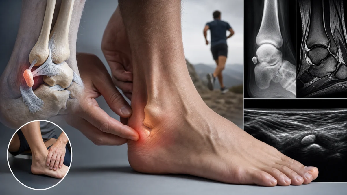
The os subfibulare lies near the lower end of the fibula on the outside of the ankle. It may originate as an anatomical variant or as a persistent fragment detached during an earlier ligament avulsion. Distinguishing these possibilities can be difficult when trauma was remote or forgotten. The fragment’s contours and earlier images provide clues; they do not replace the clinical history [1, 2].
The anterior talofibular ligament and, depending on the configuration, other lateral structures may relate to the fragment. If a ligament attachment is involved, the mechanical question is whether the ligament–fragment complex adequately stabilises the ankle. The issue is therefore more than the presence of an additional bone. Studies of unstable ankles describe different situations according to ossicle shape and associated structures [5, 7].
Pain precisely below or in front of the malleolus, reproduced during usual activities, makes this region relevant to examine. Diffuse pain or pain elsewhere encourages consideration of other explanations. An incidental finding alone does not justify intervention or a prolonged withdrawal from sport.

Recognising symptoms without reaching a premature conclusion
Symptomatic people may describe discomfort when running, pivoting or after a long walk. Some experience giving way, whereas others mainly have localised pain. These presentations are not interchangeable. An ankle may feel unreliable because of reduced muscular control, apprehension or ligament laxity; several factors may combine. General ankle-instability recommendations emphasise functional assessment [9].
After a recent sprain, pain should not automatically be attributed to the previously known fragment. A new fracture, fibular tendon injury or joint lesion may coexist. Deeper persistent pain, catching or locking also justifies appropriate investigation. Assessment checks which structure explains the current picture instead of reusing an old diagnostic label without examination.
Growing structures require particular interpretation in children and adolescents. Paediatric studies describe children with prolonged pain and instability despite conservative treatment. They do not establish that every ossicle discovered in a young athlete will become symptomatic [3, 4].
Signs that change the priority
After trauma, deformity, inability to bear weight or substantial bony pain requires prompt assessment. A cold or pale foot, loss of sensation or new weakness is urgent. A very hot, swollen joint accompanied by fever should not be treated as straightforward chronic instability. Rehabilitation should wait until the concerning cause has been assessed.
What examination and imaging can clarify
The history reviews sprains, the circumstances of the first episode, provoking activities and treatments already tried. Clarifying what “unstable” means is useful: actual giving way, pain during weight-bearing or fear of falling again? Comparison with the other side helps interpret mobility, strength and balance. Professional ligament assessment adds information when indicated [9].
Radiographs characterise the fragment and sometimes allow comparison over time. Ultrasound may examine certain ligaments and tendons; MRI may clarify neighbouring tissues, reactions at the fragment interface or another suspected injury. CT is reserved for particular bony questions or planning specialist procedures. Not every person needs all these investigations.
Studies have explored imaging characteristics associated with rehabilitation response or failure of conservative treatment. They suggest that size, position and anatomical relationships may matter. However, an association within a group cannot reliably predict an individual’s course. Imaging should guide discussion rather than become a verdict [6, 7].
| Situation | What it may suggest | Guidance for next steps |
|---|---|---|
| Ossicle without pain or giving way | Possibly an incidental anatomical finding [2] | Do not treat the image alone. |
| Focal pain and repeated giving way | Possible ligament involvement or a symptomatic interface [3, 5] | Assess clinical concordance and function. |
| Pain after a new sprain | A possible new injury despite an old ossicle | Look for concerning traumatic signs. |
| Instability despite appropriate rehabilitation | A mechanical component needs clarification [6, 7] | Discuss specialist advice. |
| Return to sport | Several capabilities need assessment [10] | Monitor pain, confidence, balance and sporting tasks. |
Clinical guide — Osteomag. This table does not replace individual assessment.
Rehabilitation centred on function
When conservative management is appropriate, the programme may combine temporary activity adjustments, strengthening and weight-bearing control. Recommendations for sprains and lateral instability support these components. Their application to an ankle with an os subfibulare must be individualised: the recommendations are not a specific trial demonstrating resolution of symptoms related to this ossicle [9].
Relative rest reduces activities that clearly maintain symptoms while preserving tolerated participation. For example, someone troubled by changes of direction may continue walking on level ground if this causes neither giving way nor sustained worsening. This is not a test of courage. It maintains useful capacity while more demanding activities are progressively rebuilt.
Strengthening may involve ankle-control muscles, the calf and other segments contributing to lower-limb balance. Exercise selection depends on pain, available mobility and movement quality. Mastering a simple exercise before adding speed, fatigue or an unstable surface is preferable. No exercise should attempt to move or fuse the bone fragment.
Balance also progresses through stages: safe supported weight-bearing, single-leg control, movement and tasks resembling actual demands. Someone who works standing up and an athlete playing a pivoting sport have different final objectives. External support may be proposed according to the circumstances; it remains an adjunct and does not remove the need to assess giving-way episodes [9].
Adjusting progression with concrete measures
Simple follow-up may record pain during activity, the next-day response and further giving-way episodes. Persistently increased pain or swelling calls for reducing load and reviewing the programme. Progression may change one variable at a time: duration, terrain difficulty, speed or repetitions. This makes the response easier to interpret.
These measures are practical tools for support, not a validated protocol specifically for os subfibulare. Their purpose is to make decisions observable. “I am better” becomes more precise when someone can add: “I walk longer without swelling and feel more secure on stairs.”
What role can osteopathic support play?
A consultation may explore movement habits, compensations and difficulties during activities. Once concerning signs have been assessed, cautious support for comfort and mobility in related regions may fit within the care pathway. It should remain coordinated with rehabilitation and the recommendations of the professional responsible for diagnosis.
There is no strong evidence that osteopathic treatment corrects an os subfibulare, independently restores an impaired ligament or consistently avoids an operation. Specific publications mainly concern anatomy, imaging and surgical outcomes in selected people. Explaining these limitations helps establish realistic objectives: improving function, managing activity demands and recognising when different expertise is needed [1, 3, 4].
Support must not delay referral when giving way persists or pain prevents progression. Repeating an intervention without measurable functional improvement is insufficient. An agreed review point allows discussion of next steps rather than automatic continuation of the same care.
When should specialist advice be discussed?
Persistent localised pain, repeated instability or failure of a well-conducted conservative programme may lead to an orthopaedic opinion. Some paediatric series report improvement after fragment removal and ligament reconstruction or repair. They concern patients meeting particular criteria; they do not prove that surgery is preferable for everyone with an ossicle [3, 4].
The strategy depends partly on fragment size, its attachments and the stability required. A review of symptomatic avulsions describes several approaches, while studies of large fragments also report fixation techniques. This explains why discussion cannot be reduced to “removing the little bone”. Expected benefits, risks and subsequent rehabilitation should be explained individually [1, 8].
Persistent difficulty does not necessarily mean that initial treatment was useless. It may reveal a mechanical component requiring another solution or an associated injury needing clarification. Bringing earlier images and a summary of tolerated activities facilitates specialist discussion.
Returning to activities with greater confidence
For return to sport, the PAASS framework proposed after a lateral ankle sprain considers several dimensions: pain, ankle impairments, athlete perception, sensorimotor control and functional performance. It may inform a broader assessment, but has not been validated as a condition-specific return-to-sport criterion for symptomatic os subfibulare [10].
Return should therefore not depend solely on reassuring radiographs or a date on the calendar. Sporting demands, any giving-way episodes and the response after a session also matter. The person should understand which activities are permitted, which require adaptation and which symptoms justify another consultation.
Living with an anatomical variation does not automatically require giving up activities. Coherent assessment and progressive objectives help distinguish an ankle needing rehabilitation from a situation requiring specialist treatment. The guiding principle remains actual function over time rather than the fragment’s presence alone.
Osteopathic support plan: key points
- Connect pain and giving way with examination, not radiographs alone.
- Refer promptly for concerning traumatic, neurological or circulatory signs.
- Temporarily adapt activity while progressively rebuilding strength and balance.
- Monitor pain, swelling and the response after exercise.
- Address comfort and function without promising to correct the bone fragment.
- Review objectives and seek specialist expertise when progress remains limited.
When reviewing progress, separate three questions: is pain less troublesome, does the ankle give way less often, and are important activities easier? These answers need not change together. Keeping them distinct makes the next discussion more precise and avoids treating a reassuring image as the only measure of recovery.
References
- [1] Surgical outcome of avulsion fractures of the distal fibula: a systematic literature review. PMID: 34820695.
- [2] Os subfibulare. Investigation of an accessory bone. PMID: 10546424.
- [3] Surgical treatment of ankle instability in children with os subfibulare. PMID: 37246999.
- [4] Chronic symptomatic os subfibulare in children. PMID: 23965709.
- [5] Comparison of symptomatic unstable ankle with and without os subfibulare. PMID: 35144867.
- [6] Radiologic findings for prediction of rehabilitation outcomes in patients with chronic symptomatic os subfibulare. PMID: 28664488.
- [7] Morphological Characteristics of Os Subfibulare Related to Failure of Conservative Treatment of Chronic Lateral Ankle Instability. PMID: 31665918.
- [8] Outcomes of Osteosynthesis of Os Subfibulare for Chronic Lateral Ankle Instability With Large Os Subfibulare. PMID: 39831545.
- [9] Ankle Stability and Movement Coordination Impairments: Lateral Ankle Ligament Sprains Revision 2021. PMID: 33789434.
- [10] Return to sport decisions after an acute lateral ankle sprain injury: introducing the PAASS framework—an international multidisciplinary consensus. PMID: 34158354.