Recognizing bone involvement, understanding imaging and protecting hip function
Hip pain is often attributed to tendinopathy, osteoarthritis or mechanical stiffness. Yet some progressive groin pain may reflect osteonecrosis of the femoral head, a condition in which part of the bone loses its blood supply and may gradually collapse.
Osteonecrosis is not a ‘locked’ joint that needs mobilizing. Early diagnosis matters because the options for preserving the hip depend strongly on the stage of the disease. The osteopath’s role is mainly to recognize the pattern, avoid aggressive techniques and refer when the presentation is compatible.
Anatomy: from living bone to subchondral fragility
What is osteonecrosis of the femoral head?
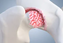
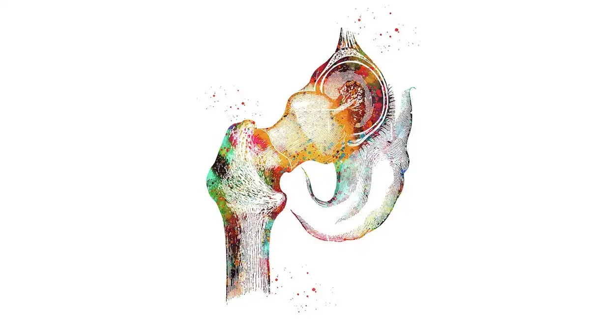
Osteonecrosis is the death of bone tissue caused by a disruption of its blood supply. In the hip, it affects the head of the femur.
Over time, the subchondral bone may lose its strength and the joint surface may collapse.
The femoral head is covered with cartilage and articulates with the acetabulum of the pelvis. Immediately beneath the cartilage, the subchondral bone helps distribute the loads of walking. Osteonecrosis initially affects bone viability; mechanical damage and secondary osteoarthritis may follow. Circulation depends on a vascular network, not a simple ‘blocked pipe’ that could be reopened by external pressure. Repair and resorption around the affected area contribute to the loss of strength. This explains why relatively moderate pain can coexist with a lesion that requires precautions. [1] [2]
Risk factors: taking a history without jumping to conclusions
What are the known risk factors?
Classic factors include:
- high-dose or prolonged systemic corticosteroid exposure;
- heavy alcohol consumption;
- hip trauma;
- certain blood disorders;
- transplantation;
- certain autoimmune diseases;
- particular treatments or circumstances.
In some cases, no clear cause is identified.
Why are corticosteroids important?
Substantial glucocorticoid exposure is one of the main nontraumatic risk factors.
The osteopath therefore needs to ask about past and current treatments, not just the medications being taken today. [3]
Does alcohol automatically cause osteonecrosis?
No. The risk is mainly associated with heavy, prolonged consumption, and not everyone who is exposed develops the disease.
This factor should be considered in its clinical context, not used as a moral judgment.
What about locally injected corticosteroids?
The risk profile depends strongly on the cumulative dose, route and context. A previous injection should not automatically be presented as a definite cause.
Why can osteonecrosis be found on both sides?
In forms associated with corticosteroids, alcohol or certain systemic diseases, both hips may be affected. An asymptomatic hip may sometimes show changes on imaging.
Orthopedic follow-up may therefore consider the opposite side, depending on the context.
Sickle cell disease is a specific context to look for in the medical history. Smoking is also associated with osteonecrosis in observational studies. These associations do not establish a definite cause in an individual, nor do they allow prognosis to be inferred from a single exposure. Prescribed treatments must never be stopped without consulting the medical team. [4] [5]
Symptoms and clinical examination: looking for a coherent pattern
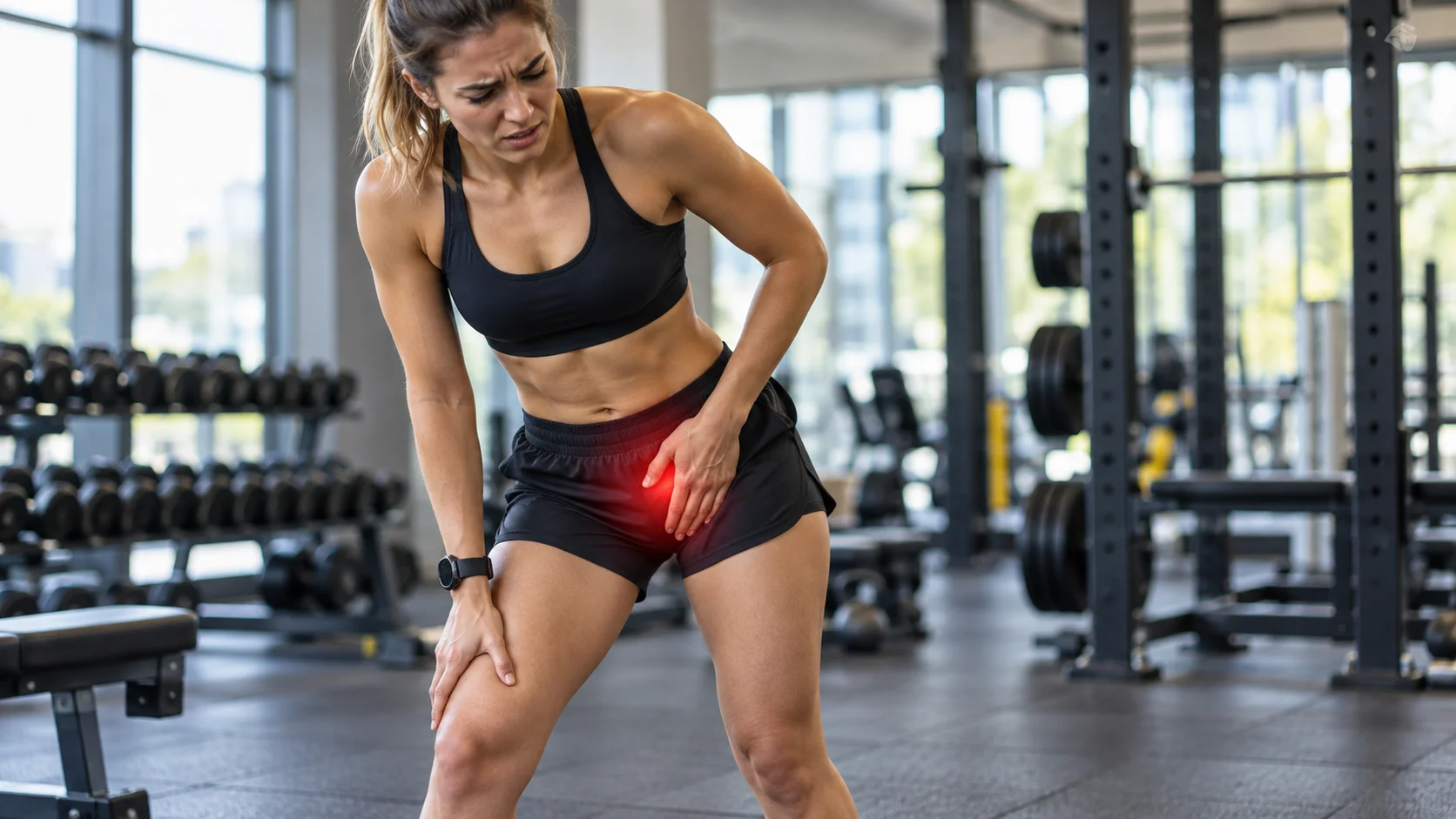
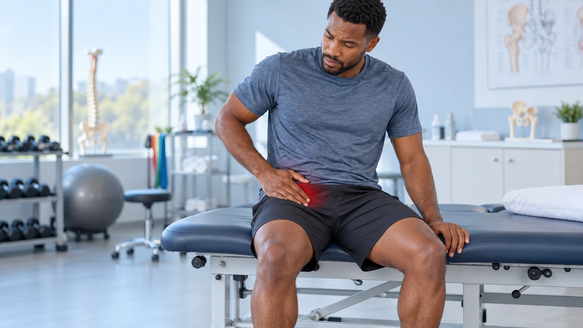
What type of pain can occur?
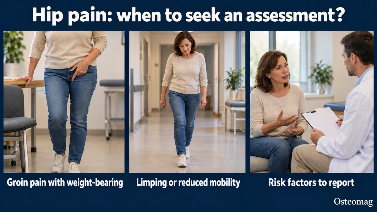
Pain is often felt:
- in the groin;
- at the front of the thigh;
- in the buttock;
- sometimes toward the knee.
It may initially occur with weight-bearing and then become more persistent.
Why can knee pain be misleading?
The hip can refer pain to the knee. A person may therefore receive several knee treatments when the main source is the hip.
Which movements often become limited?
Internal rotation, flexion and some weight-bearing movements may become painful or restricted.
However, no single limitation confirms osteonecrosis.
Why does hip pain in a younger person sometimes deserve closer attention?
Osteonecrosis can affect relatively young adults, unlike advanced osteoarthritis, which becomes more common with age.
Progressive, unexplained pain in an adult with risk factors warrants a low threshold for imaging.
Why is limping important?
A progressive limp may reflect pain, loss of mobility or collapse of the femoral head.
It must be interpreted alongside the overall course and risk factors.
Why can treating the back be misleading?
Hip pain can be confused with low back pain, and vice versa. Partial improvement in the lower back should not distract from persistent weight-bearing groin pain.
Why is pain that eases at rest not always reassuring?
In the early stages, pain may mainly be triggered by weight-bearing and remain mild at rest. Improvement when the patient sits down therefore does not rule out a bone lesion.
Why should function be documented?
Follow-up can track:
- walking;
- stairs;
- weight-bearing;
- hip rotation;
- rising from a chair;
- limping.
These measures do not replace imaging when it is indicated.
The interview clarifies onset, progression, tolerated weight-bearing, night pain and activities that have become difficult. Observing gait and a few comfortable movements documents function; multiplying provocative tests does not make the assessment more reliable. Osteoarthritis, tendinopathy, stress or insufficiency fracture, transient osteoporosis, joint infection and referred pain must remain part of medical reasoning. The osteopath identifies features that justify this assessment; the osteopath does not diagnose osteonecrosis. [1] [6]
Imaging and stage: information that changes management
Which imaging is most useful?
The systematic review informing the ARCO guideline, published in 2026, reports pooled MRI sensitivity of approximately 91% and specificity of approximately 96%. These estimates come from selected studies and populations: they do not mean that MRI is perfect, or that a particular pain can be attributed to an image without clinical examination. Radiographs can remain normal initially. [6] [7]
Why is a normal radiograph not always enough?
Radiographic changes may appear after the process has begun. If clinical suspicion remains high, MRI can detect lesions earlier.
What is the ARCO classification?
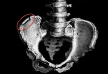
The ARCO classification revised in 2019 distinguishes four stages. In stage I, radiographs are normal but MRI or bone scintigraphy shows an abnormality. In stage II, radiographs become abnormal without fracture or flattening. In stage III, a fracture of the subchondral bone or necrotic area appears; depression of the femoral head distinguishes IIIA from IIIB. Stage IV includes osteoarthritic changes, including joint-space narrowing. Classification relies on medical interpretation of imaging, not a manual test. [8]
Why should a manual practitioner know the stage?
Because a hip with weakened bone and collapse must not be treated as a simple capsular restriction.
Aggressive mobilization does not restore necrotic bone.
Why do lesion size and location matter?
Two cases of osteonecrosis at the same stage do not necessarily carry the same risk of collapse. Larger lesions or those in heavily loaded areas may have a different prognosis.
This information comes from imaging, not palpation.
Disease stage and lesion extent are two complementary dimensions. Location-based classifications may help the orthopedic team assess prognosis, but they must not be confused with the 2019 ARCO stages. A retrospective classification study provides prognostic information, not a universal rule for surgical indications. [9]
Orthopedic management and coordinated rehabilitation
Why is early diagnosis important?
Before collapse, some surgical strategies aim to preserve the femoral head. When the structure has already collapsed and osteoarthritis is advanced, joint replacement may become necessary.
The stage therefore has a major influence on management.
What medical or surgical treatments are available?
Depending on the stage, options may include:
- monitoring and risk-factor management;
- load modification;
- core decompression;
- femoral head-preserving procedures;
- grafts or biological techniques in certain centers;
- joint replacement when the joint is severely affected.
Why does the evidence on treatments vary?
The 2026 ARCO review emphasizes the heterogeneity of lesions and interventions. Some joint-preserving outcomes are favorable, but certainty is often very low and comparisons are insufficient. A decompression, grafting or support technique therefore offers no individual guarantee. Surgical trends observed in large databases also describe practices and populations; by themselves, they do not establish the superiority of a treatment. [6] [10]
What is the role of core decompression?
Core decompression is a surgical procedure used in certain early stages to reduce intraosseous pressure and support preservation of the femoral head. Indications depend on the stage and lesion characteristics.
This procedure illustrates why early diagnosis can make a real difference to prognosis.
What is the role of exercise?
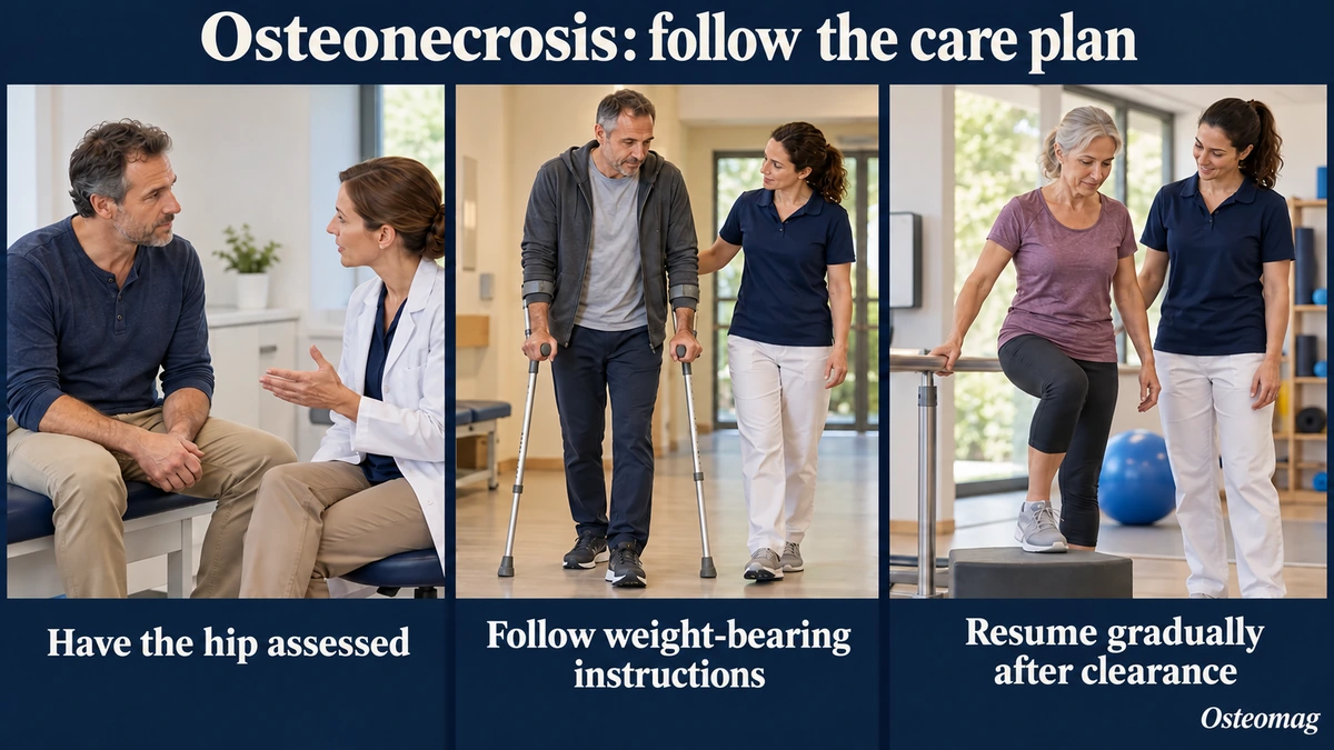
Activity depends on the stage and orthopedic recommendations. In some phases, loading needs to be modified to protect the femoral head.
After treatment, rehabilitation aims to restore strength, mobility and walking.
Why can simply advising someone to ‘walk more’ be inappropriate?
If a pre-collapse lesion requires temporarily reduced loading, greatly increasing walking without a diagnosis could be counterproductive.
The orthopedic context must take priority.
Why must the return to activity after surgery be coordinated?
After decompression or another procedure, weight-bearing progression depends on the technique and the surgeon. A manual practitioner must not increase loading simply because pain seems mild.
Osteopathy: a possible adjunct, not bone repair
Can gentle mobilization help?
When there are no warning signs and the diagnosis and restrictions have been clarified, a gentle intervention for secondary muscular pain may be discussed for comfort. Its specific benefit in osteonecrosis is not established. It must not provoke hip symptoms, change weight-bearing instructions or delay orthopedic management.
Does pain relief with manual care rule out osteonecrosis?
No.
Context, muscle relaxation or sensory modulation may temporarily reduce pain even when the bone lesion remains.
What role can osteopathy have after orthopedic treatment?
After clearance, it may possibly help with some secondary pain, mobility or comfort, always within active rehabilitation.
Why should the osteopath avoid promises about ‘restoring blood supply’?
A manual technique around the hip may change sensation and soft-tissue mobility, but there is no solid basis for claiming that it restores the blood supply to a necrotic femoral head.
How can the muscles around the hip be managed?
Pain in the gluteus medius, adductors or psoas may develop secondary to limping. Gentle treatment of these tissues may improve comfort, provided the orthopedic specialist’s weight-bearing restrictions are respected.
A useful way to explain this to the patient
‘Your hip pain and risk factors warrant imaging before we consider this simple mechanical stiffness. Osteonecrosis is managed very differently from a muscular problem, especially when it is detected early.’
Red flags and referral
Which signs should prompt referral?
These include:
- unexplained progressive groin pain;
- a substantial history of corticosteroid exposure;
- hip trauma;
- progressive loss of range of motion;
- increasing limping;
- persistent weight-bearing pain;
- normal radiographs despite strong suspicion.
When should another hip emergency be considered?
Acute pain with a complete inability to bear weight, fever, significant trauma or a suspected fracture requires urgent assessment. Osteonecrosis is not the only serious cause of groin pain.
| Situation | Priority | What manual care must not do |
|---|---|---|
| Progressive groin pain with risk factors | Medical assessment and imaging according to the context | Repeat sessions on the assumption of a simple blockage. |
| Normal radiographs but persistent suspicion | Contact the physician again to discuss further assessment | Conclude that all bone involvement has been ruled out. |
| Known osteonecrosis or after surgery | Follow the weight-bearing and rehabilitation plan | Increase loading because pain has decreased. |
| Acute pain, fever, trauma or inability to bear weight | Urgent assessment according to the signs | Delay triage to test a technique. |
Conclusion
Osteonecrosis of the femoral head is a less common but important cause of hip pain, often in adults younger than those with typical osteoarthritis.
The osteopath’s main role is to recognize risk factors, progressive groin pain and loss of function. MRI is the most effective examination when suspicion is high, and manual care must never delay orthopedic referral.
References
- [1] Hines JT, Jo WL, Cui Q et al. Osteonecrosis of the Femoral Head: an Updated Review of ARCO on Pathogenesis, Staging and Treatment. J Korean Med Sci. 2021;36(24):e177. doi:10.3346/jkms.2021.36.e177.
- [2] Shah KN, Racine J, Jones LC et al. Pathophysiology and risk factors for osteonecrosis. Curr Rev Musculoskelet Med. 2015;8(3):201-9. doi:10.1007/s12178-015-9277-8.
- [3] Birla V, Vaish A, Vaishya R. Risk factors and pathogenesis of steroid-induced osteonecrosis of femoral head – A scoping review. J Clin Orthop Trauma. 2021;23:101643. doi:10.1016/j.jcot.2021.101643.
- [4] Leandro MP, De Sá CKC, Filho DPS et al. Association and Risk Factors of Osteonecrosis of Femoral Head in Sickle Cell Disease: A Systematic Review. Indian J Orthop. 2022;56(2):216-225. doi:10.1007/s43465-021-00469-4.
- [5] Wen Z, Lin Z, Yan W et al. Influence of cigarette smoking on osteonecrosis of the femoral head (ONFH): a systematic review and meta-analysis. Hip Int. 2017;27(5):425-435. doi:10.5301/hipint.5000516.
- [6] Parikh RR, Mirzaei A, Butler ME et al. Diagnosis and Treatment of Nontraumatic Osteonecrosis of the Femoral Head: A Systematic Review and Meta-Analyses for the ARCO Clinical Practice Guideline Development Workgroup. Med Sci (Basel). 2026;14(1):107. doi:10.3390/medsci14010107.
- [7] Zhang YZ, Cao XY, Li XC et al. Accuracy of MRI diagnosis of early osteonecrosis of the femoral head: a meta-analysis and systematic review. J Orthop Surg Res. 2018;13(1):167. doi:10.1186/s13018-018-0836-8.
- [8] Yoon BH, Mont MA, Koo KH et al. The 2019 Revised Version of Association Research Circulation Osseous Staging System of Osteonecrosis of the Femoral Head. J Arthroplasty. 2020;35(4):933-940. doi:10.1016/j.arth.2019.11.029.
- [9] Yue J, Guo X, Wang R et al. Reliability and repeatability of 2021 ARCO classification and its guiding significance in treatment of nontraumatic osteonecrosis of the femoral head. BMC Musculoskelet Disord. 2023;24(1):469. doi:10.1186/s12891-023-06587-4.
- [10] Buddhiraju A, Khanuja HS, Hegde V et al. Epidemiology, Management, and Systematic Review of Surgical Trends for Patients Who Have Osteonecrosis of the Femoral Head. J Arthroplasty. 2025;40(10 Suppl 1):S112-S119.e1. doi:10.1016/j.arth.2025.06.002.