Understanding an anatomical hypothesis without diagnosing compression from posture
Introduction – One possible region, several causes to examine
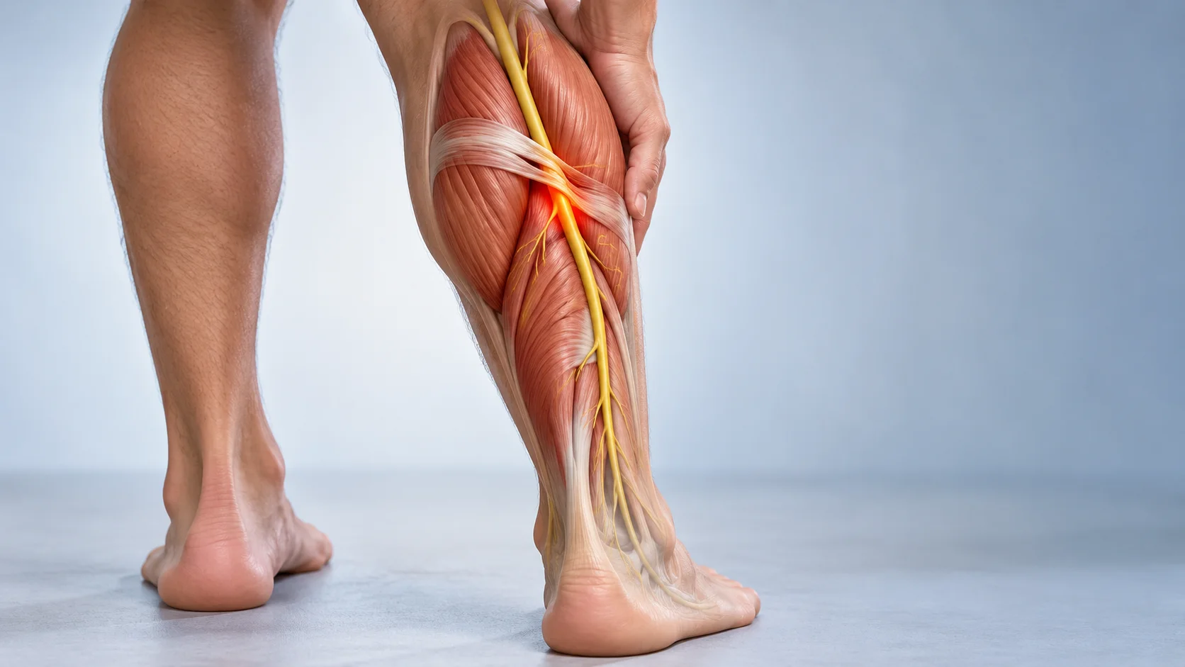
Pectoralis minor is an unobtrusive muscle, often overshadowed by pectoralis major. Its proximity to the brachial plexus cords and axillary vessels explains its relevance in some presentations of arm pain or tingling. In selected situations, the retropectoral region may contribute to compression. But a heavy arm, reduced strength or muscle tension cannot locate the cause: several diseases and musculoskeletal disorders may produce similar symptoms [1] [4] [6].
Repetitive activities, overhead movements, an injury and individual context may guide the clinical history. They do not prove that modern posture inevitably shortens this muscle or makes it a ‘weak link’. The aim is to understand symptoms, their consequences and hypotheses to investigate rather than identify a guilty structure [2] [8].
Anatomy and the muscle’s functional role
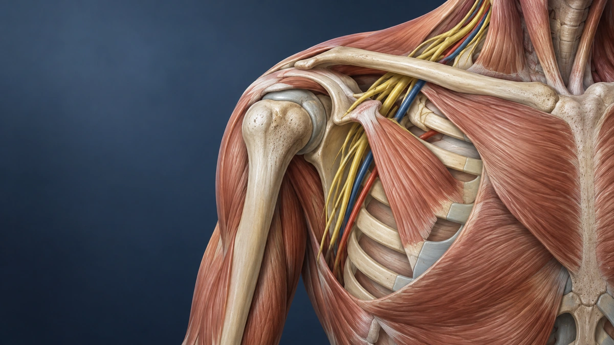
Pectoralis minor originates from the third, fourth and fifth ribs near their costal cartilages and inserts on the scapula’s coracoid process. This attachment makes it an important stabilizer and mover of the shoulder. Its main function is to depress and bring the scapula forward while contributing to anterior scapular tilt.
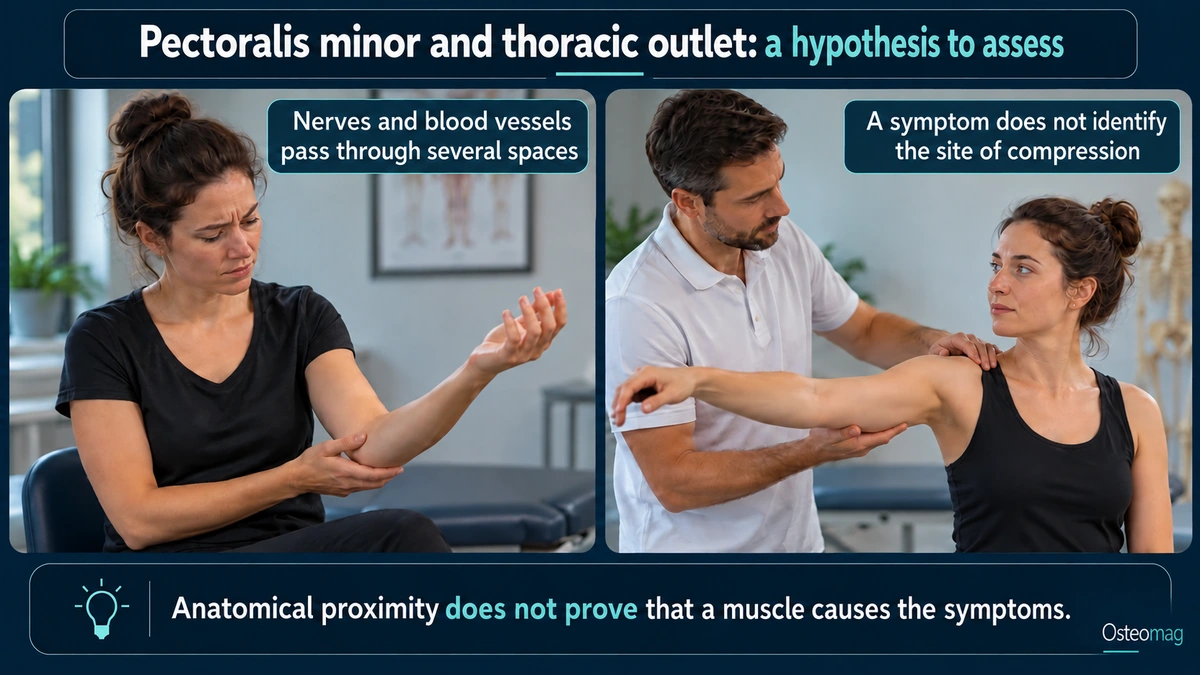
It may also assist inhalation when the scapula is stabilized by helping elevate the ribs. This accessory role does not show that upper-chest breathing or stress makes the muscle pathological. Respiratory recruitment depends on effort, demand and clinical context.
The brachial plexus cords and axillary vessels pass beneath pectoralis minor. This anatomical proximity makes a local contribution plausible in some cases, but increased tone, tenderness or shortening is insufficient to prove nerve or vascular compression. Anatomy must be related to history, examination and relevant additional investigations [4] [10].
Connection with the thoracic outlet
The thoracic outlet is the passage between the neck and upper limb used by the nerves, arteries and veins that supply and control the arm. Three main spaces may be sites of compression: the interscalene space, costoclavicular space and space beneath pectoralis minor. The last is our focus here.
Pectoralis minor syndrome describes possible compression in the retropectoral region. It may present with pain, paresthesias and activity-related discomfort; its boundaries with neurogenic thoracic outlet syndrome remain debated. Neurogenic, venous and arterial forms must be distinguished: heaviness or numbness alone does not prove vascular involvement [1] [4] [10].
In osteopathy, knowing these relationships primarily helps avoid overlooking a differential diagnosis or necessary referral. Manual care may complement comfort, but cannot confirm compression or guarantee muscular decompression.
Discoveries over time: clarifying sites without simplifying causes
The modern thoracic outlet concept developed around presentations previously grouped under different names, including those associated with a cervical rib. Peet and colleagues proposed the term thoracic outlet syndrome in 1956. This historical grouping does not imply that all arm pain has the same mechanism [2].
Later work distinguished several possible regions: the interscalene space, costoclavicular region and retropectoral space. Reviews by Sanders and Annest, then Ahmed and colleagues, describe pectoralis minor’s possible contribution. Their value is in reminding us that a local hypothesis must be compared with other causes—not in providing a universal percentage of patients or a diagnosis through stretching alone [4] [10].
Consolidating criteria remains a clinical endeavour. Society for Vascular Surgery standards and the INTOS consensus clarify descriptions and decisions for neurogenic forms. Expert consensus improves consistency in practice, but is not a trial demonstrating a manual technique’s effectiveness [1] [9].
Pectoralis minor and thoracic outlet syndrome
Mechanisms of neurovascular compression
Thoracic outlet syndrome encompasses possible nerve or vessel involvement between the base of the neck and the axilla. In the retropectoral region, relationships between pectoralis minor, nerve cords and axillary vessels may contribute. Symptoms may vary with activity, but this positional dependence does not prove a single mechanism. Cervical radiculopathy or a shoulder condition may also be influenced by movement [1] [4] [6] [10].
Differences from other thoracic outlet forms
TOS is not limited to pectoralis minor. Several possible narrowing sites are distinguished:
- Interscalene space: between the anterior and middle scalene muscles, it contains the brachial plexus trunks and subclavian artery. The subclavian vein passes in front of the anterior scalene, outside this space.
- Costoclavicular space: between the clavicle and first rib, the space may narrow during shoulder movement, particularly if the clavicle is low or unstable.
- Cervical rib or bony abnormality: an additional rib or bony growth may make the space less flexible and maintain compression.
- Beneath pectoralis minor: our focus here, where a retropectoral contribution may be investigated according to the clinical presentation.
Distinguishing regions and types of involvement guides reasoning. A positional test, Adson test or stretch may reproduce a symptom without confirming or locating compression on its own. Agreement with the overall examination and possible diagnoses must be assessed [2] [9].
Contributing factors: posture, increased tone and repetitive microtrauma
Several elements may be explored in the history without treating them as automatic causes:
- Prolonged activities and positions: investigate their actual relationship with symptoms without concluding that a forward shoulder necessarily compresses structures.
- Repeated muscle work: load, recovery and capacity matter; a perceived contraction is not proof of compression.
- Repeated effort or trauma: clarify the timeline without inferring fibrosis from palpation.
- Breathing and effort: accessory recruitment may be observed, but cannot explain arm symptoms alone.
- Anatomical variations: some may contribute and should be assessed medically when indicated.
These factors should be investigated within a complete clinical assessment. Conservative management primarily aims for a gradual return to activity, scapular control, thoracic mobility and adapted strengthening. Manual techniques may sometimes help pain or mobility in the short term, but have not been shown to ‘release’ neurovascular compression and do not replace medical assessment when symptoms are significant.
Clinical assessment and differential diagnosis
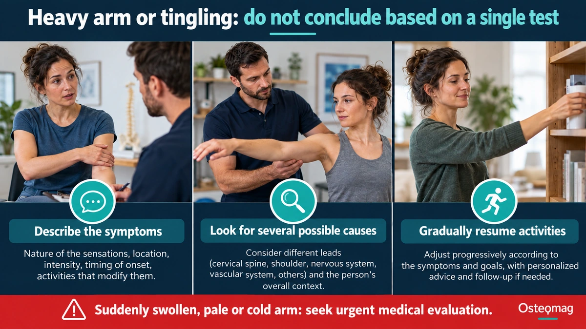
A heavy arm: distinguishing priorities
| Observation or situation | Cautious interpretation and possible next step |
|---|---|
| Activity-related pain or tingling | Clinical examination and differential diagnosis; a provocative position is insufficient. |
| Progressive weakness or muscle wasting | Specialist assessment without waiting through a prolonged programme. |
| Sudden swelling or bluish colour | Urgent medical assessment for a possible vascular cause. |
| A suddenly cold or pale hand | Urgent assessment; do not first treat it as muscle tension. |
The professional clarifies symptom distribution, onset, associated positions and effort, trauma and progression. They examine the neck, shoulder, sensory and motor function and vascular signs within their competence. Symptoms may originate from a cervical root, peripheral nerve, plexus, shoulder or vascular disease. No posture test replaces this comparison [1] [2] [6] [9].
The physician selects investigations according to the question: vascular studies if thrombosis or arterial involvement is plausible, neurological investigations or imaging if the presentation warrants it. Results must be interpreted alongside symptoms; positional variation or an anatomical image is insufficient to define the problem [1] [8] [9].
Thoracic outlet syndrome, including a form possibly related to pectoralis minor, remains a complex clinical diagnosis. Positional tests may reproduce symptoms, but do not suffice to confirm compression or reliably locate the responsible site. A structured assessment must also investigate cervical radiculopathy, brachial plexus involvement, carpal or cubital tunnel syndrome, shoulder involvement, myofascial pain or a vascular cause [1] [2] [3] [4].
True vascular symptoms—swelling, persistent colour change, a cold hand, pain with exertion or newly visible superficial veins—require particular attention. Depending on the signs, a physician may request imaging or vascular and neurological tests. Investigations are not routinely useful in every case, but are essential when thrombosis, arterial involvement or progressive neurological involvement is possible.
What can be done?
The INTOS consensus generally proposes conservative management before a surgical decision for neurogenic forms, but substantial weakness or atrophy may accelerate specialist referral. An arbitrary interval should not be awaited when the condition is worsening. The Cochrane review highlights the lack of robust trials for several treatments; manual decompression’s effectiveness is not established [3] [5] [9].
A retrospective cohort of surgically treated patients reports improvements but also residual functional limitations. These results concern surgical selection, not proof of osteopathy’s benefit. Rehabilitation selects a useful task, temporarily adjusts provocative activities and progresses according to tolerance without forcing a stretch that increases tingling [7].
For a non-urgent neurogenic presentation, conservative care is generally the first step: temporarily adapting symptom-triggering activities, gradually improving shoulder-girdle endurance and strength, thoracic and cervical mobility, and a progressive return to overhead actions. Literature emphasizes individualized rehabilitation; programmes’ exact effectiveness varies and evidence remains heterogeneous [2] [5].
Manual treatment or osteopathy may be integrated as an adjunct for comfort and return to movement, provided promises of instant ‘decompression’ or structural correction are avoided. The useful aim is function: reduce irritability, regain capacity and help the person resume activities with an active plan.
When should you seek prompt care?
Seek care promptly if an arm or hand suddenly becomes swollen, bluish, pale or cold, or for chest pain, breathlessness, progressive weakness, persistent loss of sensation, major trauma or unusual night pain. Arm pain accompanied by cardiac symptoms, feeling unwell or sweating requires urgent assessment.
Scientific references
- [1] Illig et al. Reporting standards of the Society for Vascular Surgery for thoracic outlet syndrome. J Vasc Surg. 2016;64(3):e23-e35. doi:10.1016/j.jvs.2016.04.039.
- [2] Hooper et al. Thoracic outlet syndrome: a controversial clinical condition. Part 1: anatomy, and clinical examination/diagnosis. J Man Manip Ther. 2010;18(2):74-83. doi:10.1179/106698110X12640740712734.
- [3] Hooper et al. Thoracic outlet syndrome: a controversial clinical condition. Part 2: non-surgical and surgical management. J Man Manip Ther. 2010;18(3):132-138. doi:10.1179/106698110X12640740712338.
- [4] Sanders et al. Pectoralis Minor Syndrome: Subclavicular Brachial Plexus Compression. Diagnostics (Basel). 2017;7(3):46. doi:10.3390/diagnostics7030046.
- [5] Povlsen et al. Treatment for thoracic outlet syndrome. Cochrane Database Syst Rev. 2014;2014(11):CD007218. doi:10.1002/14651858.CD007218.pub3.
- [6] Ferrante et al. The thoracic outlet syndromes. Muscle Nerve. 2012;45(6):780-795. doi:10.1002/mus.23235.
- [7] Peek et al. Long-Term Functional Outcome of Surgical Treatment for Thoracic Outlet Syndrome. Diagnostics (Basel). 2018;8(1):7. doi:10.3390/diagnostics8010007.
- [8] Li et al. Thoracic Outlet Syndrome: A Narrative Review. J Clin Med. 2021;10(5):962. doi:10.3390/jcm10050962.
- [9] Chim et al. Consensus Recommendations for Neurogenic Thoracic Outlet Syndrome from the INTOS Workgroup. Plast Reconstr Surg Glob Open. 2024;12(8):e6107. doi:10.1097/GOX.0000000000006107.
- [10] Ahmed et al. Pectoralis minor syndrome – review of pathoanatomy, diagnosis, and management of the primary cause of neurogenic thoracic outlet syndrome. JSES Rev Rep Tech. 2022;2(4):469-488. doi:10.1016/j.xrrt.2022.05.008.