

Achilles tendonitis is an inflammation of the Achilles tendon, located at the back of the ankle, caused by excessive stress, overuse or injury. It manifests itself as pain, swelling and tenderness in the affected area.
Introduction
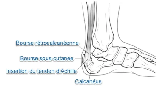
Achilles tendinitis, also known by the synonym Achilles tendinitis, is a condition frequently observed among running enthusiasts, particularly among athletes who intensively use the calf muscles. This muscle group, made up of the gastrocnemius and soleus muscles, is closely linked to the calcaneus by the Achilles tendon.
When running, these muscles are actively engaged to lift the foot off the ground, placing repeated stress on the Achilles tendon. Insufficient recovery time, combined with continued effort, can initially lead to inflammation of the paratenon, an areolar fatty tissue separating the Achilles tendon from its covering. It is crucial to note that complete rupture of the Achilles tendon, a serious injury, usually occurs following a sudden and intense load.
The Achilles tendon, resulting from the convergence of fibers from the gastrocnemius and soleus muscles, crosses the knee and ankle joints, making it more vulnerable to injury than tendons that only cross a single joint.
An important anatomical feature lies in the poor blood supply to the Achilles tendon, particularly in its distal part. This characteristic makes it the area most exposed to overuse tendon injuries. It is essential to emphasize that Achilles tendon injuries, contrary to what one might think, are not primarily of an inflammatory nature (tendinitis), but rather involve collagen degeneration (tendinosis) as the primary pathology. .
Achilles tendinosis often occurs in runners who suddenly increase the intensity or duration of their sessions. It is also common among middle-aged individuals who play sports like tennis or basketball sporadically on weekends. Understanding these risk factors and the underlying mechanisms is essential for the prevention and effective management of Achilles tendonitis.
Two types of Achilles tendinosis
- Non-insertional
- Insertional
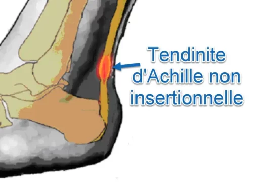
Non-insertional Achilles tendonitis
Non-insertional Achilles tendinosis, also called non-insertional Achilles tendinopathy, is characterized by damage and degenerative changes of the Achilles tendon, but unlike insertional Achilles tendinosis, the alterations occur in the central part or middle of the tendon, far from its insertion at the heel bone (calcaneus). Here are some characteristics of non-insertional Achilles tendinosis:
- Location of Injuries: Injuries affect the middle or central part of the Achilles tendon, rather than the area where the tendon attaches to the heel bone.
- Degenerative Changes: As with insertional Achilles tendinosis, non-insertional tendinosis is associated with degenerative changes to the tendon, such as changes in collagen, nodules, or calcifications.
- Pain Along the Tendon: Pain is usually felt along the Achilles tendon, away from the heel bone. It can be chronic and increase with physical activity.
- Stiffness and Swelling: Symptoms such as stiffness and mild swelling may accompany non-insertional Achilles tendinosis.
- Pain When Walking or Running: Activities that stress the Achilles tendon, such as walking, running or jumping, can cause or worsen pain.
- Sensitivity to Touch: Palpation of the tendon along its medial portion may cause pain, indicating increased sensitivity.
- Morning Pain: Some individuals may experience increased pain upon rising in the morning or after a period of inactivity, due to tendon stiffness.
- Structural Alterations Visible on Imaging: Imaging tests, such as ultrasound or MRI, can show structural changes characteristic of non-insertional Achilles tendinosis.
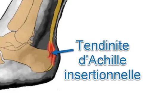
Insertional Achilles tendonitis
Insertional Achilles tendinosis, also called insertional Achilles tendinopathy, is a condition characterized by damage and degenerative changes at the insertion of the Achilles tendon to the heel bone (calcaneus). Unlike tendinitis, which involves inflammation, tendinosis is rather associated with structural alterations and degeneration of tendon tissue. Here are some characteristics of insertional Achilles tendinosis:
- Location of Lesions: Lesions specifically affect the area where the Achilles tendon attaches to the heel bone (calcaneus).
- Degenerative Changes: Unlike tendinitis, tendinosis is associated with degenerative changes to the tendon. There may be alterations in collagen, nodules, or even calcifications in the tendon.
- Tendon Insertion Pain: Pain is usually felt at the back of the heel, at the Achilles tendon insertion site. This pain can be chronic and worsen with physical activity.
- Stiffness and Swelling: In addition to pain, insertional Achilles tendinosis can cause stiffness and slight swelling in the affected area.
- Pain When Walking or Running: Symptoms may worsen during walking, running, or other activities that strain the Achilles tendon.
- Sensitivity to Touch: The tendon insertion area may be tender to touch, and palpation may cause pain.
- Morning Pain: Some individuals may experience increased pain upon rising in the morning, or after a period of inactivity, due to tendon stiffness.
- Structural Alterations Visible on Imaging: Imaging tests, such as ultrasound or MRI, can reveal structural changes characteristic of insertional Achilles tendinosis.
Causes
- Overload and Intense Activities: Overuse of the Achilles tendon from intensive physical activities, such as running, jumping, or other high-impact sports, can lead to tendinitis.
- Sudden Changes in Physical Activity: Sudden changes in the level of physical activity, such as sudden increases in running distance or exercise intensity, can overwork the Achilles tendon.
- Improper Exercise Technique: Improper exercise technique, including incorrect repetitive movements or poor posture during physical activity, can contribute to the development of Achilles tendonitis.
- Inappropriate Footwear: Footwear that does not provide adequate support, is worn, or is not suitable for the physical activity being performed can increase the risk of Achilles tendinitis.
- Biomechanical Abnormalities: Abnormalities in the biomechanics of the foot, such as excessive pronation, flatfoot, or other variations, can place additional pressure on the Achilles tendon.
- Age: Natural aging can cause a decrease in the flexibility and elasticity of the tendon, increasing the risk of tendinitis.
- Posture Problems: Poor posture or muscular imbalances can contribute to excessive stress on the Achilles tendon.
- Traumatic Injuries: Traumatic injuries, such as sprains or fractures of the foot or ankle, can increase the risk of developing Achilles tendinitis.
- Underlying Medical Conditions: Certain medical conditions, such as arthritis, diabetes, or infections, may increase susceptibility to Achilles tendonitis.
- Smoking: It has been suggested that smoking may be a risk factor, as it may reduce vascularity and affect tendon healing.
Symptoms
- Pain: Pain is one of the most common symptoms. It is usually felt along the Achilles tendon, near the heel bone. The pain may be mild at first and become more severe over time.
- Morning Stiffness: Some individuals may experience stiffness in the Achilles tendon, particularly in the morning upon waking. Stiffness may also be present after periods of inactivity.
- Swelling: Inflammation of the tendon can cause slight swelling in the affected area. However, the swelling may not be as obvious as in other inflammatory conditions.
- Sensitivity to Touch: The area around the Achilles tendon may be sensitive to touch. Direct pressure or rubbing can make the pain worse.
- Exacerbating Overheating: Pain may intensify during or after physical activities, particularly when the Achilles tendon is stressed. This may include walking, running, stair climbing, or other movements that put pressure on the tendon.
- Discomfort during Plantar Flexion: When you perform plantar flexion (tips of the toes downward) movements, pain may increase.
- Cracking or Squealing: Some individuals report cracking or screeching sensations when they move their Achilles tendon. This may be associated with abnormal friction of the tendon structures.
Pathophysiology
The pathophysiology of Achilles tendonitis involves degenerative and inflammatory changes to the Achilles tendon, which is the tendon connecting the calf muscles to the heel bone. Here are the main stages of the pathophysiology of Achilles tendonitis:
- Overuse or Microtrauma: Achilles tendonitis is often associated with overuse of the Achilles tendon. Repetitive activities, excessive loads, sudden changes in exercise intensity or frequency, or microtrauma can contribute to tendon damage.
- Initial Inflammatory Response: Microtrauma causes an initial inflammatory response in the tendon. This inflammation is a normal reaction of the body to tissue damage.
- Alteration of the Tendon Structure: Repetition of microtrauma can lead to structural alterations of the tendon. Collagen, which makes up the majority of the tendon, can undergo degenerative changes, leading to decreased strength and elasticity of the tendon.
- Inadequate Healing: In response to injury, the healing process may be disrupted. Instead of normal tissue regeneration, degenerative changes may predominate, leading to inadequate healing of the tendon.
- Formation of Abnormal Tenocytes: Tenocytes, the cells responsible for tendon regeneration, can undergo alterations. Instead of regenerating normally, tenocytes may adopt an abnormal shape, contributing to tendon degeneration.
- Increased Mechanical Load: Structural alterations of the tendon can increase the mechanical load on the Achilles tendon, which can worsen existing injuries and lead to continued inflammation.
- Formation of Nodules and Calcifications: In some cases, nodules or calcifications may form in the affected tendon, contributing to pain and loss of flexibility.
- Inflammatory Cell Proliferation: The persistent inflammatory response can lead to the proliferation of inflammatory cells, worsening inflammation and associated symptoms.
Differential diagnosis
- Plantar Fasciitis: Inflammation of the plantar fascia, the connective tissue under the foot, can cause heel pain similar to Achilles tendonitis.
- Retrocalcaneal Bursitis: Inflammation of the bursa between the Achilles tendon and heel bone can produce symptoms similar to tendinitis.
- Tarsal Tunnel Syndrome: Nerve compression in the tarsal tunnel, located on the inside of the ankle, can cause heel pain.
- Morton’s neuroma: Abnormal growth of nerve tissue between the toes can cause pain similar to Achilles tendonitis.
- Calcaneus Stress Fracture: A stress fracture of the heel can present with painful symptoms similar to tendinitis.
- Foot or Ankle Arthritis: Some forms of arthritis, such as rheumatoid arthritis or osteoarthritis, can cause pain and inflammation in the heel area.
- Posterior Tibial Tendinopathy: Inflammation of the posterior tibial tendon, which supports the arch of the foot, can also cause heel pain.
- Ankle Cyst: The presence of a cyst, especially a Baker’s cyst, can cause similar symptoms in the heel area.
- Posterior Compartment Syndrome: Increased pressure in the posterior compartment of the calf can cause pain in the heel area.
- Calcification of the Achilles Tendon: The formation of calcium deposits in the Achilles tendon can lead to painful symptoms.
Prevention
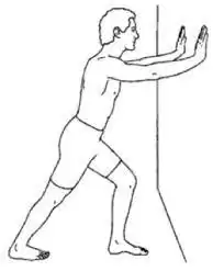
- Calf stretch
- Start of each day.
- Before and after workouts.
- Progress the intensity of your exercises in a way that does not traumatize the muscle
- Combine high and low impact exercises (eg: basketball and swimming).
- Choose shoes whose impact on the ground is well absorbed. It is important that the shoe has arch support present.
- Using an in-shoe heel lift: Slightly elevating your heel can relieve strain on the tendon and provide a cushion that reduces the force on your Achilles tendon.
Reference
- Carr AJ, Norris SH. The blood supply of the calcaneal tendon. J Bone Joint Surg Br. 1989;71(1):100–101.
- Kraushaar BS, Nirschl RP. Tendinosis of the elbow (tennis elbow). Clinical features and findings of histological, immunohistochemical, and electron microscopy studies. J Bone Joint Surg Am. 1999;81(2):259–278.
- Ahmed IM, Lagopoulos M, McConnell P, Soames RW, Sefton GK. Blood supply of the Achilles tendon. J Orthop Res. Sep 1998;16(5):591–596.


