

Introduction
The mobility and stability of our feet are often aspects of our daily lives that we pay little attention to, until an orthopedic condition such as calcaneus stress fracture suddenly catches our eye. This microscopic fracture, located in the heel bone, can have significant repercussions on quality of life, particularly in individuals engaged in regular physical activities. Through the exploration of the causes, symptoms, treatments and medical advances related to calcaneus stress fracture, this text aims to provide clarity and raise awareness of this often misunderstood condition.
Let’s first dive into the complex anatomy of the foot to understand the nuances of this fracture. Often caused by overuse, calcaneus stress fractures typically occur in individuals who frequently place stress on their feet, whether through running, prolonged walking, or other strenuous physical activities. Athletes and fitness enthusiasts are particularly prone to this injury, highlighting the importance of prevention and proper management.
Calcaneus stress fracture symptoms may be subtle at first, manifesting as a dull pain in the heel, which usually intensifies with physical activity. It is crucial to recognize these early signs to avoid worsening of the injury and long-term complications. Modern diagnostics, such as magnetic resonance imaging (MRI) and computed tomography (CT), play a vital role in confirming stress fracture and help healthcare professionals develop treatment plans tailored to each individual patient. patient.
Regarding treatment strategies, rest and modification of physical activity are often recommended to allow for adequate healing. Using appropriate footwear and adjusting training techniques can also help prevent recurrence. In more severe cases, medical intervention may be necessary, including immobilizing the foot using a cast or orthopedic devices.
The continued advancement of medical research opens up promising future prospects in the understanding and management of stress fracture of the calcaneus. Innovative approaches, such as extracorporeal shock wave therapy, are currently being studied to evaluate their effectiveness in promoting healing.
In conclusion, stress fracture of the calcaneus, although discreet, deserves special attention because of its implications on mobility and stability of the feet. Raising awareness of this condition, combined with preventive practices and appropriate treatments, can play a crucial role in improving the quality of life of affected individuals.
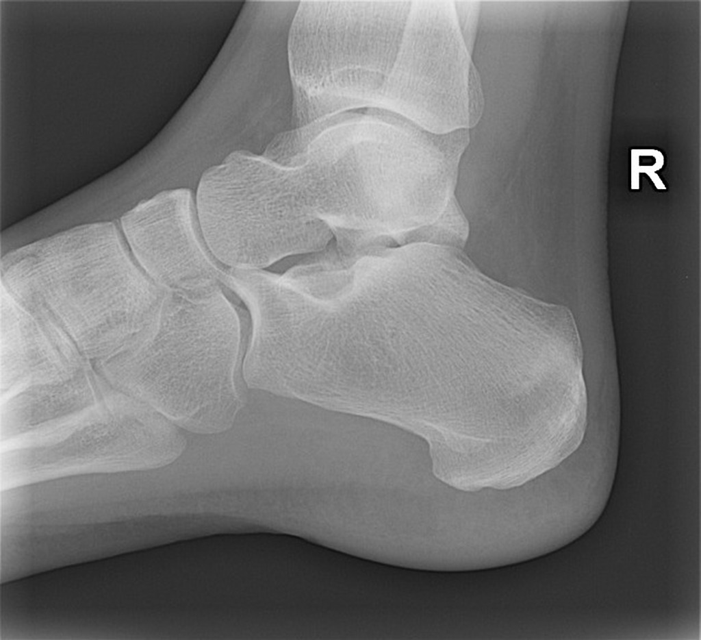
Anatomy of the calcaneus
The calcaneus is the largest bone in the foot, located at the heel. Here is a brief description of the anatomy of the calcaneus:
- Body of the calcaneus: This is the posterior part of the bone, which forms the majority of the heel.
- Posterior process: A bony prominence at the back of the calcaneus, also called the calcaneal tuberosity. The calf muscles are attached to it.
- Subtalar facet joint: A joint surface located on the lower part of the calcaneus, which forms a joint with the talus, another bone of the foot.
- Anterior process: A small bony prominence located at the front of the calcaneus.
- Sustentaculum tali: A cross-shaped structure located medially on the subtalar articular facet, which supports the weight of the body and transfers pressure to the bones of the foot.
All of these structures contribute to the stability of the foot and the transmission of forces during walking and running. The calcaneus is crucial to the biomechanics of the foot and plays an essential role in human locomotion.
Definition of Calcaneus Stress Fracture
Calcaneus stress fracture is an injury that occurs when there is a build-up of repeated or excessive stress on the heel bone (calcaneus), exceeding its ability to recover. Unlike acute fractures resulting from sudden impact, stress fractures develop gradually following overuse. This condition is common among athletes, especially those who participate in high-impact activities, such as running or jumping.
The nature of microfractures in the calcaneus during a stress fracture is important to understanding this injury. Here is a detailed explanation:
- Repetitive Strain Injury: Repetitive, high-impact activities can create microtrauma on the heel bone. These repeated stresses can exceed the bone’s ability to fully regenerate between periods of activity.
- Bone fatigue: Repetitive microtrauma causes an accumulation of fatigue within the calcaneus. The bone may begin to show signs of weakness and microfractures due to constant repetition of stress.
- Inflammatory reaction: Microfractures trigger a local inflammatory reaction. Immune system cells and bone cells attempt to repair the damage, but if overuse continues, the healing process can be disrupted.
- Formation of microfractures: Excessive stress can lead to the formation of microfractures in the calcaneus. These fractures are often very small and difficult to detect by conventional imaging methods.
Causes and risk factors
Several underlying causes and risk factors may contribute to the development of stress fractures of the calcaneus, particularly in individuals engaged in repetitive physical activities. Here are some of these causes and risk factors:
- Overuse: One of the main causes of stress fractures of the calcaneus is overuse due to repetitive or excessive physical activities. This can include running, jumping, ballet, basketball, and other high-impact sports.
- Changes in intensity: A sudden increase in the intensity or duration of physical activities can increase the risk of stress fractures. Athletes who rapidly increase their training volume without a sufficient adaptation period are more likely to develop these fractures.
- Improper training techniques: Improper training techniques, such as changes in running technique, poor cushioning in athletic shoes, or incorrect landing technique when jumping, can contribute to excessive stress on the calcaneus.
- Training surface: Training on hard or uneven surfaces, such as concrete, can increase pressure on the heel and increase the risk of stress fractures.
- Inadequate shoes: Athletic shoes that are inadequate in terms of support, cushioning, or stability can contribute to excessive stress on the calcaneus.
- Biomechanical Abnormalities: Individuals with biomechanical abnormalities, such as excessive pronation or supination, may be more likely to develop stress fractures of the calcaneus.
- Low bone density: Low bone density, often due to conditions such as osteoporosis, can make bones more vulnerable to stress fractures.
- Hormonal factors: In some populations, hormonal changes, such as those associated with menstruation in women, can influence bone density and contribute to the risk of stress fractures.
- History of stress fractures: Individuals who have previously had stress fractures elsewhere in the body are more likely to develop stress fractures in the calcaneus.
- Age: Adolescents and young adults, due to bone growth and changes in bone density, may be more prone to stress fractures.
It is important to note that these risk factors do not guarantee the development of stress fractures, but they can increase the likelihood. Preventing stress fractures often involves a holistic approach, including pr
Characteristic symptoms :
- Heel Pain: Heel pain is one of the most prevalent symptoms. It can be localized on the underside or back of the heel and intensify during walking, running or any physical activity requiring the foot.
- Worsen with Activity: Pain tends to get worse during or after high-impact activities, such as running, jumping, or even just prolonged walking.
- Local Swelling: Swelling may develop in the heel, resulting from the inflammatory response to the stress fracture. This swelling may be accompanied by local redness.
- Sensitivity to Touch: The area surrounding the stress fracture may become sensitive to touch. Palpation of the heel can trigger or worsen the pain.
- Nighttime Pain: Some individuals may experience increased pain at night, particularly when lying down. This may be related to increased blood circulation and pressure on the heel during rest.
- Lameness: To alleviate pain, people with stress fractures of the calcaneus may adopt a limping gait, avoiding putting too much weight on the affected heel.
- Rigidity: A certain stiffness in the heel may appear, limiting joint mobility.
Pathophysiology of Calcaneus Stress Fracture:
The pathophysiology of calcaneus stress fracture involves complex mechanisms resulting from repeated stress on the bone, exceeding its normal healing capacity. Key steps in pathophysiology include:
- Repetitive Strain Injury: Stress fracture of the calcaneus usually develops due to repetitive strain injury caused by high impact activities. These repetitive forces exceed the calcaneus’ ability to absorb shock and regenerate quickly.
- Overload of the Bony Trabeculum: Repeated stresses generate an overload on the bony trabecular meshwork of the calcaneus, the internal part of the bone which forms a lattice structure. This overload can lead to progressive degradation of the trabecular meshwork.
- Inflammatory Reaction: In response to microtrauma, a local inflammatory reaction occurs, characterized by the release of inflammatory mediators. This inflammation can contribute to pain, swelling, and other symptoms associated with stress fractures.
- Formation of Fracture Lines: Continuous stress causes the formation of fracture lines or cracks in the calcaneus. Initially, these lines may be thin and not very visible on conventional x-rays.
- Alterations in Bone Mineral Density: Repeated stress can cause changes in local bone mineral density. Areas of localized osteoporosis may develop, compromising the strength and integrity of the calcaneus.
- Adaptations of the Trabeculum: In response to overload, the bony trabecular meshwork may show adaptations, such as enlargement, in an attempt at localized strengthening. However, these adaptations may not be enough to prevent stress fracture progression.
- Risk of Complex Fracture: If stress persists without an adequate period of rest or if the condition is not diagnosed and treated, the stress fracture may progress to a more complex fracture, requiring further medical intervention.
Early diagnosis
Early diagnosis of calcaneus stress fractures is crucial for effective management and prevention of complications. Here are commonly used diagnostic methods, including medical imaging and clinical examinations:
- Physical examination: A healthcare professional can perform a thorough physical examination to assess symptoms and take a medical history. This may include questions about recent physical activities, the nature of the pain, its location, duration, and other associated symptoms.
- Stress tests: Some doctors may perform stress tests by applying controlled pressure to the calcaneus to reproduce pain. This can help differentiate stress fractures from other conditions.
- Magnetic resonance imaging (MRI): MRI is often one of the most sensitive ways to diagnose stress fractures of the calcaneus. It can detect metabolic changes and microfractures before they are visible on standard x-rays.
- Bone scan: Bone scan involves injecting a radioactive tracer into the bloodstream, which is then detected by a special camera. This technique can reveal areas of increased metabolic activity, indicating stress fractures.
- X-rays: Although standard X-rays are not always able to detect early stress fractures, they can be helpful in ruling out other conditions and observing more advanced signs of fractures.
- Positron Emission Tomography (PET): PET is a medical imaging technique that can also be used to assess the metabolic activity of bones and identify stress fractures.
It is important to note that stress fractures may sometimes not be visible on x-rays immediately after symptoms begin. Therefore, if a stress fracture is suspected and initial x-rays are normal, more advanced imaging tests, such as MRI or bone scan, may be necessary for an accurate diagnosis.
Differentiation from other conditions
Distinguishing calcaneus stress fracture from other foot conditions, such as plantar fasciitis or sprains, can sometimes be difficult due to similar symptoms. However, certain clinical features, physical examinations, and imaging studies can help differentiate these conditions. Here are some points to consider:
- Stress fracture of the calcaneus:
- Pain: Pain is often localized to the heel, particularly at the posterior part of the heel. It can be exacerbated during physical activity and decrease with rest.
- Possible swelling: Swelling may be present, but it is generally less pronounced than in severe sprains.
- Activity History: A history of repetitive physical activities or a sudden increase in intensity may be an indicator.
- Plantar Fasciitis :
- Pain: Pain is often felt in the heel, near the insertion of the plantar fascia onto the heel bone.
- Morning pain: The pain is often more intense in the morning, with the first step out of bed.
- Stiffness: Stiffness in the foot can accompany plantar fasciitis.
- Foot sprain:
- Mechanism of Injury: Foot sprains often result from a traumatic injury, such as excessive twisting or flexion of the foot.
- Swelling and bruising: Sprains are usually associated with immediate swelling and bruising.
- Pressure pain: Pain is often exacerbated when pressure is applied to the injured area.
- Additional tests :
- X-rays: X-rays can help rule out obvious fractures, but they may not reveal early stress fractures.
- MRI: MRI is particularly useful for detecting stress fractures in the calcaneus, showing signs such as metabolic changes and microfractures.
- Bone scan: Bone scan can also be used to detect areas of increased metabolic activity, indicating stress fractures.
When in doubt, it is essential to consult a healthcare professional, such as an orthopedist or podiatrist, for an accurate diagnosis. Additional tests, such as MRI or bone scan, may be recommended to confirm a calcaneus stress fracture. Appropriate treatment will depend on the specific diagnosis, and it is important to follow the healthcare professional’s advice for optimal recovery.
Conservative treatments
Conservative treatments, also called non-surgical approaches, are often favored for the management of calcaneus stress fractures. These approaches aim to promote healing by reducing stress on the bone. Here are some elements commonly included in conservative treatments:
- Rest: One of the key elements of treatment is rest. This involves temporarily reducing or avoiding high-impact physical activities that could worsen the stress fracture. Weight loading on the affected foot should be minimized.
- Modification of activities: Adapting physical activities according to the condition is essential. This may involve changing exercise types, reducing intensity, or opting for low-impact activities like swimming or cycling.
- Applying ice: Applying ice can help reduce inflammation and relieve pain. It is recommended to apply ice for 15-20 minutes at regular intervals, making sure to use protection to avoid ice burns.
- Elevation of the foot: Elevating the foot can help reduce swelling. This can be achieved by using cushions or placing the foot on an elevated support when resting.
- Nonsteroidal anti-inflammatory drugs (NSAIDs): Anti-inflammatory medications such as ibuprofen may be prescribed by a healthcare professional to relieve pain and reduce inflammation.
- Osteopathy and Physiotherapy: Osteopathy or physiotherapy sessions may be recommended to help restore mobility, strengthen surrounding muscles and promote healing. Professionals can also provide advice on rehabilitation.
- Appropriate footwear: Wearing appropriate footwear with good support and cushioning is crucial to reducing stress on the foot. Orthopedic insoles may also be recommended.
- Splint or walking boot: In some cases, a splint or walking boot may be prescribed to partially immobilize the foot and promote healing.
It is important to note that recovery time may vary depending on the severity of the stress fracture and the individual response to treatment. A multidisciplinary approach, involving collaboration between doctors, osteopaths, and physiotherapists, can be beneficial for optimal recovery. If in doubt, it is recommended to consult a healthcare professional for a personalized treatment plan.
Recovery time
Recovery time for stress fractures of the calcaneus can vary depending on several factors, including the severity of the fracture, compliance with rest and treatment measures, the general health of the individual, and the individual response to healing. . Stress fractures of the calcaneus are often characterized by a gradual healing process. Here are some key steps in the recovery process and general timelines:
- Acute phase (1 to 6 weeks):
- During this period, complete rest is generally recommended to allow the bone to begin to regenerate.
- The use of suitable shoes, splints or walking boots may be necessary to limit weight loading on the affected foot.
- Pain reduction (6 weeks to several months):
- As the acute phase dissipates, the pain may gradually decrease.
- High-impact physical activities should be avoided, and the return to normal activities should be gradual.
- Rehabilitation and muscle strengthening (2 to 6 months):
- Rehabilitation under the supervision of a health professional, such as a physiotherapist, can begin.
- Muscle strengthening exercises, especially for the foot, ankle and calf muscles, can be introduced gradually.
- Return to normal activities (6 months to 1 year):
- Fully resuming normal activities may take several months.
- The healthcare professional can provide advice on how to return to physical activities without risk of relapse.
- Long-term follow-up:
- Even after returning to normal activities, regular follow-up may be recommended to ensure recovery is complete and to address any lingering concerns.
Prevention of stress fractures
Preventing stress fractures of the calcaneus involves measures to reduce excess stress on the bone and promote overall foot health. Here are some tips and preventive measures:
- Gradual progression of activities:
- Avoid a sudden increase in the intensity or duration of physical activities. Opt for a gradual progression to allow the bones and tissues to adapt.
- Suitable shoes:
- Wear sports shoes appropriate for your physical activity. Make sure they provide good support, adequate cushioning and fit your foot correctly.
- Proper training techniques:
- Learn and practice proper training techniques, especially for high-impact activities such as running. This may include paying close attention to how you land on your feet.
- Muscle strengthening:
- Strengthen the muscles of the foot, ankle and calf. Good muscle strengthening contributes to the stability and protection of bones.
- Regular stretching:
- Incorporate regular stretching into your routine to keep muscles and tendons flexible, which can reduce stress on the calcaneus.
- Improved bone density:
- Adopt a healthy lifestyle that promotes bone health, including a balanced diet rich in calcium and vitamin D, which are essential for bone density.
- Monitoring symptoms:
- Pay attention to signs of fatigue or pain in the heel. If you experience persistent pain, consult a healthcare professional.
- Variation of activities:
- Vary the types of physical activities to reduce stress on specific areas of the foot. This may include alternating between running, swimming, and other low-impact activities.
- Planning rest periods:
- Build rest periods into your workout routine to allow your body to recover and prevent overuse.
- Professional consultation:
- Consult a healthcare professional, such as a podiatrist, for personalized advice on preventing foot-related injuries.
By following these tips and taking a proactive approach to your foot health, you can reduce the risk of developing stress fractures in your calcaneus. It’s also important to know your limits, respect your body’s signals, and consult a healthcare professional if you have concerns or persistent symptoms.
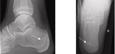
Radiographic Signs of Calcaneus Stress Fracture:
Radiographic signs of calcaneus stress fracture can sometimes be subtle, requiring more sensitive imaging techniques than conventional x-rays. However, certain characteristics can be observed, including:
- Fine Fracture Lines: X-rays may reveal fine fracture lines or cracks in the bone structure of the calcaneus. These lines may not be immediately obvious and may require special attention from the radiologist.
- Areas of Localized Osteoporosis: Areas of localized osteoporosis, characterized by reduced bone density, may be visible on x-rays. This may be an indirect indication of the presence of a developing stress fracture.
- Enlargement of the Bony Trabeculum: In some cases, enlargement of the bony trabecular meshwork may be observable, indicating an adaptive response to increased stress on the calcaneus.
- Changes in Bone Mineral Density: Changes in the bone mineral density of the affected area may be detected, reflecting the breakdown of normal bone structure.
There is an ill-defined sclerotic band across the superior medial aspect of the calcaneal tuberosity representing a stress fracture.
Conclusion
In conclusion, stress fracture of the calcaneus represents a delicate and often underestimated condition that can affect the quality of life of individuals, particularly those engaged in repetitive physical activities. The insidious nature of these heel microfractures highlights the importance of early diagnosis and an appropriate treatment approach.
Understanding the causes, symptoms, and risk factors associated with calcaneus stress fracture is essential for an accurate diagnosis. Conservative treatment methods, such as rest, physiotherapy, and activity modification, play a crucial role in managing this condition, promoting full recovery.
Prevention also takes center stage, highlighting the need for careful management of physical activities and particular attention to bone health. Advances in medical research continue to provide new insights into the prevention, diagnosis, and treatment of calcaneus stress fractures, paving the way for more effective approaches and improved outcomes.
By addressing these aspects, it becomes possible to offer comprehensive support to individuals affected by calcaneus stress fracture, promoting optimal recovery and minimizing the risks of recurrence. Awareness of this condition and continued research are crucial to improve the clinical management and quality of life of affected patients.


