

Definition of osteosarcoma
Osteosarcoma is a type of bone cancer that develops from bone cells. It is the most common primary bone cancer, usually affecting long bones such as the femur, tibia, and humerus. This cancer is most common in adolescents and young adults but can also affect other age groups. Symptoms include persistent bone pain, swelling, and sometimes spontaneous fractures. Diagnosis is based on imaging tests and a biopsy. Treatment often involves a combination of surgery, chemotherapy, and radiation therapy. The prognosis depends on several factors, including the stage at diagnosis and response to treatment.
Epidemiology
Osteosarcoma is a relatively rare form of cancer, but it accounts for about 20% of bone cancers. This malignant tumor primarily affects adolescents and young adults, usually between the ages of 10 and 30. The average age at diagnosis is about 18. However, it can also develop in younger children and older adults.
In terms of incidence, osteosarcoma is more common in males than in females. Adolescents have a higher incidence than younger children. Furthermore, there is a slight racial predominance, with a higher incidence in whites than in blacks.
Regarding the sites of development, osteosarcoma has a predilection for long bones, particularly around the knees. The most common sites of osteosarcoma include the femur, tibia, and humerus.
It is important to note that although osteosarcoma is relatively rare compared to other types of cancer, it is an aggressive form of bone cancer that requires specialized medical care. Epidemiological data help understand the prevalence of this disease and guide diagnostic and treatment strategies.
Causes and risk factors
- Genetic Factors: Genetic mutations may predispose some people to developing osteosarcoma. Alterations in tumor suppressor genes, such as TP53 and RB1, have been associated with increased susceptibility.
- Hereditary Factors: Although the majority of osteosarcoma cases are not inherited, there may be a genetic predisposition in some families. People with a family history of bone cancer may have a slightly higher risk.
- Environmental Factors: Certain environmental factors may play a role, although their specific impact is not always clear. Exposure to ionizing radiation, for example, has been linked to an increased risk of osteosarcoma.
- Age-Related Factors: Osteosarcoma most often occurs in growing adolescents and young adults. Rapid growth during puberty may contribute to the formation of abnormal bone cells.
- Pre-existing Genetic Disorders: Certain genetic conditions, such as Li-Fraumeni disease, are associated with an increased risk of cancers, including osteosarcoma.
- Repeated Bone Trauma: Although rare, repeated bone trauma or chronic fractures may be linked to osteosarcoma. However, most cases are not associated with prior injuries.
Symptoms of osteosarcoma
- Bone Pain: Pain in the affected area is the most common symptom. It may begin insidiously and become more intense over time. The pain may be present even at rest and may be mistaken for an injury or muscle pain.
- Swelling: A swelling or lump in the affected area may develop. This may be caused by tumor growth.
- Joint Stiffness: If the tumor affects a joint, it may cause stiffness or limitation of movement in that area.
- Easy Fractures: Due to the weakening of the bone by the tumor, bones may be more prone to fracture, even with minor trauma.
- General Fatigue: Some patients may experience fatigue or unintentional weight loss.
- Fever: A fever can sometimes accompany osteosarcoma, although this is less common.
Pathophysiology of osteosarcoma
Osteosarcoma is a type of bone cancer that arises from primitive bone cells. The pathophysiology of osteosarcoma involves genetic and molecular alterations that lead to the abnormal growth of bone cells. Here is a simplified explanation of the pathophysiology of osteosarcoma:
- Genetic Alterations: Osteosarcomas are often associated with genetic alterations, including mutations in tumor suppressor genes and chromosomal abnormalities. Mutations frequently affect the TP53 and RB1 genes, which are key regulators of cell growth.
- Mesenchymal Stem Cells: Osteosarcoma typically arises from multipotent mesenchymal cells, which have the ability to differentiate into various cell types, including bone cells. Abnormalities in the cell differentiation process can lead to the formation of tumor cells.
- Abnormal Cell Proliferation: Osteosarcomatous tumor cells multiply uncontrollably, forming a tumor mass within the bone. This rapid proliferation can lead to the formation of an expansive and invasive tumor.
- Abnormal Bone Matrix Production: Tumor cells often produce an unorganized, immature bone matrix that differs from normal bone structure. This tumor matrix is typically less organized and may contain necrotic areas.
- Primary Tumor Formation: The primary tumor develops in the bone, usually in long bones, such as the arms or legs. Osteosarcomas can also occur in flat bones, but this is less common.
- Metastatic Potential: Osteosarcoma has a high potential for metastasis, often to the lungs. Tumor cells can spread through the bloodstream, forming distant metastases.
Osteosarcoma diagnosis
Diagnosing osteosarcoma involves several steps, combining imaging tests and biopsy confirmation. Here are the main steps in the osteosarcoma diagnostic process:
- Medical Examination and History: The physician begins by taking a detailed medical history from the patient and performs a thorough physical examination. Symptoms such as bone pain, swelling, and tenderness are assessed.
- X-rays: X-rays are usually the first imaging test performed. They can show bone abnormalities, such as areas of bone destruction or abnormal masses.
- Computed tomography (CT) scan: A CT scan may be performed to obtain more detailed three-dimensional images of the affected area. This helps assess the extent of the tumor and its impact on surrounding structures.
- Magnetic resonance imaging (MRI): MRI is used to obtain more precise images of the soft tissues surrounding the tumor, allowing its extension into these tissues to be assessed.
- Bone scan: A bone scan may be performed to identify other areas of the skeleton that may be affected by metastases.
- Biopsy: Definitive confirmation of osteosarcoma requires a biopsy. This involves removing a sample of tumor tissue using a needle or surgery. The biopsy is then examined under a microscope by a pathologist to determine the presence of cancer cells and the specific type of osteosarcoma.
- Staging: Once the diagnosis is confirmed, the doctor may perform staging to determine the stage of the disease. This often involves additional imaging tests, such as chest CT scans, to assess for the presence of metastases.
Prevention and awareness
Prevention of osteosarcoma:
- Adopt a healthy lifestyle:
- Maintain a balanced diet rich in calcium.
- Encourage regular physical exercise to strengthen bones.
- Avoid carcinogenic substances:
- Limit exposure to ionizing radiation, especially in children.
- Avoid smoking and reduce exposure to potentially harmful chemicals.
- Regular medical monitoring:
- People with a family history of bone cancer may benefit from regular monitoring.
- Promoting workplace safety:
- Implement workplace safety practices to reduce risks from carcinogens.
Osteosarcoma Awareness:
- Public information:
- Raise awareness of the symptoms of osteosarcoma, such as persistent bone pain and swelling.
- Provide information on risk factors and preventive measures.
- Screening campaigns:
- Encourage participation in early screening campaigns, particularly among those at risk.
- Education in schools:
- Integrate bone health education programs into schools to raise awareness among young people.
- Partnerships with healthcare professionals:
- Collaborate with healthcare professionals to disseminate accurate information about osteosarcoma.
- Use of social media:
- Use social media to share survivor stories, medical information, and prevention tips.
- Community Events:
- Organize community events, conferences and workshops on bone health.
Type of osteosarcoma
Osteosarcoma is a form of bone cancer that develops from bone cells. There are several types of osteosarcoma, each with distinctive characteristics. Here are some of the main subtypes of osteosarcoma:
- Conventional osteosarcoma:
- This is the most common type of osteosarcoma.
- It mainly affects long bones such as the arm and leg.
- It is characterized by the anarchic production of immature bone.
- Telangiectatic osteosarcoma:
- This subtype is less common.
- It has abnormal blood vessels (telangiectasia) within the tumor.
- Giant cell osteosarcoma:
- It has particular histological characteristics with multinucleated giant cells.
- It usually occurs in the knees.
- Clear cell osteosarcoma:
- This type is characterized by tumor cells with clear cytoplasm.
- It is rarer than conventional osteosarcoma.
- Intramuscular osteosarcoma:
- It develops in the muscles adjacent to the bones.
- It is one of the less common forms of osteosarcoma.
- Extraskeletal osteosarcoma:
- It forms outside the skeleton, often in soft tissues.
- It is rare and can be more difficult to diagnose.
Tumor location
Osteosarcoma tends to develop mainly at the ends of long bones, i.e., the arms and legs. The most common sites include:
- Femur: The thigh bone, especially near the knee, is the most common location for osteosarcoma.
- Tibia: The leg bone, especially near the knee, can also be affected.
- Humerus: The upper arm bone, near the shoulder and elbow, is another common site of this tumor.
Although these areas are more prone to osteosarcoma, it can also develop in other bones in the body. In some cases, it can occur in flat bones, such as those in the pelvis or skull, although this is less common.
Early identification of the tumor and its specific location are crucial for treatment planning and determining prognosis. Therefore, imaging tests, such as X-rays, computed tomography (CT) scans, or MRIs, are often used to precisely locate the tumor and assess its extent before deciding on a treatment plan.
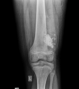
Radiographic sign
The characteristic radiographic sign of osteosarcoma is often called the “lytic shadow zone.” This is an area of destructive appearance on radiographs, corresponding to destruction of bone tissue by the tumor.
Here are some radiographic features of osteosarcoma:
- Lytic shadow area: An area where the bone appears destroyed or lytic, forming a gap in the normal bone structure. This may appear as a “hole” or a clear area on the X-ray.
- Periosteal reaction: Often, a periosteal reaction is seen, where the periosteum (the outer layer of bone) reacts to the presence of the tumor by forming new bone growth. This can give a “sunshine” appearance on X-rays.
- Sharp transition between healthy and affected tissue: There may be a sharp transition between the area affected by the tumor and the surrounding healthy bone tissue.
- Elevation of the bone cortex: The bone cortex, the outer layer of bone, may be raised or distorted by tumor growth.
- “Stepped cloud” appearance: Sometimes a “stepped cloud” appearance may be observed, resulting from the presence of areas of calcification within the tumor.
These radiographic features are not specific to osteosarcoma and can also be seen in other bone pathological conditions. However, when these signs are associated with clinical symptoms and other imaging findings, they can point to the diagnosis of osteosarcoma. To confirm the diagnosis, more advanced imaging techniques, such as computed tomography (CT) and MRI, as well as biopsies, may be necessary.
Conclusion
In conclusion, the subject of osteosarcoma is vast and complex, but it offers an important opportunity to deepen our understanding of this form of bone cancer. By exploring aspects such as causes, symptoms, treatments, and medical advances, it becomes clear that research continues to play a crucial role in improving diagnostics and treatment options.
The lives of those affected by osteosarcoma are profoundly impacted, both physically and emotionally, highlighting the importance of comprehensive care. Advances in treatment and awareness efforts are essential to provide better prognosis and improve patient quality of life.
Ultimately, prevention remains a key objective, and public awareness can play a key role in early recognition of symptoms and support for those affected. Sharing experiences through testimonials also helps humanize the disease and strengthen solidarity within the community.
Therefore, while recognizing the challenges associated with osteosarcoma, it is important to highlight the progress made, support continued research, and work collectively to improve the lives of those affected by this disease.


