

Introduction
Osteoporosis and Paget’s disease are two worrying metabolic bone disorders, significantly impacting the structure and quality of the human skeleton. While osteoporosis remains the most common metabolic bone disease, Paget’s disease stands out as the second most common pathology in this category. Like osteoporosis, Paget’s disease disrupts the bone remodeling process, but in a unique and specific way.
Paget’s disease, characterized by dysregulation of bone remodeling, presents a distinct pattern of abnormality. It begins with an increase in bone resorption, orchestrated by excessive osteoclast activity. This resorption phase is followed by new bone formation, but unlike a normal process, this regeneration is excessive and of poor quality. The rapidity of renewal is such that the quality of new bone formation is neglected, leading to bone hypertrophy and deformation.
This excessive and deforming deposition far exceeds the structural needs of the bone, leading to painful and arthritic consequences. Paget’s disease thus imposes an additional burden on the musculoskeletal system, impairing joint function and generating pain that affects the quality of life of affected individuals.
While osteoporosis and Paget’s disease share a disruption of bone remodeling, each pathology presents distinct characteristics requiring a specific approach to diagnosis and management. Understanding the underlying mechanisms of these metabolic bone diseases is essential to develop appropriate treatment strategies, aimed at alleviating symptoms, preventing complications and improving patients’ quality of life. In this exploration of metabolic bone diseases, Paget’s disease emerges as a unique clinical challenge, requiring particular attention to better understand its implications and better treat those who suffer from it.
History of Paget’s disease
Sir James Paget (1814-1899) remains an eminent figure of the 19th century, marking medical history with his exceptional contribution to the understanding and characterization of Paget’s disease, also called Osteitis Deformans. Born in Great Yarmouth, England, Paget left a lasting legacy as a visionary physician, researcher and teacher.

His fame was built around his reports and in-depth studies on this progressive bone disorder. Paget’s disease, first described by Paget himself in 1877, is a condition characterized by bone deformity resulting from excessive and disordered growth of the bone. Through his insightful clinical observations, Paget contributed significantly to the identification and understanding of this disease, laying the foundation for future research.
Sir James Paget’s dedication to the advancement of medicine was palpable in his multiple roles. He practiced as a surgeon, professor, and pathologist, establishing his reputation as one of the medical leaders of his time. His work greatly influenced the understanding of bone diseases and laid the foundation for future medical advances in the field of orthopedics.
Paget’s impact is not limited to the characterization of the eponymous disease. He was also a major contributor to the understanding of tumors, nipple diseases, and he described for the first time a rare genetic disorder, Ondine’s disease. These multiple contributions consolidated Paget’s place among the pioneers of medicine.
His scientific rigor and commitment to teaching left a lasting legacy. Paget was a respected professor at University College London and St Bartholomew’s Hospital, where he shared his knowledge with many medical students. His academic work, including his lectures on Surgical Pathology, has been widely praised for its clarity and educational impact.
Sir James Paget’s legacy lies not only in his ability to identify and describe complex pathologies, but also in his continuing influence on medical research and medical education. Paget’s disease is named after him, but its impact extends far beyond, touching various fields of medicine and inspiring future generations of researchers and health professionals.
Thus, Sir James Paget remains a giant in the world of medicine, leaving behind a legacy that transcends boundaries of time. His dedication to research and teaching continues to shape the contemporary medical landscape, emphasizing the importance of insight, scholarship, and commitment to human health.
Summary
Paget’s disease is usually localized and most commonly occurs in the pelvis, skull, spine, and legs. Science does not know the cause of this disease. In some cases, the disease runs in families and multiple genes have been linked to the disease. The cause may be environmental, scientists are studying the possibility that a slow-acting virus could cause Paget’s disease.
Causes of Paget’s disease
There is still some uncertainty about the causes of Paget’s disease, but it is generally thought to be due to a combination of genetic and environmental influences.
Genetic factors
- Play a key role in predisposition to Paget’s disease.
- People who develop Paget’s disease are thought to inherit variations in one or more genes that regulate osteoclast activity , leading to increased bone resorption.
- This is thought to be responsible for the increased bone turnover typical of the disease.
- The most important predisposing gene for Paget is called Sequestosome 1 (SQSTM1) .
- SQSTM1 abnormalities have been identified in 40% to 50% of people with a family history of Paget’s disease and also in 5% to 10% of people without a family history.
- Children of people with Paget’s disease are about seven times more likely to develop Paget’s disease than people who do not have a family history.
Environmental factors
- Also play a role in Paget’s disease, as evidenced by the fact that in recent decades the frequency and severity of the disease have declined in many countries, and are more pronounced in regions that previously had a higher prevalence high, such as the United Kingdom.
- Various environmental triggers have been suggested, including dietary calcium or vitamin D deficiency, exposure to environmental toxins, repetitive mechanical stress on bones, skeletal trauma, and slow viral infections.
- Despite this, researchers have yet to discover which environmental factors influence the development of Paget’s disease.
Symptoms of Paget’s disease
Many people with Paget’s disease have no symptoms and never develop complications. In many cases, individuals do not know they have this condition.
For those experiencing symptoms
- Pain , which can occur in any bone affected by the disease or result from arthritis, a complication that develops in some patients.
- Headaches and hearing loss , which can occur when Paget’s disease affects the skull.
- Pressure on the nerves, which can occur when the disease affects the skull or spine.
- Increase in head size, tilt of a limb or curvature of the spine, which may occur in advanced cases.
- Hip pain, which can occur when Paget’s disease affects the pelvis or thigh.
- Damage to joint cartilage , which can lead to arthritis.
Pathophysiology of Paget’s disease
The pathophysiology of Paget’s disease, also known as osteitis deformans, is characterized by a disruption of the normal bone remodeling mechanism, leading to excessive and disordered bone growth. This condition, first described by Sir James Paget in the 19th century, mainly affects the long bones of the skeleton, such as the pelvis, femur, skull and vertebrae. The pathophysiology of Paget’s disease involves several key stages:
- Excessive resorption phase: The disease begins with a phase of excessive bone resorption. Osteoclasts, the cells responsible for breaking down bone tissue, become overactive, leading to increased bone breakdown. This resorption phase is characterized by abnormal osteoclast activity, leading to faster bone destruction than normal.
- Distorted bone formation phase: In response to excessive resorption, new distorted bone formation begins. Osteoblasts, which are responsible for forming new bone, also become hyperactive. However, the rate of turnover is accelerated so much that the quality of new bone formation is compromised. It becomes less dense, more porous and more subject to structural anomalies.
- Excessive bone deposition: New bone formation exceeds the structural needs of the bone, resulting in excessive bone deposition. This overgrowth can lead to hypertrophy of the affected bones, causing characteristic deformities, including thickening, bowing, and enlargements.
- Local and systemic complications: Bone deformation and hypertrophy can lead to local complications, such as fractures, joint deformities, and nerve compression. Additionally, increased calcium release from affected bones may lead to metabolic abnormalities, such as hypercalcemia, with potential systemic consequences.
- Local inflammatory response: Disruption of bone tissue triggers a local inflammatory response, characterized by the infiltration of immune cells and the release of inflammatory mediators. This can contribute to clinical symptoms such as pain, warmth and redness in the affected area.
Complications
The potential for complications will depend on which bones are affected by Paget’s disease and may require medical or surgical intervention.
Examples of complications resulting from damage to specific bones:
- Skull. Paget’s disease of the skull can cause headaches, hearing loss, ringing in the ears, and a change in the shape and/or size of the skull (i.e., enlarged head, broad forehead).
- Spine. Paget in the spine can lead to enlargement and deformity of the affected vertebrae, which can cause curvature of the spine, pain and pressure on the nerve roots with tingling, weakness and numbness in the legs.
- Femur. Paget in the femur (thigh) can cause local pain at the site of Paget’s disease or related to osteoarthritis of the hip joint. Fissile (partial) fractures in the bone can lead to a complete fracture.
- Basin . Paget in the pelvis can be painful and accelerate osteoarthritis of the hip.
- Tibia. When the lower leg is affected, the bone may feel hot, painful, and tilted. Osteoarthritis can develop in the knee joint and fissure fractures can occur which can lead to a complete fracture.
- Deformity : Paget’s disease can cause bone enlargement and deformity. For example, if the skull is affected, the enlarged bone is sometimes first noticed when the individual realizes that their head is larger and their forehead may be wider than normal. Long-standing illness over several years can cause the curvature of the weight-bearing bones in the leg to become distorted.
- Deafness: If the skull is involved, hearing loss may occur.
- Fracture : There is an increased risk of fracture, particularly in the long bones of the arm and leg. Fractures may initially be incomplete (stress fractures or fissure fractures), which pose a high risk of complete fracture. Cleft fractures primarily, but not exclusively, affect weight-bearing bones, such as the thigh bone (femur).
- Osteoarthritis : Paget’s disease can predispose to the development of osteoarthritis in adjacent joints.
- Neurological complications: Neurological complications can occur, often from bony overgrowth leading to compression. For example, widening of the vertebrae of the spine can produce pressure on the nerves causing pain, leg weakness, or sciatica.
- Increased vascularity: If a bone fracture or surgery were to be undertaken, active Paget’s disease could lead to excessive blood loss. This is because blood flow increases in areas where Paget’s disease is active.
- Heart disease: Paget’s disease does not directly affect the heart, but if it is found in many bones, is very active and uncontrolled, the heart may have to work harder to pump extra blood to the affected bones. Although heart failure due to this increased blood flow has been reported, it is extremely rare.
Radiographic sign of Paget’s disease
- Circumscribed osteoporosis: Paget’s disease can manifest itself in the form of circumscribed osteoporosis which spreads into an oil spot or as a thickening with asymmetrical condensation (flaky form). It can be complicated by a basilar impression or hypertrophy of the eye rims.
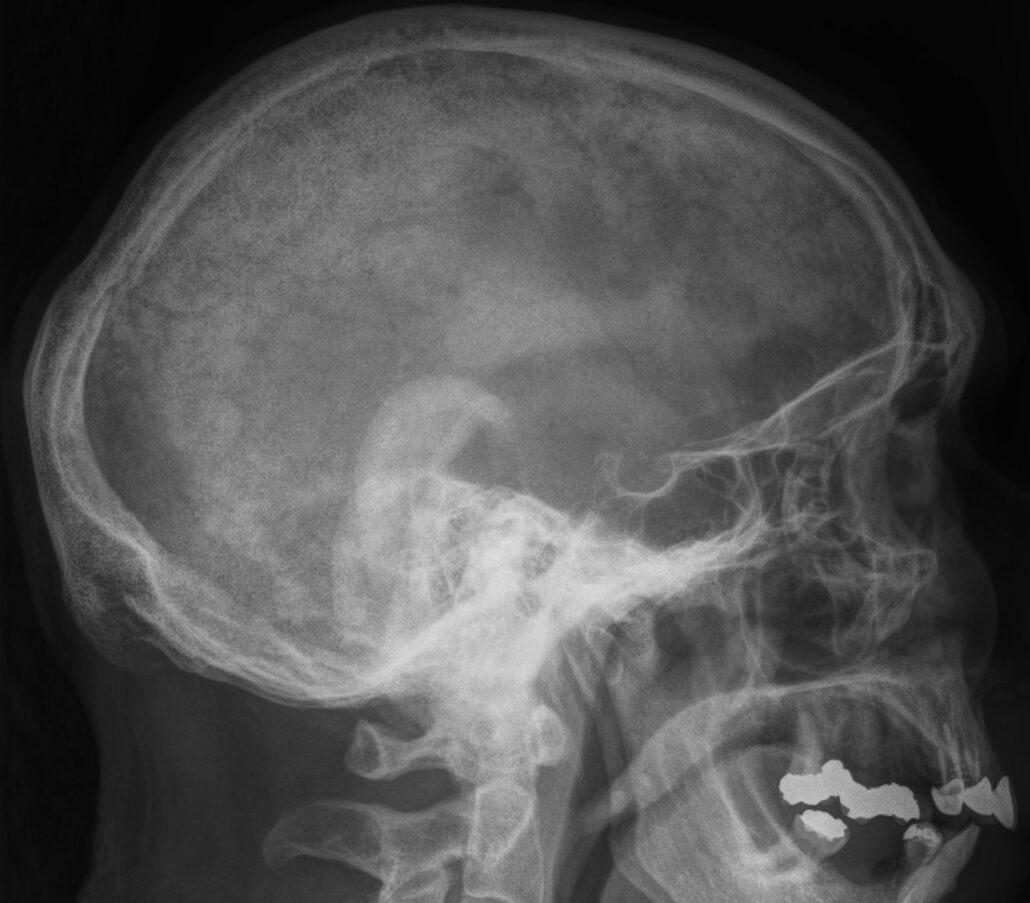
This lateral X-ray of the skull highlights the structural details that can help identify bone disorders, such as Paget’s Disease of Bone. This chronic condition, characterized by abnormal bone remodeling, often affects the skull, leading to thickening, enlargement, and irregularities in the cranial structure.
In cases of Paget’s disease, the normal balance between bone resorption and formation is disrupted, resulting in areas of excessive bone growth and weakened structure. On X-rays, these changes may appear as regions of increased density (sclerosis) or areas of lytic lesions (bone loss). In the skull, these abnormalities can manifest as a characteristic “cotton wool” appearance due to mixed sclerotic and lytic changes. The affected bones may also become deformed, thickened, or misshapen, potentially leading to complications.
Paget’s disease of the skull can cause clinical symptoms such as headaches, hearing loss (due to compression of cranial nerves), or neurological issues. Early diagnosis through imaging, such as X-rays, is critical for managing the disease effectively. Treatment typically involves medications like bisphosphonates to regulate bone turnover and alleviate symptoms.
This image underscores the importance of diagnostic imaging in detecting and managing conditions like Paget’s disease, which significantly impacts bone health and quality of life.
Case courtesy of Assoc Prof Frank Gaillard, Radiopaedia.org . From the case rID: 7477
2. At the level of long bones: V osteolytic, possibility of curvature
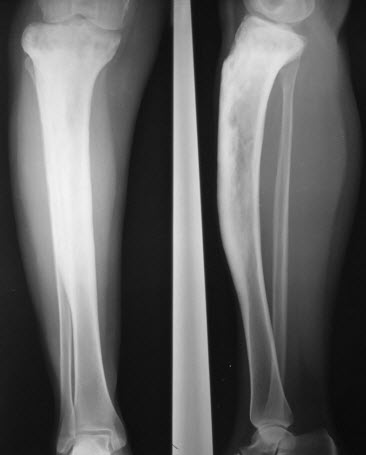
This X-ray of the lower leg highlights the significant changes seen in Paget’s Disease of Bone, a chronic skeletal disorder characterized by abnormal bone remodeling. The condition often affects weight-bearing bones, such as the tibia, leading to thickened, weakened, and misshapen structures.
In Paget’s disease, the natural process of bone resorption and formation becomes unbalanced. The excessive breakdown of bone is followed by rapid, disorganized bone formation, resulting in enlarged and structurally abnormal bones. On X-rays, these changes may present as areas of mixed lytic lesions (bone loss) and sclerosis (excessive bone formation), a hallmark feature of Paget’s disease. The bones may appear denser but less structurally sound, increasing the risk of fractures or deformities.
In the lower limb, Paget’s disease can lead to bowing or deformity of the tibia, causing gait abnormalities, joint stress, and chronic pain. The affected bone may also become thickened, as seen in this X-ray, potentially leading to complications such as osteoarthritis or compression of adjacent soft tissues.
Early diagnosis and treatment are essential in managing Paget’s disease. Medications such as bisphosphonates help regulate bone turnover and alleviate symptoms, improving mobility and quality of life. This X-ray serves as a crucial diagnostic tool for understanding and managing this condition.
References
- Paget UK Association
- Lillemon JN, Nardos R, Kaul MP, Johnson AN, Choate A, Clark AL. Complex Female Pelvic Pain: A Case Series From a Multidisciplinary Clinic in Urogynecology and Physiatry. Female Pelvic Med Reconstruction Surg. 2019 Mar/Apr;25(2):e34-e39. [ PubMed ]
- Ralston SH, Corral-Gudino L, Cooper C, Francis RM, Fraser WD, Gennari L, Guañabens N, Javaid MK, Layfield R, O’Neill TW, Russell RGG, Stone MD, Simpson K, Wilkinson D, Wills R, Zillikens MC, Tuck SP. Diagnosis and Management of Paget’s Disease of Bone in Adults: A Clinical Guideline. J Bone Miner Res. 2019 Apr;34(4):579-604. [ PMC free article ] [ PubMed ]
- McLaughlin MB, Jialal I. StatPearls [Internet]. Stat Pearls Publishing; Treasure Island (FL): May 1, 2022. Calcitonin. [ PubMed ]
- Mantovani G, Fagotti A, Franchi M, Scambia G, Garganese G. Reviewing vulvar Paget’s disease molecular bases. Looking forward to personalized target therapies: a matter of CHANGE. Int J Gynecol Cancer. 2019 Jan 23; [ PubMed ]
- Appelman-Dijkstra NM, Papapoulos SE. Paget’s disease of bone . Best Pract Res Clin Endocrinol Metab. 2018 Oct;32(5):657-668. [ PubMed ]6.Adams C, Banks KP. StatPearls [Internet]. Stat Pearls Publishing; Treasure Island (FL): Aug 11, 2021. Bone Scan. [ PubMed ]
- Kravets I. Paget’s Disease of Bone: Diagnosis and Treatment. Am J Med. 2018 Nov;131(11):1298-1303. [ PubMed ]
- Buske C, Sadullah S, Kastritis E, Tedeschi A, García-Sanz R, Bolkun L, Leleu . Treatment and outcome patterns in European patients with Waldenström’s macroglobulinaemia: a large, observational, retrospective chart review. Lancet Haematol. 2018 Jul;5(7):e299-e309. [ PubMed ]


