
Recognize the warning signs and act without delay
Very intense neck or back pain that begins suddenly and is followed by weakness, numbness, difficulty walking or loss of bladder control may signal a rare neurological emergency: spontaneous spinal epidural hematoma. Blood accumulates in the epidural space within the spinal canal. A sufficiently large collection can compress the spinal cord or nerve roots and cause neurological deterioration, sometimes very rapidly.
The initial presentation may resemble acute neck pain, low back pain, sciatica or a herniated disc. The appearance of a new or progressive neurological deficit changes the urgency. Increasing weakness, loss of sensation, sudden walking difficulty or sphincter dysfunction should not be dismissed as ordinary manifestations of mechanical pain.
What is the spinal epidural space?
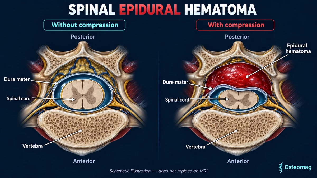
The spinal canal protects the spinal cord and nerve roots. The dura mater forms a tough covering around these structures. The epidural space lies between this covering and the walls of the vertebral canal, and contains fatty tissue and a vascular network. Bleeding into this space may form a collection. Its effect depends on the space it occupies: the spinal canal has limited capacity to expand, so an enlarging hematoma can press on neighbouring neurological structures.
Severity depends on the spinal level, the extent of the hematoma, how quickly it forms, the available space, the degree of compression and the person’s neurological condition.[1]
Why is it called spontaneous?
“Spontaneous” does not mean that there can be no contributing cause. It generally describes bleeding without major trauma or a directly responsible spinal procedure or operation. In many cases, no precise cause is identified. Associated circumstances include coagulation disorders, anticoagulant or antiplatelet use, vascular abnormalities, certain blood disorders, and, rarely, pregnancy or the postpartum period. Situations that abruptly increase venous pressure may also be reported.
Tell the emergency team about anticoagulant or antiplatelet use without changing treatment yourself. Many cases remain classified as idiopathic. The absence of a known risk factor therefore does not rule out a spinal epidural hematoma.[2] [6]
How do symptoms begin?
The classic presentation starts with sudden, unusual pain in the neck or back. It may be intense, deep, sometimes described as tearing, and either localized or radiating. A person may describe it as distinctly different from previous spinal pain. Depending on the level involved, it can extend toward a shoulder, arm, buttock or leg, or feel like a band around the chest or abdomen.
Neurological symptoms follow. They may develop very quickly or over several hours. Recognizing this sequence matters because the initial pain can look like a more common musculoskeletal problem.[6]
Which neurological signs may appear?
Symptoms depend on the location of the hematoma and the structures affected. They can include weakness in one arm or leg, weakness in both legs, or involvement of both arms and legs with some cervical lesions. Numbness, tingling, reduced sensation, new clumsiness, difficulty standing, an unstable gait, impaired coordination and changes in reflexes may occur.
Other findings include a sensory level across the trunk, reduced sensation around the perineum, urinary retention, new incontinence or loss of bowel control. Someone may initially describe a heavy leg and later become unable to walk. Progressive neurological changes are particularly concerning.[5] [6]
| Situation | Action |
|---|---|
| Sudden pain with new weakness or walking difficulty | Call 911; urgent neurological assessment. |
| Bladder or bowel changes, or reduced perineal sensation | Immediate emergency assessment; do not wait for paralysis. |
| Less pain but persistent neurological deficit | Do not interpret pain relief as reassurance. |
| Confirmed hematoma with stable or improving symptoms | Monitoring only under the specialist team’s direction. |
| New deficit during rehabilitation | Urgent medical reassessment. |
Why does the sequence matter?
Mechanical neck or back pain can be severe, so pain intensity alone does not identify this emergency. The concerning sequence is sudden spinal pain followed by a new or progressive neurological deficit. Temporary improvement in pain does not provide reassurance once weakness or loss of sensation has developed. Neurological deficits can fluctuate.
A previous disc herniation, osteoarthritis or known spinal stenosis should not automatically be assumed to explain new neurological deterioration. A person may still be able to walk early in the presentation and subsequently worsen.[6] [8]
What other conditions can resemble it?
The medical differential includes several causes of acute spinal pain with neurological deficits. A large disc herniation can cause radiating pain, weakness and substantial nerve compression. Cauda equina syndrome describes compression affecting the lumbosacral roots, with possible leg weakness, perineal sensory loss and sphincter dysfunction; it is also an emergency.
Spinal cord infarction can produce very rapid neurological deterioration. An epidural abscess may compress the cord or roots: fever or an infectious context raises concern, but their absence does not exclude infection. A tumour or another epidural mass generally follows a different course, although acute presentations can occur.
A vertebral fracture is another consideration, particularly after trauma or with bone fragility. Sudden chest or back pain accompanied by collapse or neurological symptoms can also indicate a major vascular emergency. Some cervical hematomas initially resemble a stroke, especially when weakness appears abruptly. Imaging and medical assessment distinguish these possibilities.[1] [6]
Why can manual tests not rule it out?
Palpating the neck or back cannot reveal what is happening in the epidural space. A painful orthopaedic test does not distinguish a disc herniation from a hematoma. Feeling better in a particular position does not exclude neurological compression either. With new weakness or deterioration, the priority is neurological assessment and imaging, rather than attempting to identify a supposedly restricted joint.[1] [6]
Clinical assessment without delaying referral
The medical team establishes when symptoms started, how weakness has evolved and whether bleeding-related risk factors are present. Examination assesses strength, sensation, reflexes and sphincter function. Repeated assessments can document rapid progression. In an osteopathic consultation, identifying new warning signs is enough to trigger emergency referral: the osteopath does not establish the medical diagnosis and should not prolong testing before contacting emergency services.
What is the role of MRI?
Magnetic resonance imaging is the reference examination when a spinal epidural hematoma is suspected. It identifies the collection, its location and extent, the degree of compression, the appearance of the spinal cord and possible nerve-root involvement. It also helps distinguish a hematoma from an abscess, tumour or some disc herniations.
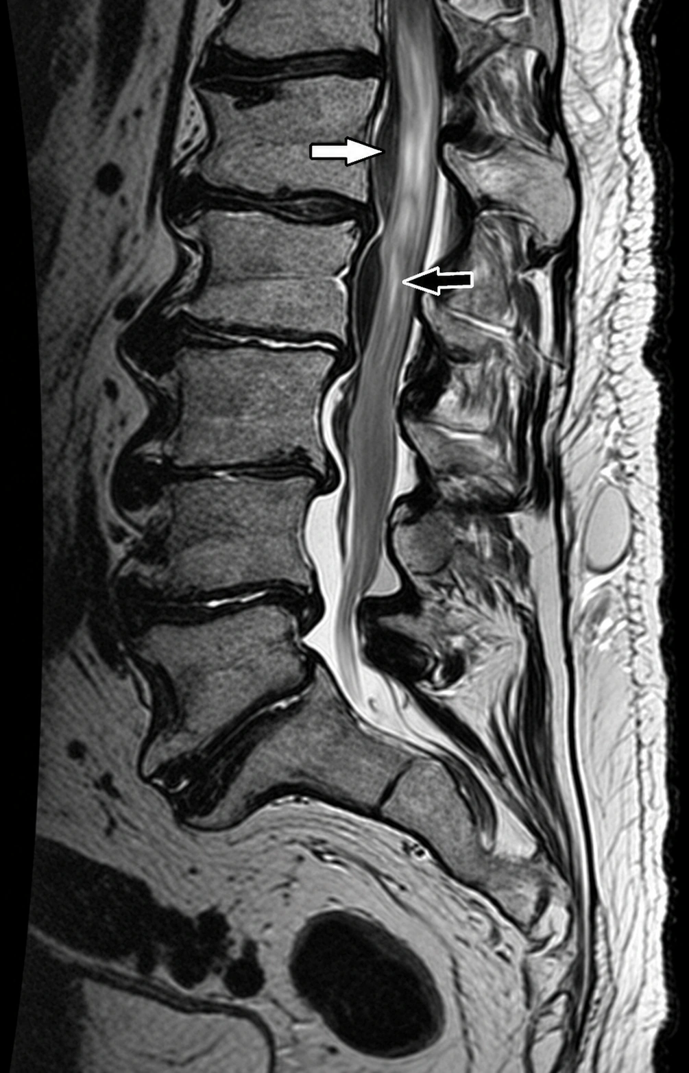
The MRI appearance of blood changes over time. Signals vary according to the age of the bleeding. The radiologist interprets multiple sequences together rather than relying on a single image.[1]
What if MRI is not immediately available?
Computed tomography can sometimes provide useful information, but is less detailed for assessing the spinal cord, roots and soft tissues. The emergency team determines the imaging approach. The essential point is not to delay assessment while a neurological deficit evolves. A manual treatment session must not precede emergency evaluation simply to see whether the symptoms improve.[1]
Can the hematoma resolve without surgery?
Yes, but this possibility requires considerable caution. Spontaneous improvement and hematoma resorption have been described. Nonoperative monitoring may be considered in carefully selected people with minimal or absent deficits, or rapidly improving symptoms. It is not a strategy for observation at home.
The decision depends on the neurological examination, MRI, symptom stability and evolution, and specialist assessment. Repeat neurological examinations and follow-up imaging form part of supervised care. Secondary deterioration can require rapid intervention. Findings from selected nonoperative cases cannot be generalized to someone whose deficits are progressing.[8] [9]
When is surgery considered?
Substantial compression associated with a significant or progressive neurological deficit frequently leads to surgical decompression and evacuation of the hematoma. The aim is to relieve pressure before prolonged compression causes irreversible injury. Studies describe neurological recovery after surgery, but evidence largely comes from retrospective series because the condition is rare.
Research on surgical timing supports rapid intervention, without establishing one universal deadline for every patient. A worsening neurological condition is not a reason to wait. Decisions depend on the entire clinical picture and the specialist team’s assessment.[4] [7] [10]
What influences recovery?
The neurological condition at the time of treatment is a major prognostic factor. A multicentre study of 105 patients found that initial deficit severity was associated with outcome. The duration of substantial paralysis before decompression was also an unfavourable factor in that population. A 2024 study likewise highlights the importance of neurological status and prompt management when deficits progress.
Studies do not all identify exactly the same predictors. Their differences reflect the rarity of the condition, limited sample sizes and differing populations. The practical message remains consistent: severe or progressive neurological deterioration requires rapid specialist evaluation.[3] [5] [7]
What happens after the acute phase?
Recovery varies considerably. After medical or surgical stabilization, rehabilitation may address strength, transfers, standing, walking, balance, coordination, endurance and independence in daily activities. Some people recover quickly; others retain sensory, motor or sphincter difficulties. Resolution of pain does not necessarily mean that neurological function has normalized.
Functional follow-up can compare getting up from a chair, standing safely, walking a set distance, climbing steps or using a hand effectively. Strength, sensation and coordination can be monitored over time. New deficits after improvement must be reported promptly.[4] [5]
What is the place of osteopathy?
During the acute phase, the role is clear: recognize the warning pattern and refer immediately. Suspend manual treatment whenever a spinal hematoma is possible. Manual care does not resorb the hematoma, move blood out of the canal, decompress the spinal cord, repair a neurological injury or replace MRI. Temporary pain relief would not exclude the emergency.
After hospital treatment, and only when the specialist team considers the condition stable, cautious manual care might accompany comfort or mobility goals. It must respect restrictions, neurological deficits and the rehabilitation programme. No evidence demonstrates that osteopathy improves hematoma resorption or neurological recovery. The purpose is possible comfort support after the critical phase, not treatment of the hematoma.
Red flags: when to call emergency services
Sudden, intense neck or back pain with new or increasing weakness, spreading numbness, walking difficulty, unexplained balance loss, impaired coordination, reduced perineal sensation, urinary retention, new incontinence or loss of bowel control requires immediate evaluation. Do not wait for complete paralysis.
The same urgency applies to acute spinal pain with fainting, significant chest pain, breathlessness, fever with neurological symptoms or other features suggesting a vascular or infectious emergency. Someone with progressive deficits or unsafe walking should not drive to the emergency department.[6] [8]

Key takeaways
This condition is rare and should not be assumed to explain every episode of acute spinal pain. However, sudden pain followed by new weakness or sensory loss demands urgent assessment. MRI defines the collection and compression; the specialist team decides between surgery and supervised monitoring. Early walking ability or pain improvement does not rule out the emergency. Rehabilitation follows neurological function and independence, with medical reassessment for any new deficit.
Scientific references
- [1] Perillo et al. (2024). Spontaneous epidural and subdural hematomas of the spine: Review of anatomy and imaging findings.
- [2] Kumar et al. (2024). Spontaneous Spinal Hematoma in Patients Using Antiplatelets and Anticoagulants: A Systematic Review.
- [3] Hsu et al. (2024). Prognostic Factors and Treatment Efficacy in Spontaneous Spinal Epidural Hematoma: A Single Center Experience and Literature Review.
- [4] Kissling et al. (2024). Clinical outcome after surgical management of spontaneous spinal epidural hematoma.
- [5] Peng et al. (2022). Prognostic Factors and Treatments Efficacy in Spontaneous Spinal Epidural Hematoma: A Multicenter Retrospective Study.
- [6] Figueroa and DeVine (2017). Spontaneous spinal epidural hematoma: literature review.
- [7] Vastani et al. (2022, online publication). Prognostic factors and surgical outcomes of spontaneous spinal epidural haematoma: a systematic review and meta-analysis.
- [8] Raasck et al. (2017). Spontaneous spinal epidural hematoma management: a case series and literature review.
- [9] Nitta et al. (2025). Outcomes of Conservative Management of Spontaneous Spinal Epidural Hematomas: A Retrospective Study.
- [10] Nakamura et al. (2023). Ultraearly Hematoma Evacuation (<12 Hours) Associated with Better Functional Outcome in Patients with Symptomatic Spontaneous Spinal Epidural Hematoma.