Deep buttock pain: assess the possibilities and rebuild activity gradually
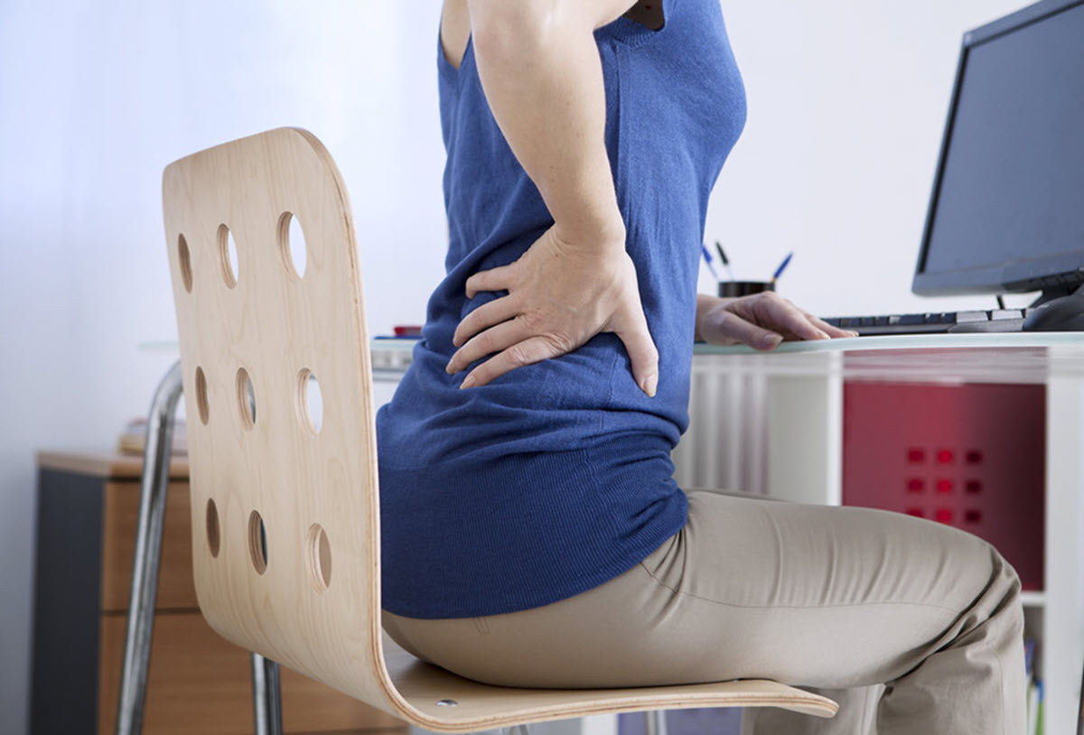
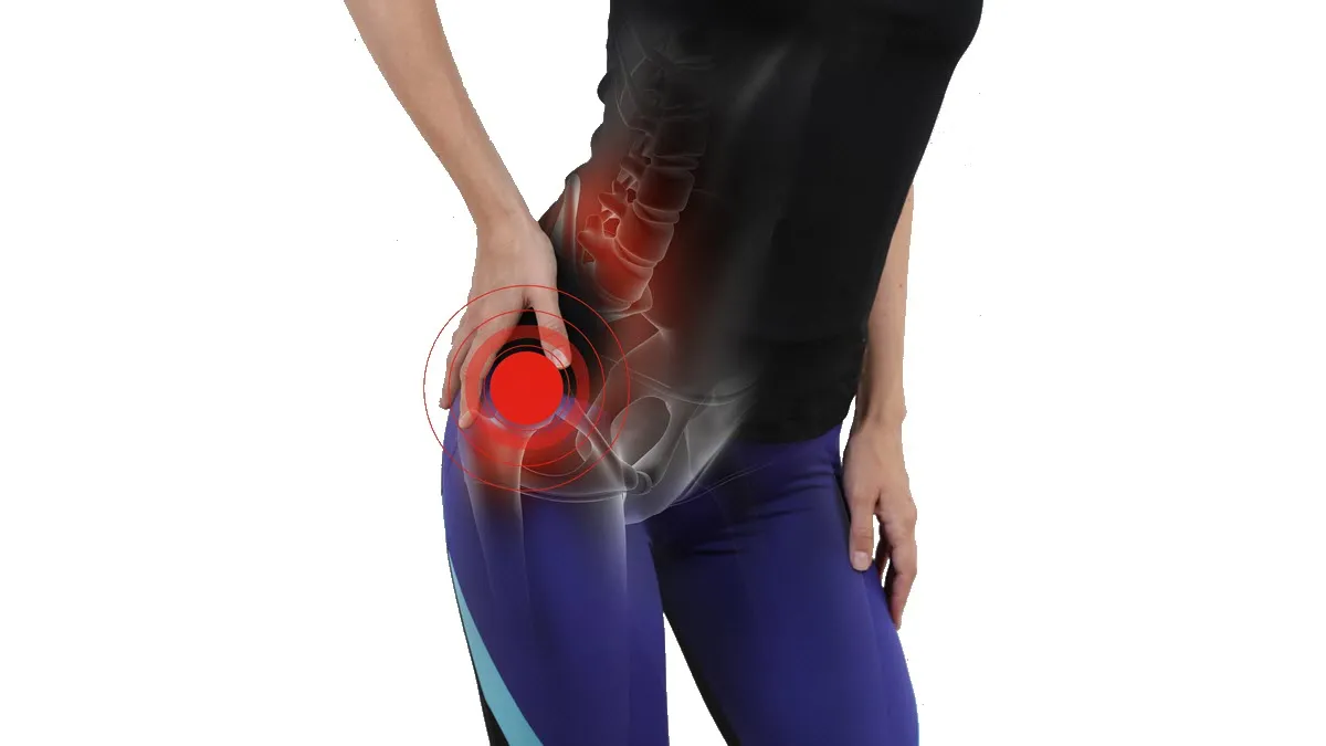
Pain deep in the buttock may be felt near the hip, sacrum or back of the thigh. It is often labelled piriformis syndrome, but the location alone does not identify the painful structure. Lumbar nerve roots, the hip joint, nearby tendons and other tissues can produce overlapping symptoms. A tender muscle or a single positive test does not establish nerve compression. [3] [4]
The practical aim is to recognise important associated signs, develop a reasonable working explanation and restore everyday function. This article describes general principles for assessment and rehabilitation. An individual plan depends on the examination, the progression of symptoms and the activities that matter to the person.
Anatomy: the muscle and the neighbouring nerve
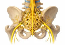
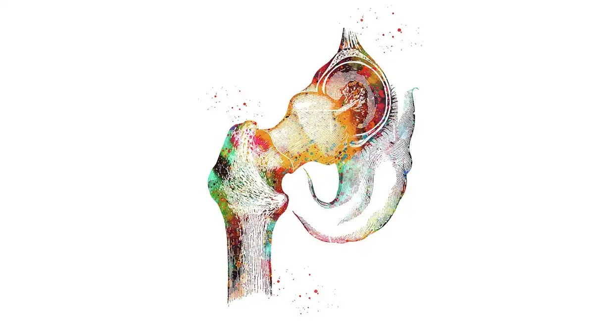
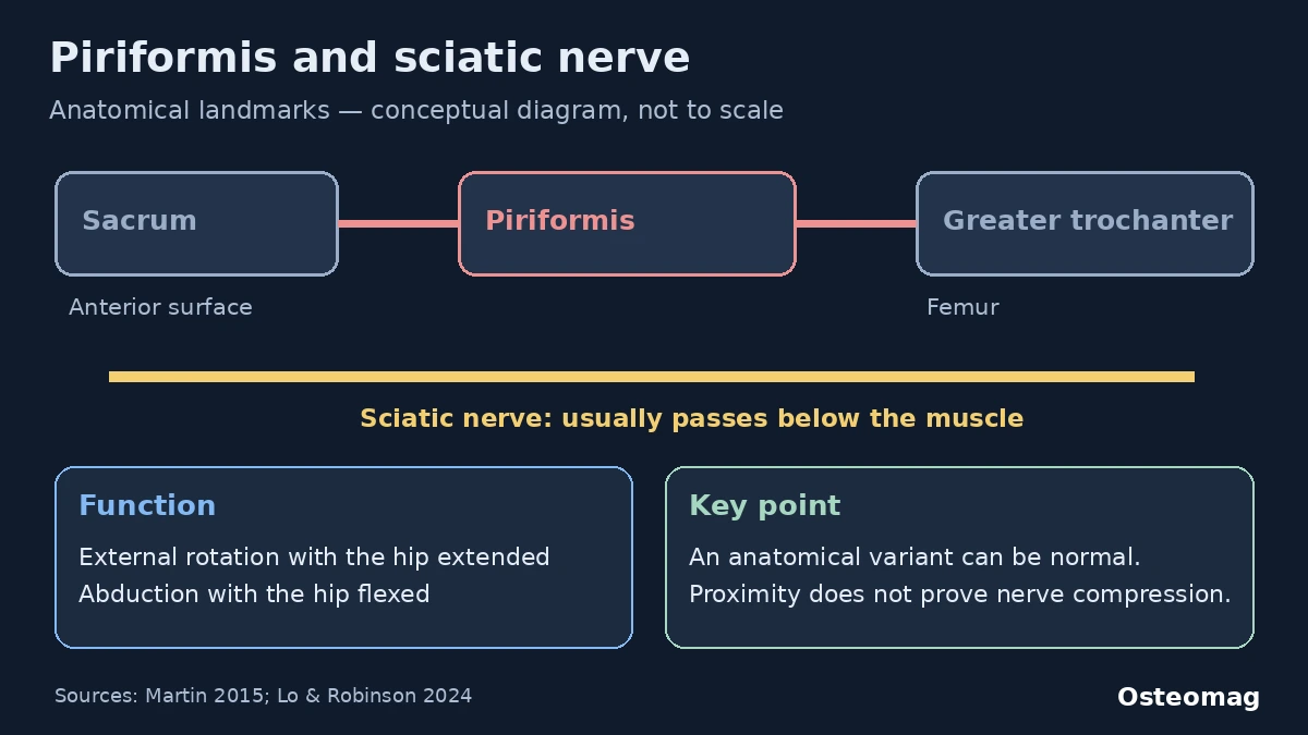
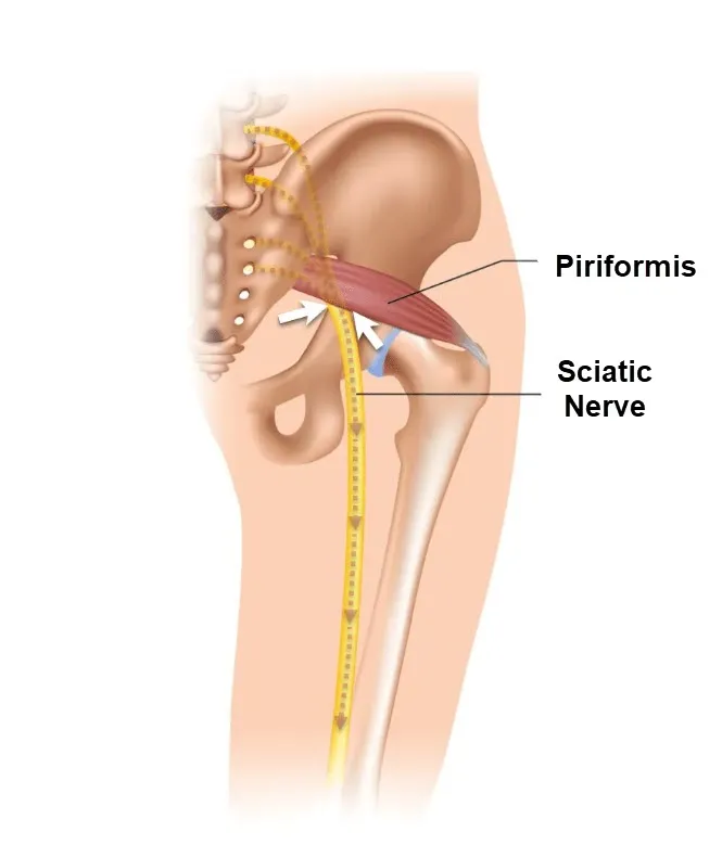
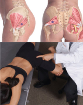
The piriformis lies deep in the buttock. It runs from the anterior surface of the sacrum to the superior border of the greater trochanter of the femur. It contributes to external rotation when the hip is extended and to abduction when the hip is flexed. Its motor supply comes from branches of the sacral plexus, classically described as the nerve to piriformis, rather than primarily from the sciatic nerve. [1] [5]
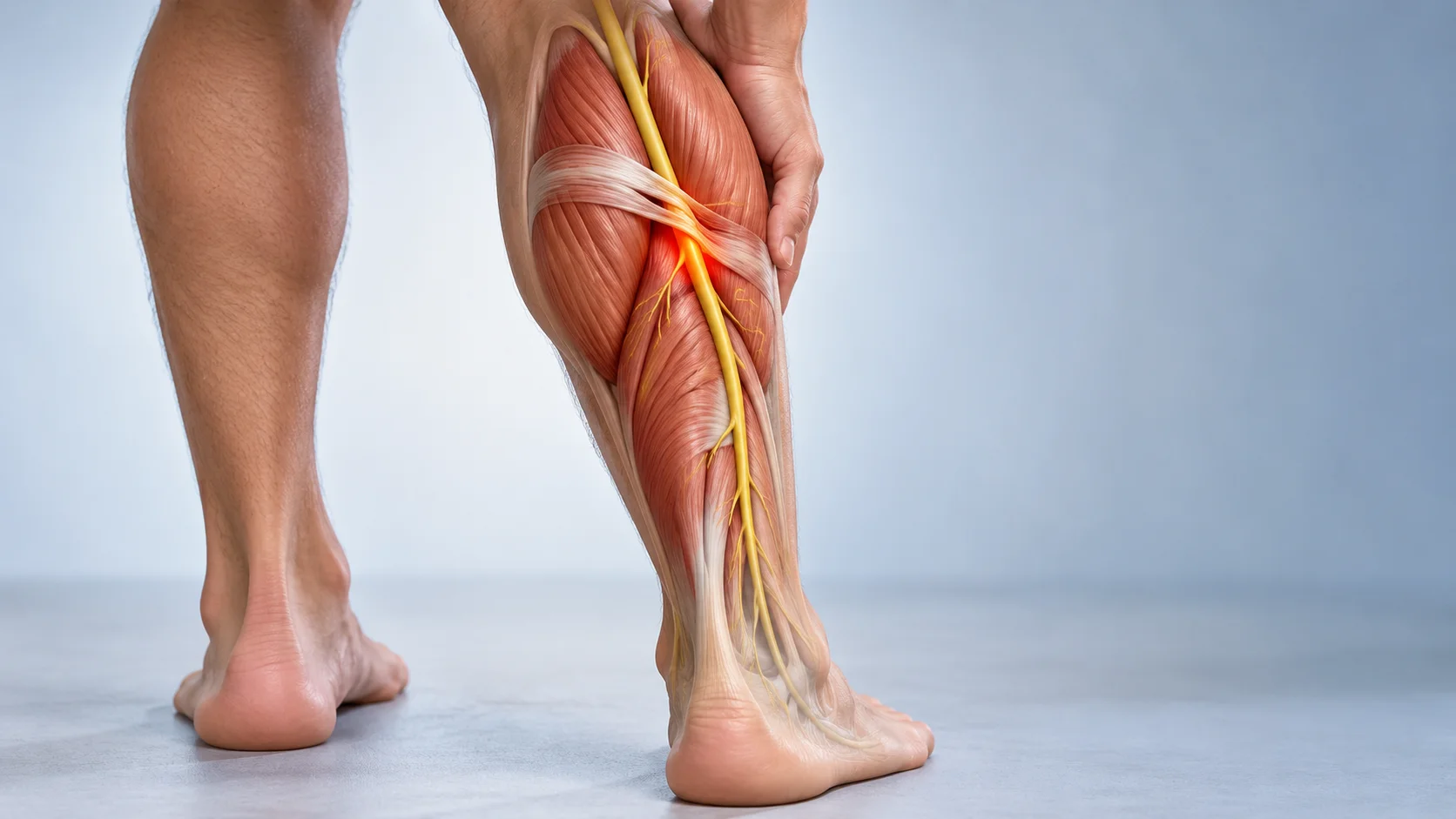
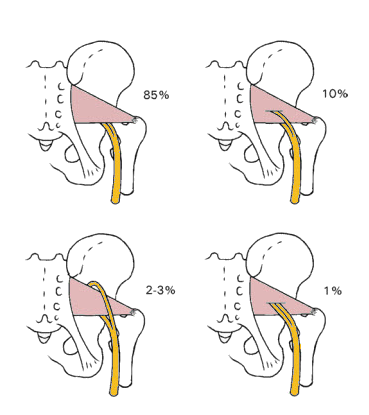
The sciatic nerve usually leaves the pelvis below the piriformis and continues down the posterior thigh. Variations occur, including configurations in which a nerve division passes through the muscle. A variant can be present without symptoms: seeing an unusual course does not prove that it causes the pain. [6]
Piriformis syndrome, deep gluteal syndrome and buttock pain
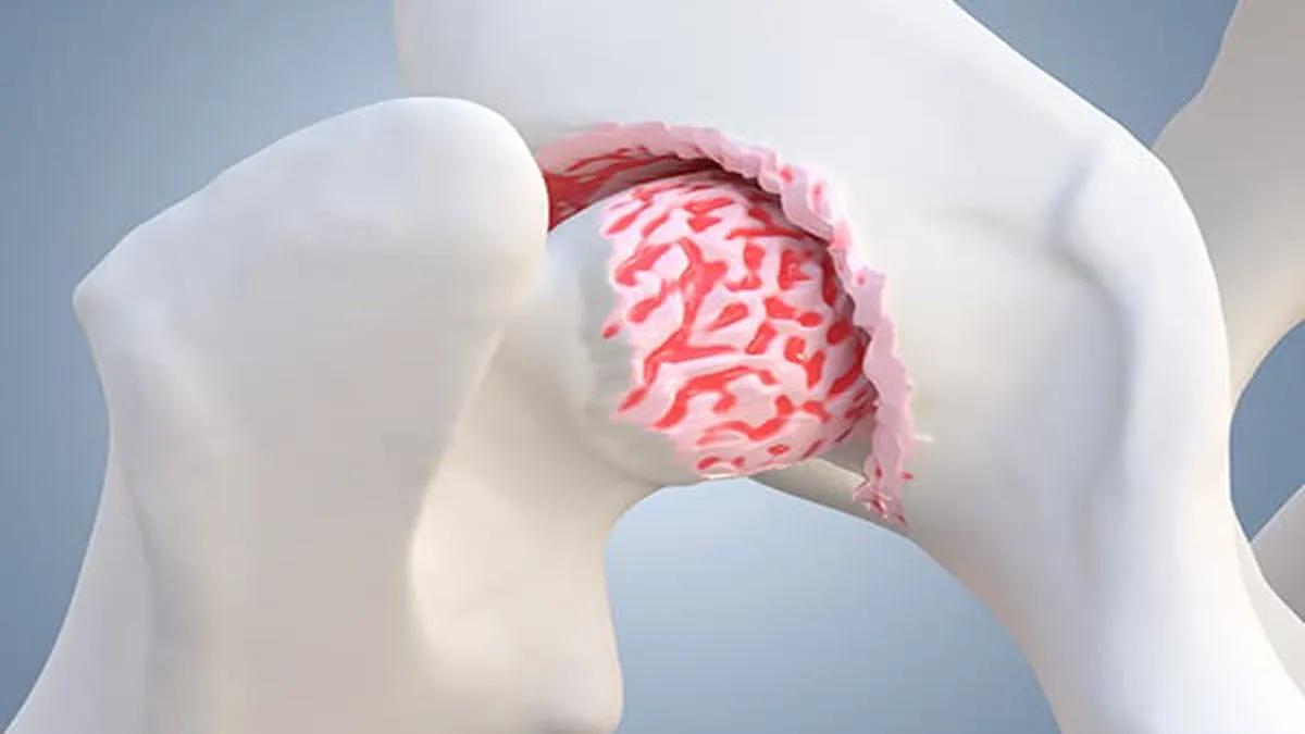
Deep buttock pain describes a symptom location. Deep gluteal syndrome more specifically refers to a non-discogenic nerve entrapment in the deep gluteal region. Piriformis syndrome is one proposed form; adjacent tissues, proximal hamstring structures and the ischiofemoral space can also be involved. The terms should not be used as interchangeable labels for every painful buttock. [1] [2] [7]
An increase in running, prolonged sitting, an unfamiliar effort or a fall may precede symptoms. Possible mechanisms include local muscular sensitivity and irritation or entrapment of nearby neural tissue. These are hypotheses to compare with other explanations. A feeling of tightness does not demonstrate a trapped nerve, and a visibly rotated leg at rest is not diagnostic. [5]
Symptoms that can guide the assessment
Commonly reported features include buttock pain, discomfort during prolonged sitting and pain reproduced by certain hip positions. Symptoms may extend into the posterior thigh. Burning, tingling, numbness or weakness raise the possibility of neural involvement and require comparison with a lumbar origin. None of these features is specific enough to confirm piriformis syndrome alone. [3] [4]
Groin pain and restricted hip rotation may suggest a hip joint problem. Pain around the ischial tuberosity during sitting or loading can suggest proximal hamstring involvement. Pain on the outer side of the hip, especially with side-lying, calls for a different examination. New neurological changes should change the plan rather than simply increase the intensity of stretching. [7]

Clinical examination and differential assessment
The clinician asks about onset, trauma, activity changes, sitting tolerance, radiation and progression. Examination compares the lumbar spine and hip, walking, muscle strength, sensation and reflexes when indicated. Palpation can locate sensitivity, but it cannot identify a neuropathy by itself. The medical differential includes lumbar radiculopathy, hip disorders, sacroiliac pain, tendon or muscle injury and less common fracture, infection or mass lesions. [7]
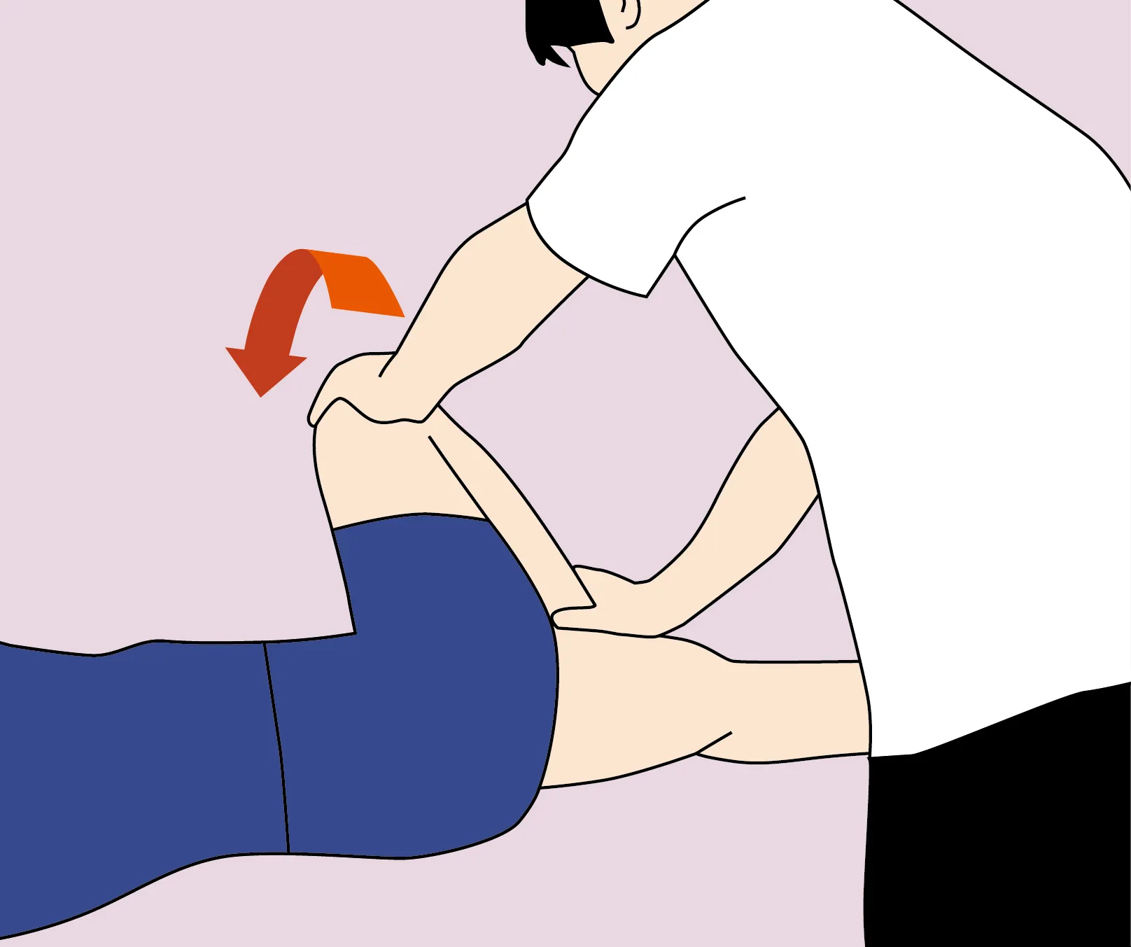
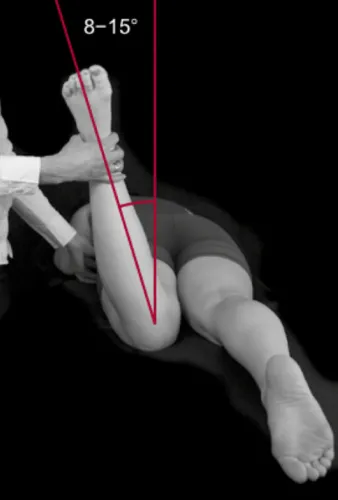
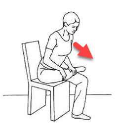
The active piriformis and seated piriformis stretch tests can contribute to assessment. FAIR describes a position combining flexion, adduction and internal rotation. Diagnostic studies have used selected groups, so their accuracy estimates should not be treated as a universal rule. Reproducing pain supports a working hypothesis only when it agrees with the wider history and examination. [11]
Existing examination illustrations

When imaging or nerve testing may help
Imaging is not automatically needed for uncomplicated buttock pain. A clinician may consider it after trauma, with atypical or persistent symptoms, or when a concerning finding needs clarification. The question is whether the result would alter management. Depending on the suspected cause, assessment may concern the lumbar spine, hip or pelvis rather than the piriformis alone. [7] [8]
MRI, ultrasound and electrodiagnostic studies each have limitations. They may help identify an alternative cause or evaluate a suspected neurological problem. There is no universally accepted test that confirms piriformis syndrome in every case, and an imaging variation must be interpreted alongside symptoms and examination findings. [6]
When to seek medical care
Emergency signs
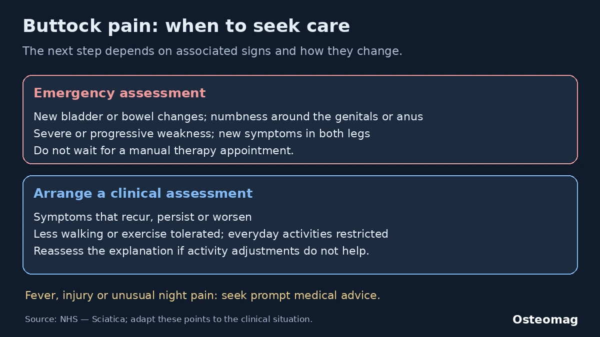
Seek immediate assessment for new difficulty starting urination, urinary retention, new loss of bladder or bowel control, or numbness around the genitals or anus. Severe or progressive weakness, foot drop or new symptoms in both legs also require urgent medical evaluation. These findings must not be explained away as piriformis tightness. Do not wait for a manual therapy appointment. [9]
Fever with significant pain, a substantial injury with difficulty bearing weight, or rapidly deteriorating general health warrants prompt medical advice. Unusual night pain and unexplained weight loss need assessment in context. They are reasons to reconsider the cause, rather than proof of one particular disease.
Non-urgent assessment
Arrange an appointment if symptoms recur, worsen, fail to improve after several weeks of activity adjustments, or restrict work, sleep, walking or leisure. A progressively lower activity threshold, such as pain starting after a shorter walk than before, is another reason to review the explanation and rehabilitation plan. [9]
Begin rehabilitation by adjusting the load
Temporarily reduce an activity that causes substantial or lasting aggravation while keeping tolerable movement in the day. Shorter walks, fewer hills or breaks during a demanding task may help. Complete inactivity is usually unnecessary unless a medical finding requires it. Sitting tolerance can be supported by varying positions, reducing uninterrupted sitting and avoiding strong local pressure. [8] [9]
Record one or two useful measures: comfortable sitting time, walking distance or recovery the following morning. These help distinguish a temporary fluctuation from a sustained decline. The aim is to find a repeatable starting point, rather than a posture that must be held perfectly throughout the day.
Choose exercises according to the response
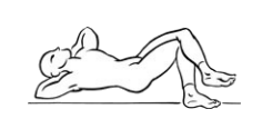
A programme may start with comfortable hip movements and low-effort contractions, then progress to strengthening the hip abductors and extensors. A bridge, supported standing movement or side-lying exercise can be adapted to current tolerance. The exact starting dose should be agreed with a clinician when symptoms are uncertain. Increase one variable at a time, such as repetitions, duration or resistance.
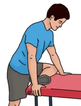
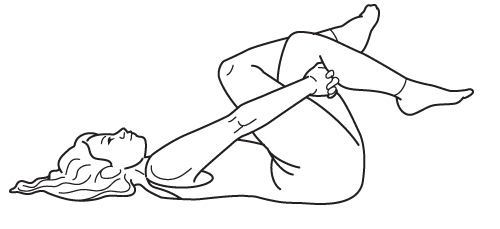
Stretching is optional. A local pulling sensation is different from spreading burning, electric pain or tingling, although the distinction is not always clear. Avoid forcing the hip into end range or repeatedly provoking symptoms to test the muscle. Reduce or stop an exercise that increases neural symptoms, leaves a lasting aggravation or produces new weakness. Rehabilitation follows general graded activity principles; no single stretch has been established as a universal cure. [5] [6]
Existing stretching illustrations
Returning to walking, running and everyday tasks
Progress when the current level is repeatable with a manageable response and acceptable recovery. A runner might begin with short walks, then walk–run intervals, before increasing running time. A person whose main difficulty is sitting may work first on tolerating shorter periods separated by standing breaks. The best progression addresses the actual task being limited.
If symptoms remain clearly worse the next day, step back temporarily and review the dose. Change distance, speed, frequency or hills separately so the response remains understandable. There is no universally valid percentage increase or fixed recovery deadline. A persistent lack of improvement should lead to reassessment, rather than indefinite repetition of the same programme.
What osteopathic care can realistically offer
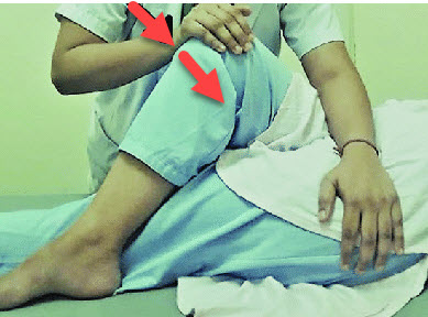
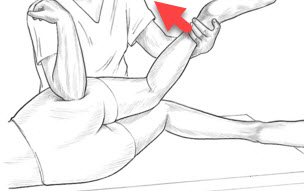
In the Quebec context, an osteopath does not make the medical diagnosis of a neuropathy. Osteopathic support may consider comfortable movement of the hip, pelvis and back and offer tolerable soft tissue care. Any benefit should be assessed through practical function and integrated with active rehabilitation. A reduction in pain after a session does not prove that a nerve was released. [10]
Claims about permanently freeing the sciatic nerve, realigning a displaced pelvis or correcting one hidden cause exceed the evidence. The older osteopathic review describes an osteopathic medical approach; it does not establish that manual osteopathic treatment cures this syndrome. NICE guidance for low back pain and sciatica supports manual care only within a package including exercise, and should not be presented as specific proof for piriformis syndrome. [8] [10]
Existing illustrations of manual care
Practical assessment guide
| Finding | Possible implication | Prudent next step |
|---|---|---|
| Local buttock pain | Several muscular, tendon or joint sources | Compare the hip, pelvis and lumbar spine |
| Radiation, tingling or numbness | Possible neural involvement | Assess strength, sensation and relevant reflexes |
| Groin pain with restricted rotation | Possible hip joint disorder | Broaden the medical assessment |
| Recurring pain or declining walking tolerance | Working explanation or load may need review | Arrange a clinical reassessment |
| Progressive weakness or new sphincter symptoms | Potentially important neurological disorder | Urgent medical evaluation |
Follow function and keep the explanation open
Select meaningful goals together: sitting through a shorter meeting, walking a familiar route or returning to an activity without lasting aggravation. Review whether these measures improve over time. Repeatedly measuring muscle tension is less useful if daily function does not change. The person can ask for a different pressure or decline a technique, and the plan should remain adjustable.
Persistent symptoms deserve a fresh assessment of the original hypothesis, neurological signs and activities actually performed. Pain can be influenced by sleep, stress and previous experiences without being imaginary. When appropriate, support for these factors can accompany physical rehabilitation. The priority is autonomy and a proportionate response to the findings, with timely referral when the pattern changes.
References
- [1] Martin HD, Reddy M, Gómez-Hoyos J. Deep gluteal syndrome. Journal of Hip Preservation Surgery. 2015;2(2):99–107. doi:10.1093/jhps/hnv029.
- [2] Kizaki K, Uchida S, Shanmugaraj A, Aquino CC, Duong A, Simunovic N, Martin HD, Ayeni OR. Deep gluteal syndrome is defined as a non-discogenic sciatic nerve disorder with entrapment in the deep gluteal space: a systematic review. Knee Surgery, Sports Traumatology, Arthroscopy. 2020;28(10):3354–3364. doi:10.1007/s00167-020-05966-x.
- [3] Hopayian K, Song F, Riera R, Sambandan S. The clinical features of the piriformis syndrome: a systematic review. European Spine Journal. 2010;19(12):2095–2109. doi:10.1007/s00586-010-1504-9.
- [4] Hopayian K, Danielyan A. Four symptoms define the piriformis syndrome: an updated systematic review of its clinical features. European Journal of Orthopaedic Surgery & Traumatology. 2018;28(2):155–164. doi:10.1007/s00590-017-2031-8.
- [5] Probst D, Stout A, Hunt D. Piriformis Syndrome: A Narrative Review of the Anatomy, Diagnosis, and Treatment. PM&R. 2019;11(Suppl 1):S54–S63. doi:10.1002/pmrj.12189.
- [6] Lo JK, Robinson LR. Piriformis syndrome. Handbook of Clinical Neurology. 2024;201:203–226. doi:10.1016/B978-0-323-90108-6.00002-8.
- [7] Park JW, Lee YK, Lee YJ, Shin S, Kang Y, Koo KH. Deep gluteal syndrome as a cause of posterior hip pain and sciatica-like pain. The Bone & Joint Journal. 2020;102-B(5):556–567. doi:10.1302/0301-620X.102B5.BJJ-2019-1212.R1.
- [8] National Institute for Health and Care Excellence (NICE). Low back pain and sciatica in over 16s: assessment and management. Guideline NG59. 2016. Recommendations.
- [9] National Health Service (NHS). Sciatica. Health A to Z. Page reviewed 3 December 2024.
- [10] Boyajian-O’Neill LA, McClain RL, Coleman MK, Thomas PP. Diagnosis and management of piriformis syndrome: an osteopathic approach. Journal of the American Osteopathic Association. 2008;108(11):657–664. doi:10.7556/jaoa.2008.108.11.657.
- [11] Martin HD, Kivlan BR, Palmer IJ, Martin RL. Diagnostic accuracy of clinical tests for sciatic nerve entrapment in the gluteal region. Knee Surgery, Sports Traumatology, Arthroscopy. 2014;22(4):882–888. doi:10.1007/s00167-013-2758-7.