Observing the tendon in motion and understanding lateral knee pain
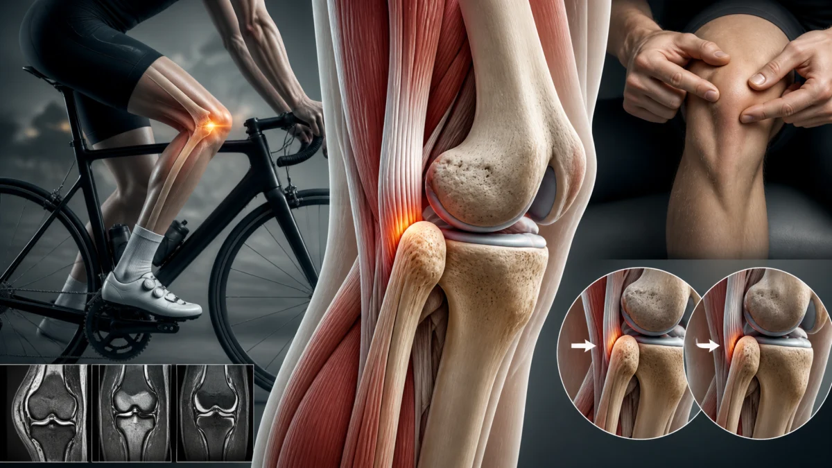
A snap on the outside of the knee can be surprising, especially when it returns with every deep bend. In some cases, the biceps femoris tendon moves abruptly near the fibular head. This snapping is a rare cause of lateral knee pain. It must be distinguished from other joint noises and more common injuries, because a sound alone is insufficient to establish a diagnosis [1, 2].
The useful question is straightforward: what movement is occurring, where does it happen and does it cause a meaningful difficulty? An incidental, painless phenomenon differs from painful snapping that limits work or sport. Available studies mainly describe cases and small series. They explain possible mechanisms without providing a universal treatment recipe.
Why can this tendon produce a snap?
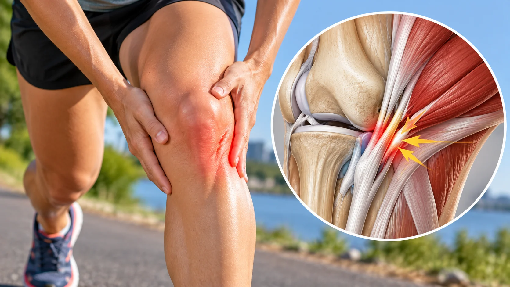
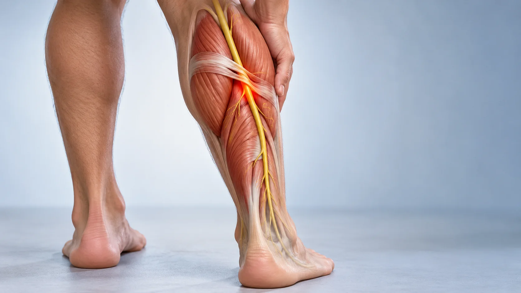
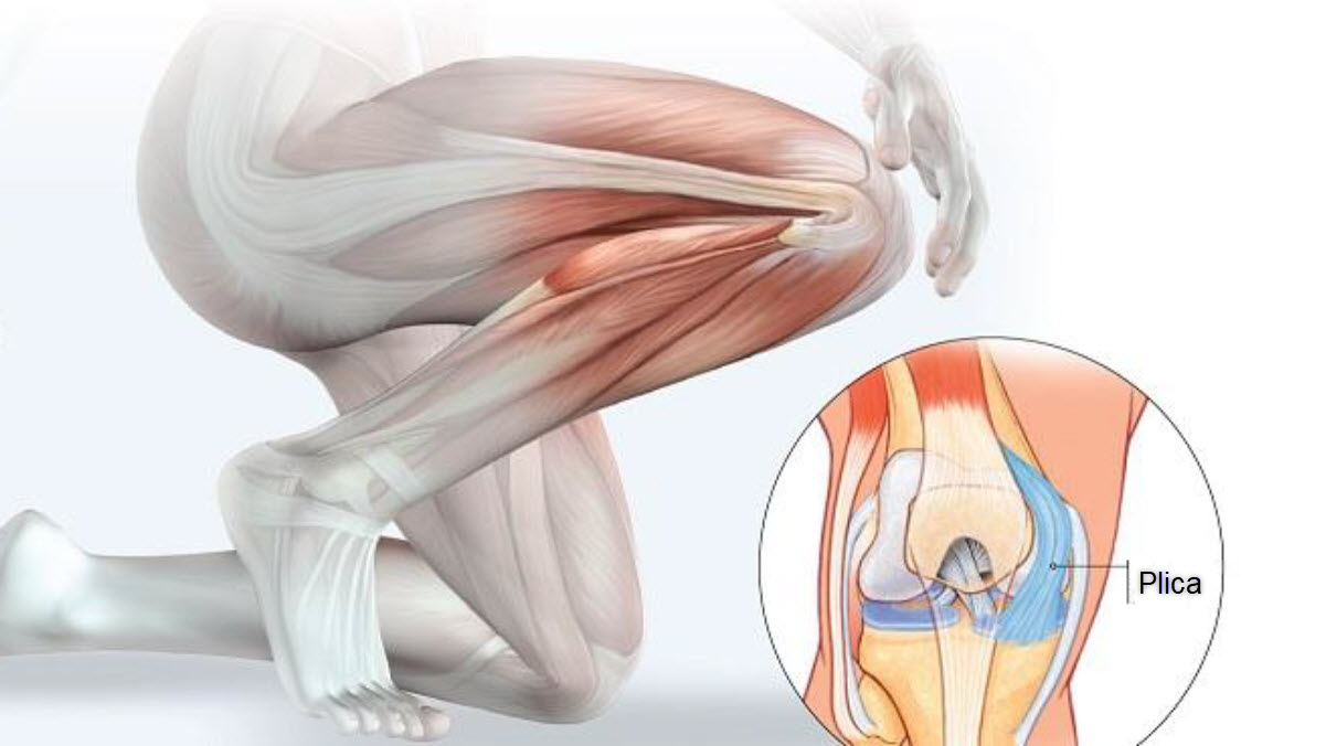
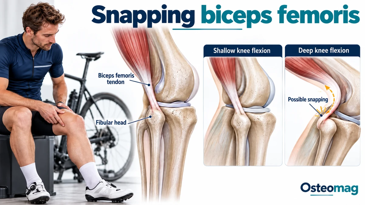
The biceps femoris is one of the hamstring muscles. Both heads contribute to knee flexion, and its distal attachment has several components around the fibula. This anatomy is more complex than a single cord fixed at one point. Certain variations in its course or attachments may favour abrupt displacement of the tendon during bending [1, 2].
Publications describe a more anterior attachment, an unusual tibial component or a particular shape of the fibular head. These factors may combine, depending on the case. Case reviews do not identify an identical mechanism in every person. Proposed classifications mainly organise the observations; they are not validated predictive tools for the general population [1, 3].
Normal anatomical configurations must also be recognised. A bifurcating distal tendon, for example, may be misinterpreted on ultrasound if the examiner is unfamiliar with that possibility. An unusual image therefore proves neither a tear nor the cause of snapping. The relationship with the symptomatic movement remains essential [6].
A sound, friction or lateral pain
The phenomenon may be perceived as a snap or as palpable movement near the fibular head. Some people describe mainly friction without an obvious sound. A series of fifteen symptomatic knees specifically distinguished snapping presentations from friction presentations. This diversity shows that the same anatomical region can produce several clinical experiences [2].
Substantial knee flexion is often involved in published cases. Cycling, squatting or certain positions may trigger symptoms, but not every cyclist with lateral knee pain has this problem. The provoking angle and degree of discomfort vary with anatomy, the task and the individual [2, 8].
The history should clarify symptom onset, possible trauma, whether one or both sides are involved and which activities are limited. Bilateral cases without trauma have been described. Conversely, onset after an injury encourages investigation of an associated lesion. Context helps formulate possibilities but does not replace observation of the movement [1, 3].
If the snapping remains painless, stable and without functional restriction, intervention is not automatically necessary. Assessment may provide reassurance and clarify signs to monitor. The objective should not be to eliminate every sound produced by a knee that functions normally.
Distinguishing other causes of discomfort on the outside of the knee
Lateral pain may arise from the iliotibial band, a meniscus, the popliteus tendon or the proximal tibiofibular joint. These structures lie at different levels and respond to different movements. Joint locking, joint-line pain or a sense of instability requires appropriate investigation rather than immediate attribution to the biceps femoris.
The common fibular nerve passes close to the fibular head. New numbness or difficulty lifting the foot should not be treated as a harmless consequence of snapping. These symptoms justify prompt assessment. This anatomical proximity also matters when a specialist intervention is considered.
After significant trauma, deformity, inability to bear weight or substantial swelling changes the priority. A red, very hot knee accompanied by fever also requires urgent assessment. In these circumstances, stop attempts to reproduce the snap until its cause has been clarified.
| Observation | Question to clarify | Guidance for support |
|---|---|---|
| Painless snapping without limitation | Mechanical variation without meaningful discomfort? | Do not automatically aim to eliminate the sound. |
| Painful movement near the fibula | Tendon snapping or friction? [2] | Relate the observed movement to the usual symptom. |
| Symptoms occurring only in movement | A dynamic phenomenon? [5] | Discuss whether dynamic ultrasound would be useful. |
| Locking or joint-line pain | Another knee structure? | Broaden assessment before reaching a conclusion. |
| Numbness or difficulty lifting the foot | Possible nerve involvement | Seek prompt assessment. |
Clinical guide — Osteomag. This table does not replace individual assessment.
Observing a dynamic phenomenon
Examination attempts to reproduce the usual movement cautiously within a tolerated range. The professional identifies the tendon, the snapping area and the relationship between displacement and pain. Repeatedly producing a painful snap is unnecessary to demonstrate that it exists. Comparison between sides may help, particularly when the phenomenon is bilateral [1, 4].
Dynamic ultrasound is particularly useful because it shows the tendon during movement. Examination at rest may miss a displacement occurring only at a particular angle. A publication on real-time ultrasound assessment illustrates this approach. Its value must nevertheless be considered alongside the clinical picture and the examiner’s expertise [5].
MRI may clarify tendon attachments, tissue condition or other possible causes of pain. It does not necessarily capture the snap because it generally describes a static position. Radiographs may be useful when a bony abnormality or consequence of trauma is suspected. Investigations are selected to answer a question, rather than simply to accumulate images.
The most relevant finding is concordance: the observed displacement matches the movement the person recognises and reasonably explains the discomfort. If concordance is absent, other possibilities must remain under consideration. A tendon whose shape differs from a standard diagram is not automatically responsible for every knee symptom.
Adapting activity and monitoring progress
When conservative management is appropriate, movements that clearly provoke pain can be reduced temporarily. Someone troubled by deep bending may explore tasks performed within a shorter range, according to the recommendations received. This adaptation aims to preserve tolerated activity without continually forcing the tendon across a painful area.
For cycling, assessment of position and training load may be useful. An adjustment should not be presented as a universal anatomical correction. Changing saddle height or foot position without assessing the whole task may transfer demands elsewhere. A professional familiar with the activity can help choose an adjustment consistent with the symptoms.
Follow-up may record session numbers, duration, pain and the response the next day. A snap that remains present but is less painful differs from one accompanied by progressive worsening. Objectives should therefore distinguish the sound, pain and functional capacity.
The specific literature does not establish that an exercise programme consistently eliminates snapping. Many publications concern operated patients, limiting conclusions about conservative treatment effectiveness. Activity adjustments and strengthening should be presented as individualised measures with follow-up rather than as a proven protocol for this rare condition [1, 2, 3].
Strengthening without repeated provocation
Rehabilitation may develop lower-limb capabilities within tolerated ranges: hip control, hamstring strength, weight-bearing and coordination, according to identified needs. Resistance and speed are added progressively. The selected exercise should allow controlled movement without a sustained worsening of symptoms.
Progression may change one variable at a time. Increasing bending depth, load and frequency simultaneously makes the response difficult to interpret. A successful stage means more than completing a set of repetitions; it should also be compatible with activities later that day and on the following day.
These general principles do not change a tendon attachment. Their purpose is to support usable capacity and document the response. If the same restriction persists despite an adapted programme, that information may help determine whether specialist consultation is needed.
Osteopathic support with realistic objectives
Support may contribute to understanding movement habits, repeated demands and compensations associated with pain. Cautious work aimed at comfort and mobility in related regions may form part of care after concerning causes have been assessed. Objectives should be explicit and compatible with rehabilitation.
There is no strong evidence that osteopathic treatment permanently changes the biceps femoris attachment, corrects the fibular head’s shape or eliminates this snapping mechanism. Surgical and anatomical observations do not demonstrate the effectiveness of an osteopathic approach. The person should understand this limitation before committing to a series of consultations [1, 2, 4].
A measurable objective may be to tolerate a working day more comfortably, return to an adapted activity or reduce associated discomfort. A promise of anatomical correction, however, exceeds the available evidence. Lack of progress should prompt reconsideration of the strategy and, when needed, referral for different expertise.
What surgical publications tell us
Older and more recent cases describe different solutions: anatomical repair, redirection of an attachment or release of an implicated component. The choice depends on the mechanism actually observed. Techniques described in one case are not interchangeable and do not automatically justify a bone or tendon procedure in another person [7, 9, 10].
The fifteen-knee series published by Catonné and colleagues reported improvement and return to activities in selected patients. Its evidence remains limited: it is a series without randomised comparison, involving particular anatomical configurations. The results do not guarantee an individual outcome or precisely quantify the benefit compared with every other option [2].
Reviews also describe precautions and possible complications according to the procedure. Specialist consultation should clarify the responsible structure, expected benefits, risks and recovery stages. The fibular nerve’s proximity and the stabilising function of the region are part of that assessment [3, 4].
Returning to the tasks that matter
Return to a demanding activity is prepared through representative tasks: the required range of motion, repetitions, resistance and duration. One painless bend does not prove that the knee will tolerate a prolonged session. The response after exercise and confidence in the movement provide additional information.
Keeping a record of adjustments tried and their effects is useful. It avoids constantly changing strategy without knowing what helps. It also supports discussion with a specialist if a substantial mechanical limitation remains.
Snapping biceps femoris chiefly requires precise identification and realistic expectations. Management is built around the actual difficulty and the observed mechanism. Understanding that a sound, an anatomical variation and pain are not synonymous helps choose appropriate measures and avoid unnecessary interventions.
Osteopathic support plan: key points
- Distinguish painless noise from snapping that genuinely limits function.
- Identify the mechanism through clinical and, when useful, dynamic assessment.
- Adapt the ranges and tasks that cause persistent pain.
- Strengthen gradually within tolerated ranges without repeated provocation.
- Do not promise to change a tendon attachment through osteopathy.
- Refer if restriction persists or neurological symptoms appear.
References
- [1] Snapping Biceps Femoris Syndrome: A Case Presentation and Review of Current Literature. PMID: 39429390.
- [2] Biceps femoris snapping and friction on the fibular head secondary to abnormal distal tendon insertion: 15 cases and a literature review. PMID: 35183755.
- [3] Bilateral snapping biceps femoris tendon: a case report and review of the literature. PMID: 30770981.
- [4] Snapping Biceps Femoris: clinical demonstration and operative technique. PMID: 29364023.
- [5] Snapping biceps femoris tendon: a dynamic real-time sonographic evaluation. PMID: 20658565.
- [6] The bifurcating distal biceps femoris tendon: potential pitfall in musculoskeletal sonography. PMID: 21795497.
- [7] Snapping biceps femoris tendon treated with an anatomic repair. PMID: 20020099.
- [8] Snapping Biceps Femoris Tendon. PMID: 30075041.
- [9] Partial tendon release for treatment of a symptomatic snapping biceps femoris tendon: a case report. PMID: 23015904.
- [10] Snapping knee from biceps femoris tendon. A case report. PMID: 2603666.