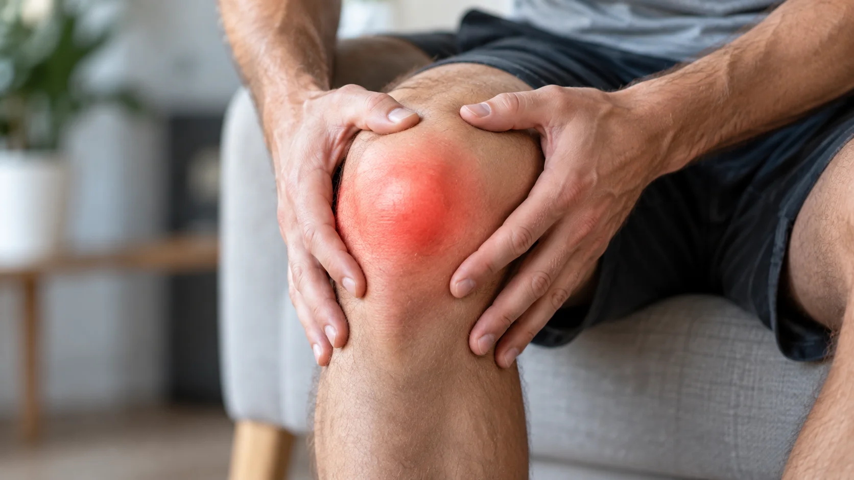
After a first episode: assessing injuries and planning recovery
A patellar dislocation often happens during a pivot, a landing or a collision. The kneecap leaves its usual track, generally towards the outside of the knee. It may move back into place spontaneously, but this does not mean that every tissue remains intact. A first dislocation deserves assessment even when the pain has already begun to settle [1].
Follow-up identifies associated injuries, factors contributing to instability and the person’s needs. It then organises protection and rehabilitation. The plan differs between an adolescent playing a pivoting sport and an adult hoping to walk comfortably again. Their shared objective is to recover enough knee function and confidence for the activities they choose.
The kneecap, the MPFL and other stabilising structures
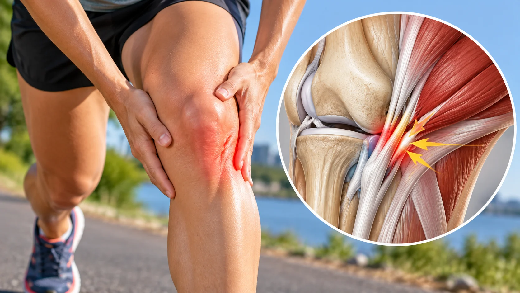
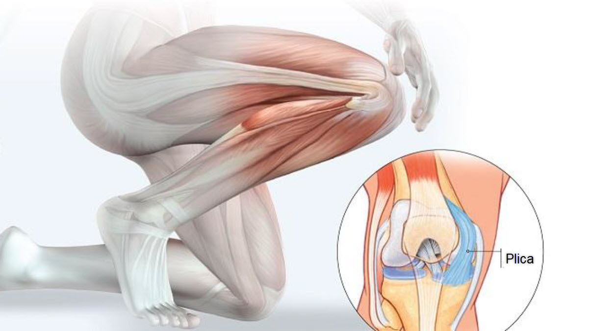
The patella is part of the knee’s extensor mechanism, between the quadriceps tendon and the patellar ligament. It improves the quadriceps’ mechanical efficiency and glides within the trochlea, a groove on the front of the femur. The shape of this groove and the height of the patella influence stability. Ligamentous tissues and muscular control also contribute.
The medial patellofemoral ligament, or MPFL, restrains lateral displacement, particularly when the knee is close to straight. It is frequently injured during a dislocation. As the knee bends, engagement of the patella within the trochlea provides more bony guidance. Stability therefore depends on several structures and their relationships, rather than on one supposedly weak muscle [2].
A subluxation is a partial displacement, whereas a dislocation involves a temporary complete loss of the usual joint contact. However, a feeling that the knee is giving way does not always prove that the kneecap moved. After an injury, pain and swelling can inhibit the quadriceps. The account of the episode and the examination help distinguish these possibilities.
Why check the cartilage and look for osteochondral fragments?
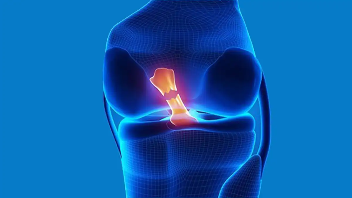
As it moves out of place or returns, the patella can strike the femur. These impacts may damage cartilage and sometimes detach a fragment containing both cartilage and bone: an osteochondral injury. A loose fragment within the knee can cause catching or locking. Persistent pain or a substantial joint effusion should therefore not be attributed solely to muscle tension [3].
Systematic reviews show that cartilage and MPFL injuries are common in the groups studied after an acute dislocation. Estimates vary with the imaging methods and the patients included. Cartilage damage does not always mean a fragment requiring an operation; these are different findings. Management depends on the type, size and location of the injury [2, 3].
A knee that swells quickly after trauma may contain blood within the joint. This observation strengthens the case for looking for an internal injury. Locking, inability to regain extension and repeated catching are also important details to report. A movement should not be forced in an attempt to make an obstruction disappear.
What should you do immediately?
If the kneecap remains visibly displaced, seek urgent medical care. While waiting, support the knee in a comfortable position and avoid putting weight on it. Do not try to push the kneecap back yourself. Deformity, very rapid swelling, a significant wound or inability to move the leg also calls for urgent assessment [1].
Even when the kneecap moves back spontaneously, prompt consultation allows the consequences of the episode to be checked. Inability to actively straighten the knee or lift the leg while keeping it straight raises concern about the extensor mechanism. A cold, pale, numb or newly weak foot after trauma requires immediate assessment of circulation and nerves.
During recovery, increasing redness and warmth of the knee with fever, unexplained deterioration or unusual calf swelling requires further advice. Sudden breathlessness or chest pain requires emergency care. These situations should not wait for an appointment intended to improve comfort or guide a return to exercise.
Examination and imaging after the first episode
The clinician asks how the injury happened, whether the displaced patella was seen, when swelling appeared and which activities remain possible. Earlier subluxations, symptoms in the other knee and the person’s age are relevant. Examination assesses movement, active extension, pain and the condition of the limb. In the acute phase, examination techniques are adapted to avoid unnecessarily provoking further apprehension.
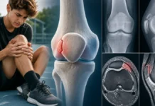
Radiographs look particularly for fractures and visible bony fragments. They also provide anatomical information. A normal radiograph does not exclude every cartilage or soft-tissue injury. MRI allows closer assessment of the MPFL, osteochondral injuries and anatomical factors associated with instability [4].
The ESSKA 2024 consensus, published in 2025, gives MRI an important role after a first dislocation, with radiographs or rapidly available MRI according to the circumstances. Imaging helps identify injuries and guide decisions. Measurements should not become isolated verdicts: they must be interpreted alongside examination findings, symptoms and goals [5].
Other injuries may partly resemble a patellar dislocation: a patellar fracture, meniscal injury, ligament sprain or injury to an extensor tendon. Patellofemoral pain may also occur without actual displacement. Making these distinctions avoids prescribing the same programme for every pain felt at the front of the knee.
Understanding the risk of another dislocation
A meta-analysis of 17 studies reported another dislocation in approximately one third of patients after a first episode. This is a group average, not a prediction of an individual’s course. Younger age, open growth plates, trochlear dysplasia, a high-riding patella and certain relationships between the tibia and trochlea were associated with more recurrences [6].
Trochlear dysplasia describes a groove whose shape is less favourable for guiding the patella. A high-riding patella may engage this guiding area later as the knee bends. These features do not mean that the knee cannot be used. They help tailor follow-up and treatment discussions, especially when several factors occur together.
Repeated subluxation symptoms or another dislocation changes the assessment. Stability, anatomy and available options should be reconsidered. A risk factor alone does not determine the need for an operation. Conversely, avoiding a second dislocation does not guarantee satisfactory function. Catching, apprehension and restrictions in activity matter as well.
| Situation | Priority | What to avoid |
|---|---|---|
| Kneecap still displaced or knee deformed | Urgent care and comfortable support | Trying to put it back yourself |
| Spontaneous return after the first episode | Injury assessment and appropriate imaging | Resuming pivots because the pain is settling |
| Locking or substantial rapid swelling | Investigate an internal injury, including osteochondral damage | Forcing bending or straightening |
| Recovery without an injury requiring intervention | Protection followed by progressive rehabilitation | Prolonged immobilisation without clear instructions |
| Persistent instability or another episode | Specialist reassessment of the treatment plan | Repeating the same programme indefinitely |
The stages depend on the injuries, individual risk and function.

Bracing and early movement: tailoring protection
After initial assessment, a brace or crutches may be used according to the injuries and instructions received. A brace does not guarantee that dislocation will not recur. In a randomised trial, a brace that substantially restricted bending for four weeks did not significantly reduce further dislocations compared with a soft brace. It was associated with greater muscle loss and reduced mobility during the first months [7].
This finding does not authorise someone to remove prescribed protection on their own, particularly after surgery or a cartilage injury. It encourages clarification of the protection’s purpose and duration. Early exercises aim to activate the quadriceps and restore permitted movement without excessive pain or a sustained increase in swelling. Weight-bearing instructions should be discussed with the team familiar with the injuries.
Daily practice may start with brief, adapted activities: walking with the necessary support, changing position and performing the prescribed exercises. Progression should avoid treating every sensation as evidence of another dislocation. A simple record of activities, swelling and giving-way episodes can help adjust the programme.
Restoring strength, control and confidence
Rehabilitation gradually develops quadriceps strength and the capabilities of the lower limb. It may incorporate hip control, balance and tasks such as stepping up or standing from a chair. A recent review of physiotherapy management proposes progression from the initial phase to functional and sporting tasks, while monitoring the knee’s response [8].
The objective is not to identify one muscle responsible for the dislocation. A small trial comparing knee strengthening with a combined knee-and-hip programme found no clear superiority on the principal functional score. Its participants had a history of non-traumatic dislocation; the findings cannot be transferred directly to every first acute injury [9].
Apprehension deserves attention. Seeing or feeling the kneecap dislocate can make movement feel unpredictable and worrying. Progressive tasks repeated under controlled conditions help restore reference points. Confidence develops alongside observed strength and stability; it does not replace investigation when the knee continues to give way.
When is surgery discussed?
A first episode does not automatically require an operation. An osteochondral fragment, persistent symptoms or a high recurrence risk may nevertheless justify an early specialist discussion. The ESSKA consensus favours individualised decisions and gives greater consideration to surgery for young patients at risk. It also states that several recommendations rely on limited evidence and expert opinion [10].
The 2023 Cochrane review found evidence suggesting fewer recurrences after surgery, but rated the certainty of the assessed outcomes as very low. Differences between techniques and populations limit universal conclusions. Discussion should therefore explain possible benefits, recovery demands and uncertainty, while giving genuine consideration to the patient’s preferences [11].
For adolescents, consensus recommendations published in 2024 remain favourable to initial non-operative treatment when there is no osteochondral fracture. Differences in emphasis between recommendations underline the importance of the individual’s profile and injuries. They cannot be resolved by applying one rule to every first dislocation [12].
Returning to running and then sport
Return is guided by mobility, minimal joint effusion, strength and task control. Walking and stairs generally precede running, followed by jumping, deceleration and changes of direction. Sport-specific movements are reintroduced progressively. A review of return to play recommends an assessment combining physical tests, sporting demands and psychological readiness [13].
Comparing the two legs can help, but good symmetry is insufficient if both limbs are deconditioned. Passing a test does not guarantee freedom from future injury. Follow-up also assesses tolerance across several sessions and the knee’s response the next day. Competition decisions form part of the plan agreed by the healthcare team and athlete.
The limited role of osteopathy
Osteopathic support may address comfort in neighbouring regions and adaptations to permitted movements. It does not reconstruct the MPFL, treat a loose fragment or change the shape of the trochlea. The references presented do not demonstrate that it prevents another dislocation. Feeling better after a consultation is not sufficient clearance to resume pivoting.
A person-centred approach can support understanding of the recovery pathway and confidence in permitted tasks. It respects prescribed protection and physiotherapy progression. Persistent instability or restriction requires reassessment rather than repeated consultations intended to correct the knee’s supposed position.
Key points
After a first dislocation, the kneecap’s return to position does not complete treatment. Assessment identifies injuries and recurrence risk; rehabilitation gradually restores capabilities. Decisions about surgery and return to sport are adapted to each situation.
References
- [1] NHS. Dislocated kneecap.
- [2] Migliorini et al. (2021). Chondral and soft tissue injuries associated to acute patellar dislocation.
- [3] Yi et al. (2024). Prevalence and site of concomitant osteochondral injuries after acute lateral patellar dislocation.
- [4] Qiu et al. (2023). MRI for osteochondral fractures and loose bodies following traumatic patellar dislocation.
- [5] Blønd et al. (2025). ESSKA 2024 formal consensus, Part 1.
- [6] Huntington et al. (2020). Risk of recurrence after a first-time patellar dislocation: systematic review.
- [7] Honkonen et al. (2022). Motion-restricting versus neoprene brace: randomized trial.
- [8] Physical therapy management of the first-time patellar instability event (2025).
- [9] Combined hip and knee strengthening compared with knee strengthening: randomized trial (2024).
- [10] Balcarek et al. (2025). ESSKA 2024 formal consensus, Part 2.
- [11] Smith et al. (2023). Surgical versus non-surgical interventions for treating patellar dislocation. Cochrane.
- [12] Parikh et al. (2024). Consensus guidelines for first-time patellar dislocation in adolescents.
- [13] Lampros and Tanaka (2022). Return to play considerations after patellar instability.