Connecting anatomy with symptoms and recognising urgent situations
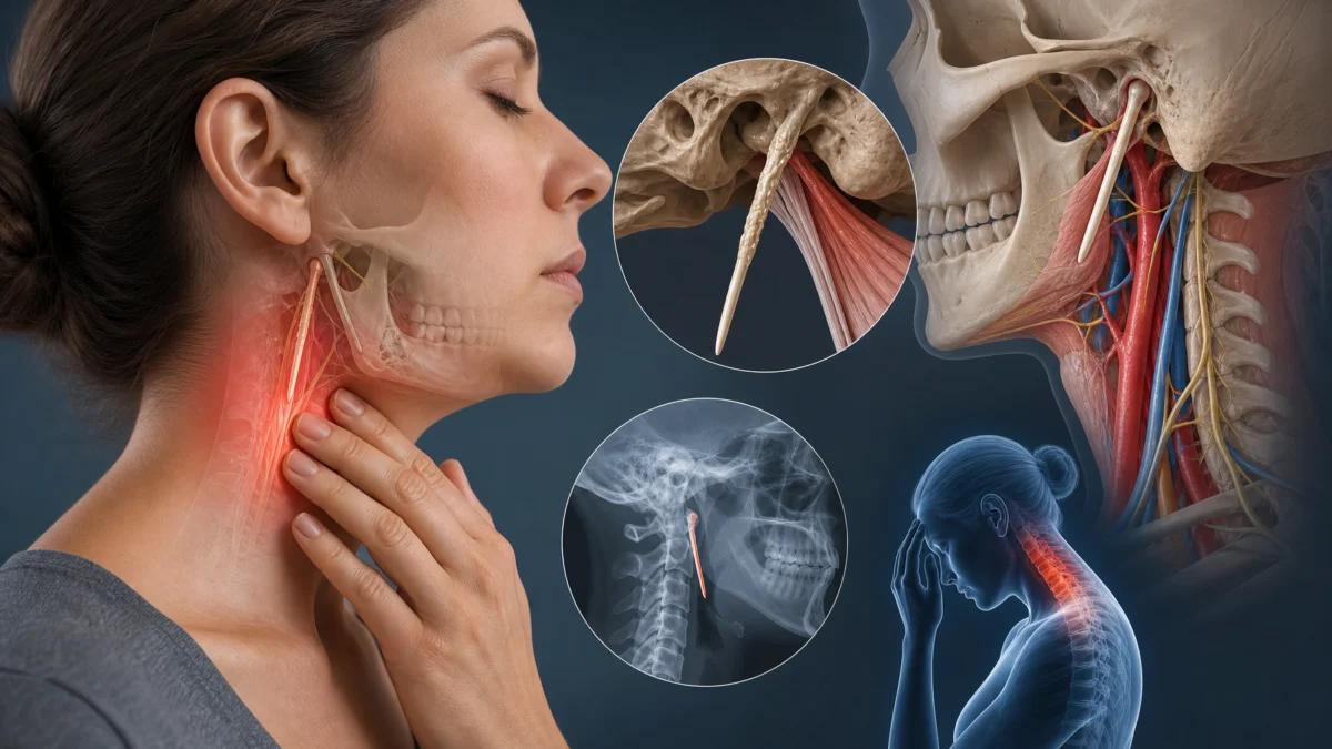
Persistent pain in the throat, beneath the jaw or towards the ear may sometimes be related to the styloid process, a bony projection of the temporal bone. Eagle syndrome is considered when symptoms and the anatomy of this region form a coherent clinical picture. A long styloid process or an ossified stylohyoid ligament found on imaging is insufficient to establish the diagnosis. This distinction prevents a common anatomical feature from being mistaken for a demonstrated cause of pain [3, 4].
There are two priorities: investigate more common explanations for throat pain and recognise situations requiring prompt referral. The vascular forms described in the literature are rare but potentially important. They should neither be dismissed nor lead to interpreting every episode of dizziness or every headache as vascular compression [1, 2].
Understanding the styloid region
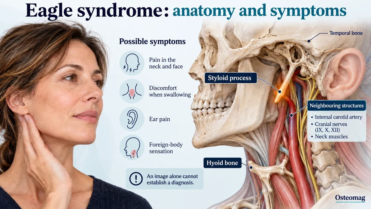
The styloid process projects downwards from the base of the skull near the ear. It provides attachment for several muscles and ligaments, including the stylohyoid ligament, which runs towards the hyoid bone. This region lies close to nerves and blood vessels in the neck. Its relationships vary between individuals according to the length, orientation and distances between structures [3].
Length is therefore only one component of the assessment. Researchers have compared CT characteristics in symptomatic people and asymptomatic controls. Other studies have examined styloid process angulation. These findings support a broader anatomical interpretation; they do not supply a threshold that independently determines the cause of symptoms in every patient [5, 6].
Ligament ossification may also change the configuration of the stylohyoid chain. Again, imaging must be related to the clinical history. Someone may have a visible abnormality without pain, whereas another person may have symptoms requiring examination of nearby structures. Diagnosis remains a process of reasoning, rather than a simple measurement on a CT scan.
Possible symptoms that are not specific to this syndrome
The so-called classic form may involve throat pain, discomfort with swallowing, a foreign-body sensation or pain radiating towards the ear and jaw. Certain movements, chewing or swallowing may change the symptoms. None of these signs is specific: they can also accompany other ear, nose and throat, dental, joint or neurological conditions [3, 4].
The person should be able to describe the location, whether symptoms are continuous or intermittent, triggering circumstances and their effect on eating, sleep or activities. A recent symptom is not interpreted in the same way as longstanding, stable discomfort. Medical history and previous investigations help organise assessment without assuming that they explain everything.
Very brief episodes of pain, discomfort mainly associated with the jaw or dental pain may suggest other possibilities. A persistent voice change, a neck lump, unexplained weight loss or progressively worsening swallowing difficulty warrants appropriate assessment. These signs should not be attributed to the styloid process before other important causes have been considered.
Vascular forms: maintaining proportionate vigilance
Published reports describe abnormal relationships between the styloid chain and certain arteries or veins. Reported arterial complications include injuries capable of producing neurological symptoms. Venous forms sometimes involve a reduced space between the styloid process and the first cervical vertebra. These mechanisms require suitable expertise and imaging; they cannot be inferred from an isolated symptom [2, 7].
A large recent synthesis of neurological presentations brings together published cases and demonstrates their diversity. Proportions observed in such a collection do not represent the frequency of vascular forms in the general population. Complex or surgically treated cases may be more likely to be published than asymptomatic incidental findings [1].
Signs requiring urgent help
Sudden weakness on one side, new difficulty speaking, abrupt loss of vision or another sudden neurological deficit requires calling emergency services, even if the symptom disappears. Unusual, severe neck or head pain accompanied by neurological signs also requires urgent care. Do not repeatedly move the neck to see whether these symptoms return.
Breathing difficulty, inability to swallow saliva or rapidly worsening swallowing also changes the priority. Support intended to improve comfort must not delay assessment. These instructions are based on the seriousness of the symptoms, regardless of whether Eagle syndrome is known or suspected.
| Situation | Cautious interpretation | Referral or next step |
|---|---|---|
| Long styloid process without symptoms | Possibly an incidental finding [3, 5] | Do not decide on treatment from the measurement alone. |
| Throat, jaw or ear pain | A non-specific clinical picture [4] | Assess ENT and dental causes and other possibilities. |
| Sudden neurological change | An urgent cause must be investigated. | Call emergency services even if the sign disappears. |
| Suspected abnormal vascular relationship | A specialist question [2, 7] | Imaging and expertise tailored to the symptoms. |
| Persistent discomfort with clinical concordance | Discussion of specialist management [8, 10] | Compare potential benefits, limitations and risks individually. |
Clinical guide — Osteomag. This table does not replace individual assessment.
How is the diagnosis established?
Clinical assessment examines the symptoms and competing explanations. Depending on the presentation, it may require ENT, maxillofacial, neurological or vascular expertise. Examination of the mouth, throat, ears, neck and neurological function is adapted to the question being investigated. Locally provoked pain is not, by itself, proof of causation [3, 4].
CT and three-dimensional reconstructions can clarify the length, orientation and bony relationships. When vascular involvement is suspected, the specialist may request a relevant vascular investigation. The choice depends on the clinical signs; additional investigation is not automatically necessary for every finding of an elongated styloid process [4, 5].
Some anatomical questions concern available space more than absolute length. The publication describing styloid–C1 juxtaposition illustrates this possibility in particular cases. It does not establish that a headache or a sense of pressure necessarily represents venous obstruction. A competent team must determine whether this relationship is clinically relevant [7].
Trying to locate the styloid process yourself by applying deep pressure inside the throat or neck is not recommended. Repeating a position that triggers unusual symptoms does not make the diagnosis more reliable. Any positional assessments belong in specialist care when they are genuinely indicated.
What can be done while assessment is underway?
Advice depends on the symptoms and the instructions received. When the situation is stable, a simple record can document the location of pain, triggering activities and effects on swallowing or chewing. It is useful to report exactly what is new, rather than recording only an overall pain score.
Temporarily adapting activities that clearly provoke discomfort may improve comfort. This should not lead to accumulating restrictions without explanation or abandoning all activity for a prolonged period. If the specialist advises against certain movements or positions, those instructions take precedence over a general exercise programme.
Difficulty eating or drinking should be reported promptly. A persistent change in food intake, weight loss or dehydration should not be treated as minor inconveniences to endure while waiting. Management should address the practical consequences of symptoms as well as the interpretation of scans.
Longstanding pain can also become worrying and exhausting. Taking this experience seriously does not require attributing every symptom to the styloid process. Explaining what is confirmed, what remains uncertain and what will be reassessed can reduce confusion and improve collaboration between professionals.
What role can osteopathy play?
Once concerning causes have been assessed and the situation permits, cautious support may address comfort, movement habits and associated jaw or neck tension. These objectives concern functional difficulties. They must remain compatible with the restrictions and recommendations of the team responsible for care.
There is no strong evidence that osteopathy shortens the styloid process, reverses ligament ossification or treats a vascular complication. Specific studies mainly concern anatomy, diagnosis and outcomes of specialist management. They do not validate an osteopathic method for correcting the syndrome [1, 3, 4].
Support should avoid deep pressure over the front or side of the neck and repeated extreme positions that reproduce concerning symptoms. It must not attempt to demonstrate compression by provoking faintness, a visual disturbance or a neurological symptom. The appearance of such signs calls for appropriate referral rather than continuation of the consultation.
A useful objective may be to tolerate a permitted activity more comfortably or understand how to adjust a troublesome demand. Lack of improvement should prompt review of the plan. Repeated consultations do not replace specialist discussion when symptoms remain significant or the diagnosis is uncertain.
Surgery: a decision based on clinical concordance
Styloidectomy involves shortening or removing the implicated part of the styloid chain, depending on the case. Approaches through the mouth or through a neck incision have been described. The choice depends on anatomy, the clinical presentation, nearby structures and the team’s experience. An asymptomatic imaging abnormality does not automatically justify an operation [4, 8].
A large recent surgical series reported improvements in selected patients. Other retrospective studies examined outcomes following a cervical approach and changes in quality of life. These observations inform discussion, but their retrospective design and lack of robust comparison limit conclusions. Average improvement within a series does not guarantee another person’s outcome [8, 9, 10].
The synthesis of published outcomes also suggests benefit in many treated cases, while relying substantially on non-randomised data. Decisions should take this uncertainty, procedural risks and the likelihood that symptoms truly relate to the styloid region into account [1].
Before surgery, the person should understand which symptoms may improve, what limitations may remain and how recovery will be organised. Pain with several contributing components may persist partly even after an anatomical conflict has been treated. This possibility deserves a clear explanation when discussing expectations.
Follow-up guided by everyday function
After an intervention, the surgical team’s instructions determine the return to eating, movement and other activities. Changes in voice, swallowing, pain and mobility should be monitored according to the proposed plan. No general timetable replaces these individual instructions.
When surgery is not selected, follow-up still helps document symptom stability and review adaptations. Improved comfort does not prove that bone length has changed. Conversely, new discomfort should not automatically be attributed to the same mechanism without reassessment.
Eagle syndrome requires careful interpretation of the relationship between anatomy and symptoms. An impressive scan may show an incidental finding; a concerning symptom may require urgent attention even without a confirmed diagnosis. Appropriate referral depends on coherent assessment, realistic functional goals and clear communication about the limits of current knowledge.
Osteopathic support plan: key points
- Relate symptoms to anatomy without diagnosing from length alone.
- Call emergency services for a sudden neurological deficit.
- Avoid deep neck pressure and provoking concerning symptoms.
- Respect the responsible team’s instructions and individual restrictions.
- Address associated comfort without promising to correct ossification or vascular involvement.
- Monitor swallowing, voice and activities, and refer again if the situation changes.
References
- [1] Neurological phenotypes and treatment outcomes in Eagle syndrome: systematic review and meta-analysis. PMID: 38948209.
- [2] Eagle syndrome and vascular complications—a systematic review. PMID: 39406633.
- [3] Eagle’s syndrome: embryology, anatomy, and clinical management. PMID: 22638594.
- [4] Eagle syndrome: A comprehensive review. PMID: 28527976.
- [5] Conventional and 3-Dimensional Computerized Tomography in Eagle’s Syndrome, Glossopharyngeal Neuralgia, and Asymptomatic Controls. PMID: 25917666.
- [6] The importance of medial-lateral styloid process angulation/coronal plane angle in symptomatic eagle syndrome. PMID: 28192866.
- [7] Styloid/C1 transverse process juxtaposition as a cause of Eagle’s syndrome. PMID: 25726731.
- [8] Eagle syndrome presentation and outcomes in a large surgical case series. PMID: 39071203.
- [9] Outcomes of Elongated Styloid Process Syndrome Treated with Minimally Invasive Cervical Styloidectomy (MICS)—A Single-Center Retrospective Study. PMID: 39518548.
- [10] Does Transcervical Styloidectomy for Eagle Syndrome Improve Quality of Life? PMID: 36174663.