
Recognizing nerve involvement and understanding when investigations are useful
A lumbar disc herniation is a localized displacement of disc material beyond the disc’s usual boundaries. It can contribute to pain radiating into the leg, but its presence on an image is not enough to explain every symptom. Diagnosis combines the history, examination, and, when indicated, concordant imaging. [1]
The distinction between low back pain, radicular pain, and neurological deficit guides care. Some situations allow conservative management, while new signs require urgent assessment. The goal is to identify what requires a medical decision and preserve useful abilities without presenting the disc as a structure to put back in place.
For organizing a return to everyday activities, also see our article on rehabilitation after lumbar disc herniation: Complementary Osteomag article.

Anatomy: the disc, spinal canal, and nerve roots
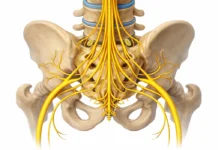
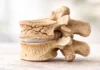
Intervertebral discs combine a central nucleus and a fibrous annulus. They help distribute forces and enable movement between vertebrae. Facet joints, ligaments, and muscles also contribute to spinal function. Disc changes depend on biological and mechanical factors, with considerable variability.
A herniation can take different forms and occur in different locations. These details affect its relationship with a nerve root. A protrusion, an extrusion, and a separated fragment do not describe the same morphology. Nevertheless, interpretation is not simply a ranking of severity. [1] [4]
In the lower lumbar region, the canal mainly contains the cauda equina nerve roots. The spinal cord generally ends higher up. A herniation at a given level may affect a root according to its position; a simplified map cannot automatically attribute each symptom to a vertebra.
Mechanisms: compression, irritation, and clinical context
A herniation may contribute to nerve irritation through its mechanical relationships and local inflammatory processes. Pain does not depend solely on measurable pressure. Symptom duration, the response to activities, and the clinical situation also matter. [1] [4]
Disc changes are common in people without symptoms. This does not invalidate the diagnosis of symptomatic herniation: it means that concordance must be sought. The side, distribution, and neurological signs should be consistent with the proposed explanation. [4]
Symptoms: three concepts to distinguish
Low back pain describes pain in the lower back. It can occur without herniation or nerve involvement. Pain extending into the buttock or thigh is not automatically sciatica. Other structures can cause referred pain.
Radicular pain follows a distribution compatible with an irritated nerve root, sometimes with burning or electric sensations. Radiculopathy refers to impaired nerve function, which may involve weakness or altered sensation or reflexes. The two can occur together, but they are not synonymous. [1]
Coughing or certain movements can increase symptoms without confirming the diagnosis on their own. A person may have substantial pain with few objective signs, or concerning weakness with relatively moderate pain. Assessment must therefore go beyond pain intensity alone.
Situations requiring urgent assessment
Differential diagnosis: do not attribute everything to the disc
Hip pain, lumbar stenosis, peripheral neuropathy, and other conditions can produce leg symptoms. Walking limitation can also have a vascular origin. Distribution and the circumstances in which symptoms ease help distinguish these possibilities. [1] [2]
A very swollen, hot, or painful leg requires assessment for a possible vascular cause. Unusual abdominal pain or declining general health changes the reasoning. Examination should not remain confined to the spine when the presentation suggests another problem.
Several conditions can coexist. A known herniation does not prevent a new hip disease or a separate neuropathy. The professional checks explanations rather than automatically retaining the diagnosis in an old record.
Clinical examination: building a coherent picture
The interview clarifies onset, radiating symptoms, progression, limited activities, and concerning urinary or sensory symptoms. The physician or relevant professional examines walking, strength, sensation, and reflexes when appropriate. Findings are compared and documented for follow-up. [1]
The straight-leg raise and other provocation tests can contribute to clinical reasoning. They are not simply a feeling of tension at the back of the thigh. Reproduction of concordant symptoms, other findings, and the test’s limitations should be considered together.
A confirmed motor deficit needs particular attention. Difficulty performing a movement because it hurts is not always neurological weakness. The professional distinguishes these situations and decides on the appropriate level of referral and monitoring.
Imaging: when a result changes the plan
MRI examines the discs and nerve structures, among other tissues. It becomes relevant with concerning signs or when persistent symptoms lead to a specialist decision. Timing depends on the presentation; a substantial or progressive deficit should not wait for an arbitrary duration. [1] [2]
A radiograph does not directly identify a disc herniation. It can answer other questions about bone or alignment. CT may be considered in certain situations, particularly when MRI is not possible or for a specific question. The choice belongs to the medical team. [2]
An image should be interpreted alongside symptoms: an abnormality on the opposite side or at an inconsistent level may not explain the complaint. Repeating MRI only to check whether comfort-focused care has “corrected” the disc is not sufficient justification.
| Finding | Cautious interpretation | Approach |
|---|---|---|
| A visible herniation without concordant symptoms | Possible incidental finding | Connect imaging with the examination and history |
| Radiating pain without an identified deficit | Several mechanisms remain possible | Assessment and monitoring of progression |
| New or progressive weakness | Neurological involvement requiring clarification | Prompt medical assessment |
| Sphincter disturbances or altered perineal sensation | Suspected urgent situation | Urgent assessment without waiting |
| Persistent symptoms substantially limiting life | The initial plan may be insufficient | Reassessment and specialist discussion |
Progression: improvement is possible without a guaranteed timeline
Some herniations decrease spontaneously on imaging. The probability varies with morphology and the populations studied. However, radiological regression and clinical improvement are not perfectly synchronized events. Resorption within a precise period should not be promised. [5]
Pain may improve while an abnormality remains visible. Conversely, difficulties may persist despite a reduction in herniation volume. Follow-up should assess function and neurological signs, not only the disc’s appearance.
Guidelines consider conservative management in the absence of cauda equina syndrome or a concerning major deficit. This involves follow-up and referral criteria rather than indefinite waiting. Abilities and restrictions are reassessed as the situation changes. [6]
Rehabilitation: adapting demands and resuming activities gradually
The program is chosen according to observed difficulties and the activities being pursued. It may include information, adapted walking, exercises, and a progression in tasks. An activity that substantially and persistently increases symptoms can be adjusted without prohibiting every back movement. [6] [7]
Strengthening and control exercises address functional needs; they do not mechanically push a herniation back into the disc. Movement preferences may guide certain exercises, with reassessment. No movement suits every presentation.
Low back pain guidelines support individualized active approaches, but their findings do not automatically apply to every radiculopathy. A deficit, recent surgery, or a specific diagnosis changes the program. The physiotherapist and physician coordinate these elements. [7] [8]
When should a specialist intervention be discussed?
Specialist advice is important when pain remains highly disabling despite appropriate management or neurological deficits develop. Surgical guidelines account for clinical and radiological concordance, progression, and risks. A visible herniation alone does not require an operation. [9]
A randomized trial in selected patients with sciatica found faster recovery with early surgery, while one-year outcomes were similar between strategies. Some people in the conservative group later underwent surgery. This finding informs shared decision-making; it does not determine an individual’s choice. [10]
The process and limits of osteopathic care
The osteopath checks warning signs, investigations, and restrictions before proposing care. They may assess permitted movements and troublesome activities without replacing medical neurological assessment. New weakness or sphincter disturbances requires appropriate referral.
Follow-up centered on function and new signs
Follow-up compares walking, sitting, sleep, or a work task alongside relevant neurological findings. Increasing independence is progress, even if imaging remains altered. Lasting deterioration or new signs requires reassessment.
When fear of movement or distress becomes substantial, referral to a psychotherapist may complement care. It does not mean that pain is imaginary. A cautious approach helps explain options, recognizes its limits, and preserves the possibility of improvement. [8]
References
- [1] Lumbar disc herniation: Epidemiology, clinical and radiologic diagnosis. WFNS spine committee recommendations, 2024. Scientific or institutional source
- [2] American College of Radiology. ACR Appropriateness Criteria: Low Back Pain, 2021 update. Scientific or institutional source
- [3] Vaishya S et al. Cauda equina, conus medullaris and syndromes mimicking sciatic pain: WFNS spine committee recommendations. 2024. Scientific or institutional source
- [4] Brinjikji W et al. Systematic literature review of imaging features of spinal degeneration in asymptomatic populations. AJNR, 2015. Scientific or institutional source
- [5] Chiu CC et al. The probability of spontaneous regression of lumbar herniated disc: a systematic review. Clinical Rehabilitation, 2015. Scientific or institutional source
- [6] The role of conservative treatment in lumbar disc herniations: WFNS spine committee recommendations. 2024. Scientific or institutional source
- [7] George SZ et al. Interventions for the management of acute and chronic low back pain: revision 2021. JOSPT. Scientific or institutional source
- [8] NICE. Low back pain and sciatica in over 16s: assessment and management, NG59. Scientific or institutional source
- [9] Role of surgery in primary lumbar disk herniation: WFNS spine committee recommendations. 2024. Scientific or institutional source
- [10] Peul WC et al. Surgery versus prolonged conservative treatment for sciatica. New England Journal of Medicine, 2007. Scientific or institutional source