Deep calf pain and plantar tingling: understanding a rare nerve hypothesis without overlooking emergencies
Soleal sling syndrome involves compression of the tibial nerve beneath a fibrous arch in the upper calf. Deep calf pain may occur together with unusual sensations in the sole of the foot. This is a rare condition, and its true frequency remains uncertain. Calf pain, even when accompanied by tingling, is insufficient to identify it. [1]
The first priority is to find a coherent explanation and exclude urgent conditions. Deep vein thrombosis, arterial disease and compartment syndrome must not be mistaken for a “tight soleus.” Diagnosis requires medical assessment; an osteopath can recognize reasons for referral and support movement after the situation has been clarified. This distinction matters because similar symptoms can require very different investigations and treatment.
Anatomy: the tibial nerve beneath the soleus
The tibial nerve arises from the sciatic nerve. It crosses the back of the knee and descends through the leg before reaching the area behind the medial ankle and the sole of the foot. Its branches contribute to muscle control in the leg and foot and to plantar sensation. The pattern of any deficit depends on the level affected and individual anatomical variations.
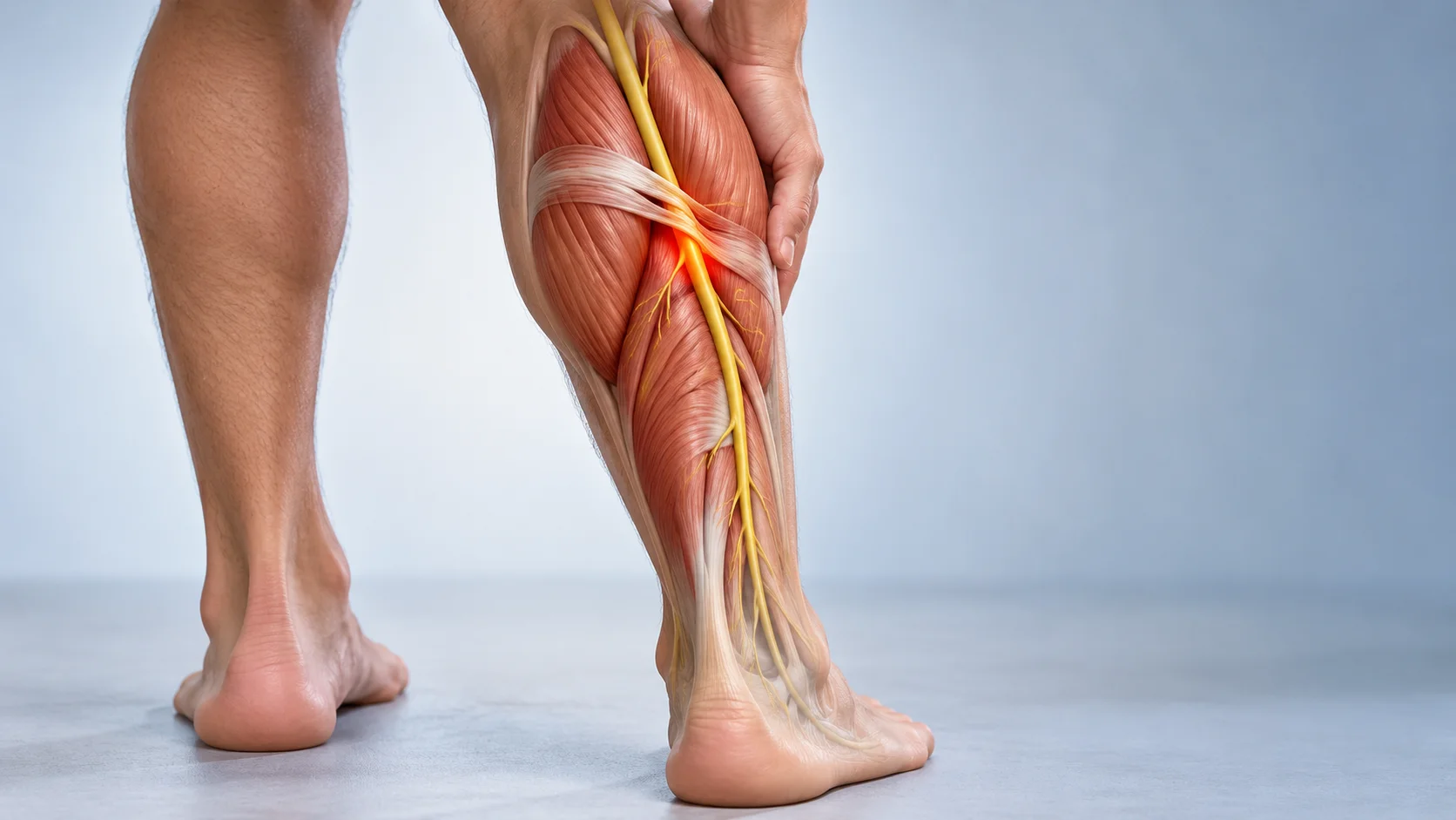
In the upper calf, the nerve passes beneath a fibromuscular arch associated with the soleus attachments. This sling is a normal structure: its presence does not imply disease. An anatomical study of 36 limbs showed that a fibrous band could exist without obvious nerve narrowing. Anatomy therefore identifies a possible compression site, without establishing that it explains every local pain. [2]
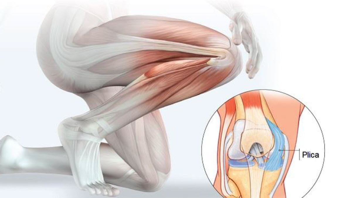
Regional blood vessels and deep muscles lie nearby. Simple external pressure does not directly examine the nerve. Feeling a “hard” area of calf tissue proves neither nerve entrapment nor a need to “release” it. A simplified anatomical illustration helps explain the possible relationship but cannot reproduce an individual’s anatomy or establish a diagnosis.
Possible symptoms and proposed mechanisms
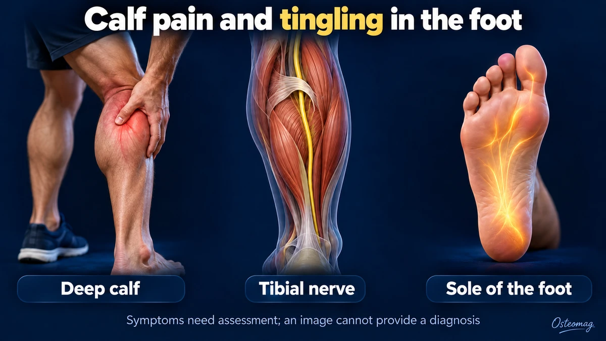
Clinical descriptions include upper calf pain, deep tenderness and burning, tingling or numbness in the sole. Symptoms may increase during walking or running. In some cases, weakness when bending the toes, particularly the great toe, draws attention. None of these findings is specific when considered alone. The distribution and evolution of symptoms are assessed together rather than treated as a checklist that confirms the condition. [1] [3]
A restrictive fibrous arch may be the compression site. Scarring after injury or a space-occupying lesion can also affect the proximal tibial nerve. Cysts, masses or vascular abnormalities sometimes require targeted investigation; they are not all located precisely beneath the sling. Cases without an obvious structural cause are harder to document. [5] [9]
Increased training load may bring symptoms to attention, but it does not prove that the muscle is compressing the nerve. Severe post-traumatic presentations described in some series cannot predict the course of intermittent symptoms in a runner. Context and progression matter more than a single mechanical explanation. [10]
Medical examination: localization and safety
The history explores symptom onset, distribution, triggers and recovery time after exercise. The physician asks about injury, recent immobilization, surgery, vascular history and known neuropathy. Back-related symptoms and changes in the other foot also contribute to the assessment. Describing when symptoms appear and how they change can be more useful than repeatedly pressing on a painful spot.
Examination may compare plantar sensation, toe strength, plantar flexion, reflexes, colour, temperature and swelling. A Tinel sign describes sensations reproduced at a distance by tapping along a nerve’s course. It may guide localization but cannot independently confirm compression beneath the soleus. [5]
Palpation cannot exclude thrombosis. Similarly, pulses felt at rest do not rule out every arterial problem triggered by exercise. There is no validated osteopathic test that establishes soleal sling syndrome. New or progressive weakness needs prompt medical assessment rather than a succession of trial treatments. The purpose of examination is both to investigate the hypothesis and to recognize findings that point elsewhere.
The main conditions to distinguish
| Possibility | Features that guide assessment | Practical implication |
|---|---|---|
| Proximal tibial nerve compression | Deep calf pain with plantar symptoms, sometimes toe weakness | Neurological assessment and targeted investigations |
| Lumbosacral radiculopathy, including S1 | Radiating symptoms, strength or reflex changes, sometimes back pain | Back pain may be absent; neurological examination is needed |
| Tarsal tunnel syndrome | Symptoms around the medial ankle and sole | Consider a more distal or coexisting nerve problem |
| Chronic exertional compartment syndrome | Reproducible exercise-related pain and tightness, often relieved by rest | Specialist assessment; pressure testing may be indicated |
| Arterial cause | Exertional pain, colour or temperature changes | Vascular assessment; emergency care for a suddenly cold painful foot |
| Thrombosis or another cause of calf swelling | Recent swelling, pain, warmth and relevant risk factors | Urgent assessment, without massage or an exercise trial |
Tarsal tunnel syndrome affects the tibial nerve at the ankle and can produce similar sensations. Ultrasound can look for local causes there, but findings at that level do not automatically validate examination of the soleal sling. [6]
Chronic exertional compartment syndrome and popliteal artery entrapment can resemble neuropathy in athletes. Pressure measurements or vascular investigations are chosen according to the clinical suspicion and have their own limitations. Muscle injury, a popliteal cyst or polyneuropathy may also be considered. Recognizing a possible nerve disorder does not remove the need to investigate these alternatives. [7] [8]
Electrodiagnostic testing, ultrasound and MRI
What electrodiagnostic testing can contribute
Nerve conduction studies and electromyography look for nerve dysfunction and its distribution. They may help distinguish tibial neuropathy from a root lesion or more widespread disease. Examining some deep muscles and localizing the precise site can be difficult. A normal result alone does not establish the absence of every intermittent compression; a specialist interprets it alongside the clinical findings. [5]
Choose imaging to answer a specific question
Ultrasound and MRI may examine the nerve and its surroundings, look for a mass or show muscle changes. Doppler ultrasound addresses a vascular question and is distinct from nerve ultrasound. Depth and operator experience affect what can be seen. A test should be selected because it can help resolve a relevant uncertainty, rather than because all calf pain requires the same scan.
Magnetic resonance neurography uses a specialized protocol and has shown corresponding abnormalities in certain surgically treated cases. This does not mean it confirms or excludes every case. Imaging must agree with the symptom distribution and examination; its usefulness is less consistent when there is no visible structural cause. [4] [9]
Conservative management: an individual plan
After urgent conditions have been excluded and medical assessment has clarified the situation, temporary activity modification may be considered for stable presentations without progressive deficit. Activities that trigger tingling are reduced, without assuming that prolonged complete rest is always needed. Maintaining tolerated activities helps limit deconditioning. The plan can change as the response becomes clearer.
Rehabilitation, including care with a physiotherapist, adjusts active movement, strengthening and walking demands. Light calf or foot exercises may be appropriate when neurological status allows. Their dose depends on the response during and after activity, without trying to push through numbness. Exercises should fit the assessment rather than being prescribed simply because the calf feels tight.
No well-validated conservative protocol is specific to this syndrome. Neurodynamic exercises should not be presented as proven decompression: if a professional selects them, they must be tailored to irritability and should not cause sustained symptom worsening. Increasing sensory loss or weakness requires reassessment rather than a stronger stretch or more repetitions.
Returning to walking and running
Follow-up can record comfortable walking time, tingling frequency, numb areas and changes in strength. These measures track function; they cannot independently identify the mechanism. A shorter distance before symptoms develop or slower recovery is a reason to review the plan. A record of the activity and its after-effects helps make these changes clear.
If progress and medical advice allow, return may move from comfortable walking to short running intervals and then a gradual increase in volume. One demand changes at a time, with adequate recovery. There is no universally validated timeline or percentage progression for this rare compression. The previous stage should be tolerable before a more demanding one is introduced.
Weakness, numbness persisting at rest or deteriorating walking should not be dismissed. After surgery, instructions from the surgeon and rehabilitation team take priority. Relief following a session or success with one exercise does not authorize unrestricted return. The wider pattern of neurological findings and everyday function remains important.
Osteopathic support and the limits of manual care
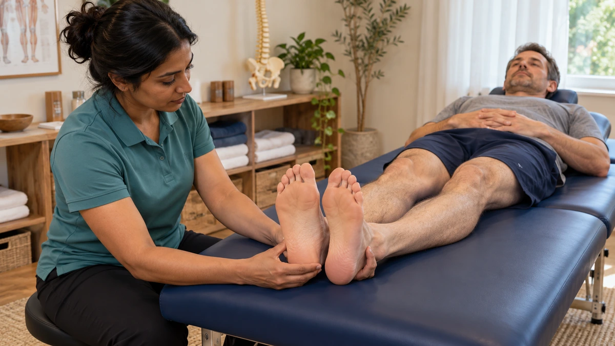
An osteopath can recognize a presentation requiring referral, explain the limits of their intervention and support comfortable movement after medical clarification. Gentle work on neighbouring tissues may be considered for comfort, without claiming to treat deep nerve compression. It should remain compatible with the rehabilitation programme and should not delay investigation.
The evidence considered here does not establish that an external osteopathic technique permanently opens the sling or restores nerve conduction. Strong deep pressure intended to “free” the nerve is unjustified. An osteopath does not make the medical diagnosis and cannot exclude thrombosis or arterial disease through manual examination. Symptom relief and treatment of an underlying cause are separate outcomes.
When is specialist assessment or surgery considered?
Persistent symptoms, lasting sensory loss, weakness or a suspected mass justify specialist medical review. Surgery may be discussed when compression consistent with the symptoms is identified and its impact, progression or structural cause warrants intervention. Calf pain alone is insufficient justification. The expected benefits and uncertainties should be reviewed for the individual presentation.
Decompression aims to free the nerve at the relevant site. A retrospective series of 49 patients reported variable outcomes across groups. The absence of a control group, different patient profiles and surgical selection limit the conclusions. Surgery should therefore not be described as a guaranteed cure or a necessity for every suspected case. [3]
Warning signs: do not wait
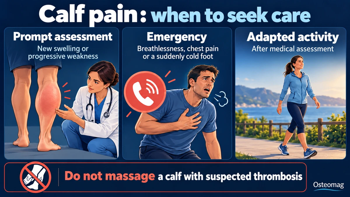
A newly swollen painful calf requires urgent assessment, especially after immobilization or surgery, without waiting for every possible sign to appear. Thrombosis cannot be confirmed or excluded by massage or an ankle movement test. [11]
Sudden breathlessness, chest pain or collapse: call emergency services. A suddenly cold, pale and painful foot is also an emergency. Increasing severe pain with a very tense calf, particularly after trauma, may indicate acute compartment syndrome: do not wait for pulses to disappear before seeking care. [8] [11] [12]
Key points
The soleal sling is a possible but rare and difficult-to-confirm site of tibial nerve compression. Calf pain with plantar symptoms deserves structured assessment. Vascular and neurological safety come before local treatment; osteopathic support remains complementary and does not replace this process.
Scientific references and guidelines
- [1] Hilbig-Vlatten L, Grauberger JN, Engmann TF, et al. Soleal Sling Syndrome: A Narrative Review. J Peripher Nerv Syst. 2025;30:e70032. PubMed.
- [2] Williams EH, Williams CG, Rosson GD, Dellon AL. Anatomic site for proximal tibial nerve compression: a cadaver study. Ann Plast Surg. 2009;62:322–325. PubMed.
- [3] Williams EH, Rosson GD, Hagan RR, Hashemi SS, Dellon AL. Soleal sling syndrome (proximal tibial nerve compression): results of surgical decompression. Plast Reconstr Surg. 2012;129:454–462. PubMed.
- [4] Chhabra A, Williams EH, Subhawong TK, et al. MR neurography findings of soleal sling entrapment. AJR Am J Roentgenol. 2011;196:W290–W297. PubMed.
- [5] Diagnostic Approach to Lower Limb Entrapment Neuropathies: A Narrative Literature Review. Diagnostics. 2023;13:3385. PubMed.
- [6] Fantino O. Role of ultrasound in posteromedial tarsal tunnel syndrome: 81 cases. J Ultrasound. 2014. PubMed.
- [7] Vogels S, Ritchie ED, Borger van der Burg BLS, et al. Clinical Consensus on Diagnosis and Treatment of Patients with Chronic Exertional Compartment Syndrome of the Leg: A Delphi Analysis. Sports Med. 2022;52:3055–3064. PubMed.
- [8] Shahi N, Arosemena M, Kwon J, et al. Functional Popliteal Artery Entrapment Syndrome: A Review of Diagnosis and Management. Ann Vasc Surg. 2019;59:259–267. PubMed.
- [9] Donovan A, Rosenberg ZS, Cavalcanti CF. MR imaging of entrapment neuropathies of the lower extremity. Part 2. The knee, leg, ankle, and foot. Radiographics. 2010;30:1001–1019. PubMed.
- [10] Tang P, He Q, Chen C, Liu X, Zhang L. Earthquake generated proximal tibial nerve compression treated by surgery. Int Orthop. 2013;37:1561–1566. PubMed.
- [11] NICE. Venous thromboembolic diseases: diagnosis, management and thrombophilia testing. NG158. Recommendations, updated 2023. Official source.
- [12] American Academy of Orthopaedic Surgeons. Management of Acute Compartment Syndrome. Clinical Practice Guideline. 2025. Official source.