A tissue capable of contributing to pain, without explaining everything
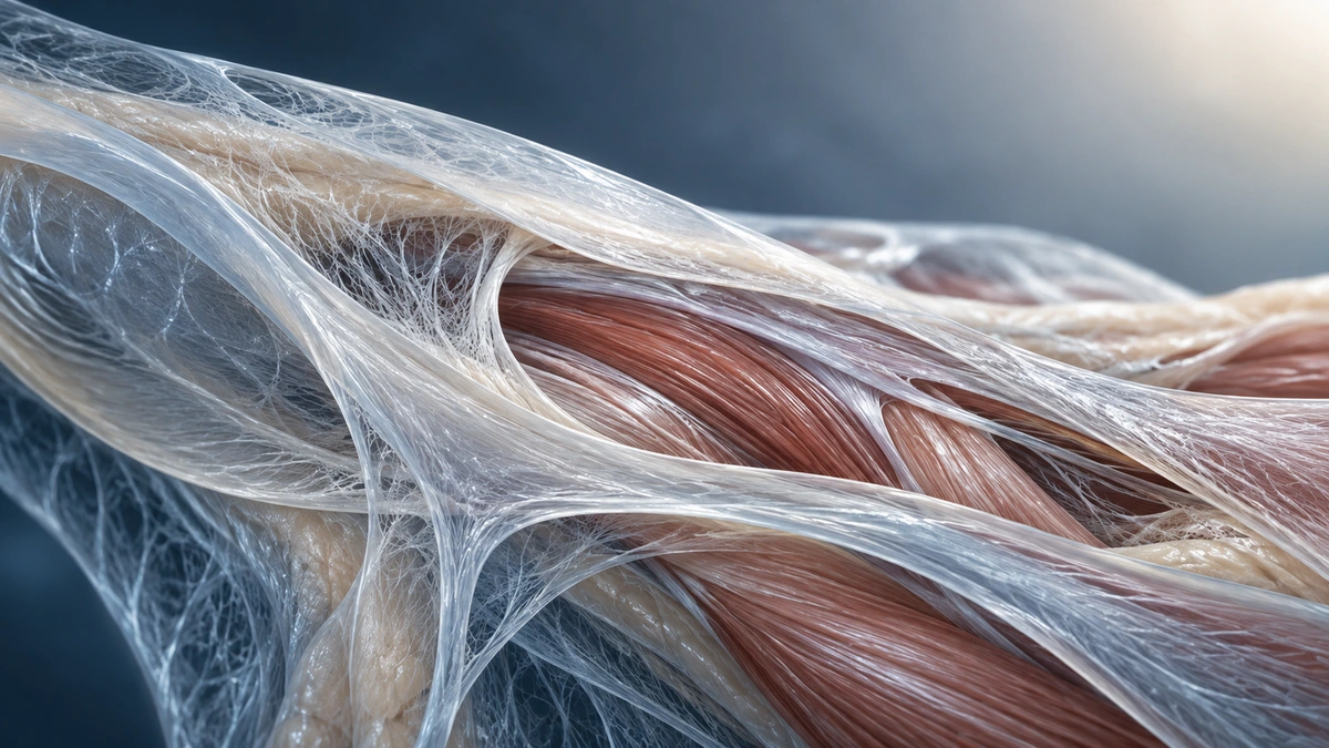
Fascia plays an important role in anatomy and in some research on movement and pain. It contains connective tissue components that support, connect, and separate structures. Several fasciae are innervated and can contribute to painful sensations. However, these findings do not support the conclusion that all pain originates from an overly tight fascial network or that a technique can release it permanently.[1] [2] [3]
To interpret studies accurately, several questions must be distinguished: which tissue is being discussed, how is it measured, in whom, and with what outcome? A cellular finding is not a therapeutic trial. A difference observed on ultrasound does not automatically demonstrate a cause of pain. A patient experiencing relief after treatment does not, by itself, prove that adhesions have been altered. These distinctions make the evidence more useful for care.
The essential takeaway
Fascia is living tissue and can contribute to pain. Nevertheless, there is no single fascial explanation for all symptoms, nor is there evidence that relief from manual therapy corresponds to the removal of a specific restriction.
What does the word fascia mean?
The word is used with different meanings. In an anatomical description, a fascia may refer to a defined sheet of connective tissue, such as the thoracolumbar fascia. The broader concept of the fascial system encompasses a continuum of loose and dense fibrous connective tissues. The nomenclature work by Adstrum and colleagues seeks to clarify these uses. Using a broad definition does not erase the differences among structures and their functions.[1]
Composition and organization vary by region. Collagen fibres contribute to mechanical strength; the extracellular matrix and cells help maintain the tissue. Some layers allow relative gliding during movement. Superficial fascia, an aponeurosis, and a deep structure do not have exactly the same properties. Referring simply to “fascia” without specifying the site can obscure this diversity.
Anatomical continuity allows interactions between neighbouring tissues, but it does not provide an automatic clinical map of pain at distant sites. A possible mechanical relationship must still be linked to an activity, symptoms, and relevant measurements. Finding a tender area at a distance is not enough to establish that it is the cause of the primary pain. Anatomy helps formulate a hypothesis; it does not validate it on its own.
Innervation: evidence for a sensory role
Tesarz’s research on the thoracolumbar fascia demonstrates sensory innervation, with differences among the layers studied. It includes observations in rats and humans. This research supports the idea that fascia can convey sensory information; it does not allow the same density of innervation to be generalized to all tissues or determine the cause of an individual’s low back pain.[2]
Experimental stimulation of the thoracolumbar fascia with hypertonic saline can provoke pain in volunteers. Schilder’s study describes characteristics that differ from those obtained in other tissues of the back. It establishes the capacity to generate pain under the experimental conditions. It does not demonstrate that typical persistent low back pain is caused in the same way or that fascia is always its primary source.[3]
Gliding and stiffness: measurements that require interpretation
Langevin’s 2011 study observed less relative gliding between layers of the thoracolumbar fascia in a group with chronic low back pain. This finding opened up interesting avenues of research. However, a comparison between people with and without pain cannot determine whether the difference precedes the pain, results from altered movement, or depends on several factors. The authors themselves consider several explanations.[4]
Ultrasound and elastography can investigate thickness, displacement, or certain mechanical properties. Results depend on the protocol, position, region, and measurement used. A different value does not necessarily indicate diseased tissue or a treatment target. Research may compare groups without yet providing a reliable threshold for diagnosing an individual in clinical practice.
Palpation addresses a different question. It describes a sensation perceived under the hand, influenced by overlapping structures, pressure, and muscle activity. The limited reliability of several manual examinations calls for caution. It is not justified to present fingers as an instrument capable of precisely measuring the microscopic properties of fascia or identifying the sole structure responsible for pain.[12]
Mechanotransduction: a biological avenue, not a promise of treatment
Mechanotransduction refers to the way cells respond to mechanical stresses through biological changes. Langevin’s research on fibroblasts notably shows changes in cell shape and cytoskeletal organization after tissues are stretched in experimental models. These findings illustrate an active cellular response rather than a completely passive material. Ex vivo or animal conditions must be distinguished from a clinical intervention.[5] [6]
These observations cannot be used to directly infer the ideal massage pressure, the number of sessions, or the resolution of pain. Moving from a mechanism to a recommendation requires demonstrating a relevant benefit in patients and comparing the intervention with appropriate options. A cellular explanation can provide a rationale for a research question without guaranteeing the success of a marketed technique.
A table to distinguish levels of evidence
| Finding | Reasonable interpretation | Unproven conclusion |
|---|---|---|
| A fascia contains sensory fibres | It can contribute to sensation and nociception | All pain comes from fascia |
| Experimental stimulation provokes pain | The tissue can be painful under this protocol | The same mechanism explains every case of chronic pain |
| Different gliding is observed on ultrasound | An association deserves further study | The difference is necessarily the cause of the symptom |
| Fibroblasts respond to stretching | Cells respond to mechanical stresses | Manual therapy heals through this specific mechanism |
| Massage improves comfort | The perceived benefit can be monitored | Deep adhesions have been broken up |
| Palpation detects resistance | An observation to be considered in context | A definitive diagnosis or a stored emotion |

Persistent pain cannot be reduced to a tissue
The IASP definition recognizes pain as a personal experience with sensory and emotional dimensions. This definition does not deny the contribution of tissues; it avoids requiring a permanently visible lesion to acknowledge that pain is real. Sleep, activity, daily demands, and nervous system responses can influence the experience and limitations.[11]
A feeling of stiffness can therefore accompany different situations: exertion, pain, protective muscle responses, lack of activity, or another condition. It does not necessarily mean that an entire fascial network has shortened. The history and examination help distinguish these possibilities. Saying that pain is multifactorial should not become a way to ignore physical symptoms or offer a vague explanation for every problem.
None of these studies validates the idea that fascia stores a particular emotion that can be detected by touch. A person may experience an emotion during treatment, but this experience does not prove the release of a memory trapped in the tissues. The practitioner can acknowledge it respectfully, without imposing an interpretation or exceeding their scope of practice. Consent remains necessary before any further contact.
Manual therapy: assessing benefit without asserting the mechanism
Wu’s review of myofascial release for chronic low back pain reports favourable results for certain outcomes, with varied studies and comparators. It does not support the claim that a technique precisely remodels fascia or outperforms all forms of care. An average statistical result must be considered in relation to its clinical importance, the quality of the trials, and the duration of follow-up.[7]
The effects of manual interventions may involve sensory, neurophysiological, and contextual mechanisms. A feeling of release may help a person move more easily without demonstrating a permanent structural change. The trial on foam rolling and the iliotibial band found no significant immediate change in stiffness under the protocol studied in healthy individuals. This result highlights the need to describe effects accurately.[10] [13]
Gentle, consensual contact can be offered for comfort and then reassessed using a relevant task. Pressure does not need to be intensely painful to be beneficial. If sessions provide only very brief relief without functional improvement, the plan should be discussed. The person should not depend on a succession of supposedly necessary releases for their body to function.
Movement and loading according to the identified problem
Plantar fasciopathy is an example in which a structure and a clinical presentation can be studied more specifically. The 2023 recommendations include stretching and resistance exercises based on individual needs, as part of a coherent care plan. This does not mean that all heel pain is fascial or that these recommendations can be applied indiscriminately to every region of the body.[8]
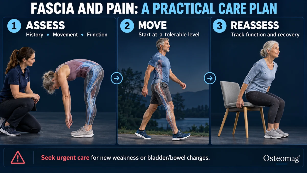
For low back pain, guidelines support different forms of exercise tailored to individual abilities and goals. Options may include strength, endurance, mobility, or functional activities. There is no single movement that can restore the entire fascial system. The key is to find a tolerable starting point, then gradually build tolerance to beneficial loads.[9]
A specific goal helps track change: walking more, getting up more easily, or returning to a sport. The response during activity and subsequent recovery guide adjustments. Improvement may occur without all sensations of stiffness disappearing. Monitoring therefore focuses on function and the person’s experience, while remaining alert to any unusual changes.
Symptoms and clinical assessment
There is no symptom that uniquely identifies fascia as the source of pain. Aching, tenderness and stiffness overlap with muscular, joint and nerve-related problems. Assessment begins with the onset, location, duration and behaviour of symptoms, including recent changes in activity, previous injury, sleep and the tasks that have become difficult. The aim is to understand the presentation and identify reasons for medical referral, rather than assign every uncomfortable sensation to a fascial restriction.
Within their scope of practice, a practitioner can observe walking, comfortable movement and a relevant task such as standing from a chair. Range of movement and tolerance to gentle loading can provide a baseline. Palpation may identify tenderness, but should be interpreted alongside the history and functional findings. It cannot establish a medical diagnosis or prove microscopic adhesions. Suspected neurological involvement requires assessment by an appropriately qualified clinician.[12] [14]
Differential diagnosis and imaging
Depending on the region, alternative explanations include muscle or tendon injury, joint disease, nerve irritation and bone stress injury. Persistent unexplained symptoms, progressive weakness or an unusual clinical course warrant medical evaluation. Imaging is selected to answer a specific clinical question. NICE advises against routine imaging for uncomplicated low back pain in nonspecialist care; specialist imaging is considered when it is likely to change management. This recommendation cannot be generalized to suspected fracture, infection or another serious condition.[15] [16]
A practical progression and the osteopath’s role
For example, someone whose walking is limited by nonspecific back discomfort might begin with a shorter, comfortable route and record how they feel during the walk and afterwards. If the activity remains manageable, duration can increase gradually. If symptoms worsen substantially or recovery becomes difficult, reduce the load and review the plan. This is an illustration, not a universal exercise prescription. Track useful outcomes such as walking tolerance, daily activities and confidence, rather than attempting to eliminate every tender point.[9]
Osteopathic care may offer consensual soft-tissue contact and help support a return to comfortable activity, as an adjunct to appropriate rehabilitation. Explain the uncertainty about mechanisms, agree on functional goals and review whether treatment adds meaningful benefit. Ongoing deterioration or lack of progress should prompt reassessment and, where appropriate, referral. Emotional distress deserves respectful support and referral to a qualified mental health professional when needed; tissue palpation is not psychotherapy.
When to investigate another cause
Highly localized bone pain after an increase in activity should raise suspicion of a stress injury rather than simple fascial tension. Numbness associated with weakness or a progressive deficit also requires assessment. Significant trauma, deformity, or an inability to bear weight warrants prompt referral. A fascial label should not delay investigation of these causes.[14] [15]
In the context of low back pain, loss of sensation in the perineal area or new problems with bladder or bowel control may signal an emergency. New pain associated with fever, a decline in general health, or unusual progression also requires an appropriate course of action. No single sign necessarily explains the entire clinical picture, but these situations require more than comfort-focused care.[14]
Key takeaways
Fasciae have important mechanical and sensory properties. Studies should be interpreted according to their protocol and population. Evidence-informed osteopathy can support comfort and function without promising to break up adhesions or release stored emotions.
Scientific references and resources
- [1] Adstrum et al. (2017). Defining the fascial system.
- [2] Tesarz et al. (2011). Sensory innervation of the thoracolumbar fascia in rats and humans.
- [3] Schilder et al. (2014). Sensory findings after stimulation of the thoracolumbar fascia with hypertonic saline.
- [4] Langevin et al. (2011). Reduced thoracolumbar fascia shear strain in human chronic low back pain.
- [5] Langevin et al. (2005). Dynamic fibroblast cytoskeletal response to subcutaneous tissue stretch.
- [6] Langevin et al. (2006). Fibroblast spreading induced by connective tissue stretch.
- [7] Wu et al. (2021). Myofascial release for chronic low back pain: systematic review and meta-analysis.
- [8] Koc et al. (2023). Heel Pain—Plantar Fasciitis: Revision 2023.
- [9] George et al. (2021). Interventions for acute and chronic low back pain.
- [10] Bialosky et al. (2009). Mechanisms of manual therapy in musculoskeletal pain.
- [11] Raja et al. (2020). The revised IASP definition of pain.
- [12] Nolet et al. (2021). Reliability and validity of manual palpation for low back pain.
- [13] Pepper et al. (2021). Foam rolling, stretching and iliotibial band stiffness.
- [14] NICE. Suspected neurological conditions, NG127.
- [15] AAOS. Stress fractures.
- [16] NICE. Low back pain and sciatica in over 16s: assessment and management, NG59.