Distinguishing imaging changes from a genuinely painful joint
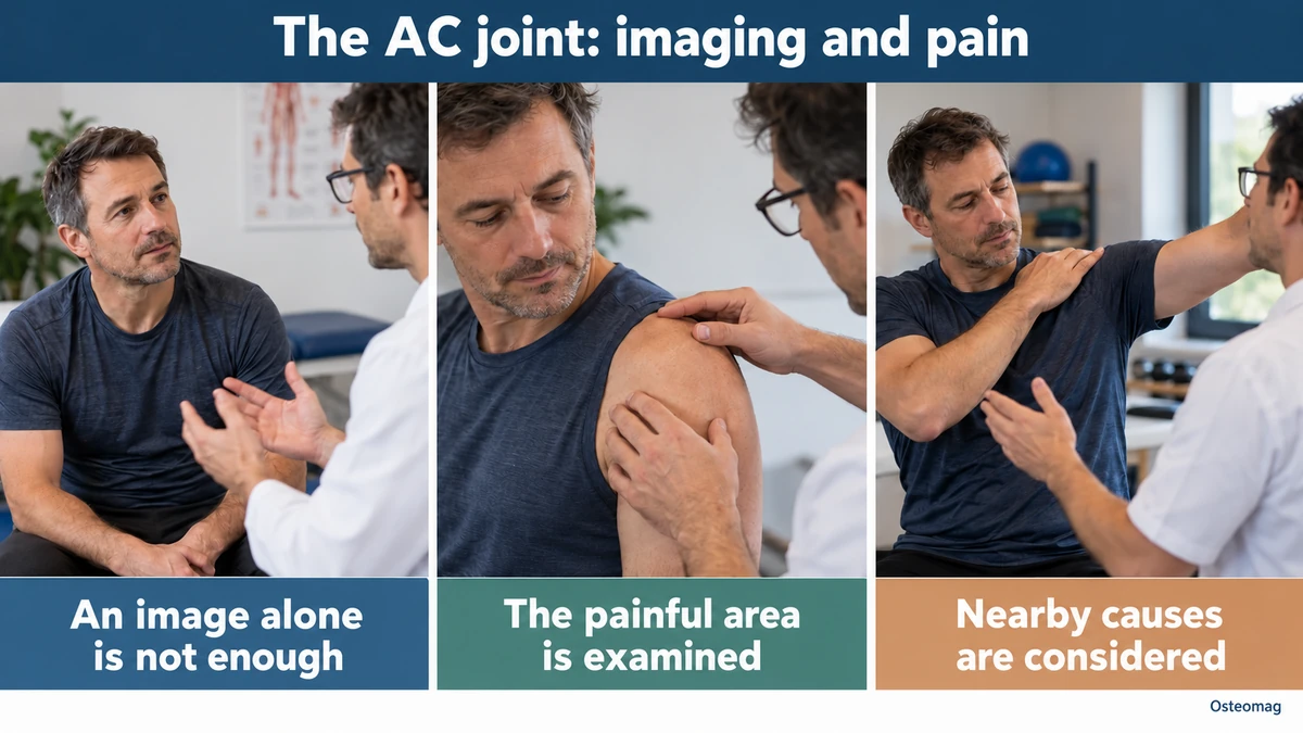
Acromioclavicular (AC) joint osteoarthritis refers to degenerative changes in the small joint at the top of the shoulder, between the lateral end of the clavicle and the acromion of the scapula. It is very common on X-rays and MRI, particularly with age, but is not always painful. This is essential: seeing AC osteoarthritis on an image does not automatically mean that this joint is causing the pain.
When symptomatic, AC osteoarthritis typically causes highly localized pain on top of the shoulder, often aggravated by movements that bring the arm across the chest—for example, reaching towards the opposite shoulder, doing push-ups, sleeping on the affected side or performing certain strength-training exercises. Diagnosis therefore depends on agreement between symptoms, clinical examination and imaging—and sometimes a diagnostic injection.
Where is the acromioclavicular joint?
The AC joint connects the outer end of the clavicle to the acromion, the part of the shoulder blade forming the top of the shoulder. It is a small synovial joint that contributes to fine adjustments of the scapula during arm movement.
It does not produce large ranges of motion like the glenohumeral joint, but must allow small rotations and translations so that the scapula can adapt to upper-limb movement. The acromioclavicular capsule and the acromioclavicular and coracoclavicular ligaments stabilize it.
Why does this joint develop osteoarthritis?
Like other synovial joints, the AC joint can develop cartilage thinning, subchondral bone remodelling, osteophytes and joint-space narrowing over time. Repeated loading probably plays a role, particularly in activities placing substantial loads on the end of the clavicle.
Previous acromioclavicular trauma can also contribute to post-traumatic osteoarthritis. An old AC sprain or dislocation may alter local mechanics and leave degenerative changes years later. However, a 2024 systematic review of the consequences of previous AC dislocations found that the relationship between the initial treatment and later osteoarthritis remained uncertain. [1]
In people who do strength training, another condition may resemble osteoarthritis: distal clavicle osteolysis. It is particularly associated with repeated loading such as bench presses or overhead movements and should be distinguished from primary osteoarthritis.
A common imaging finding that is often silent
One of the most common errors is attributing pain to osteoarthritis simply because it appears on imaging. A 2022 systematic review showed that signs of AC osteoarthritis are very common in people not seeking care for shoulder pain. In the included studies, approximately 48% of anatomical specimens and up to 70% of asymptomatic shoulders examined by MRI showed changes consistent with AC osteoarthritis. [2]
Another systematic review published in 2025 confirms that AC joint abnormalities are very common in asymptomatic shoulders. Imaging must therefore always be interpreted alongside the clinical history and examination. [3]
What are the most typical symptoms?
The most suggestive presentation is pain directly on top of the shoulder, over the AC joint. The person can often indicate the painful area with a single finger.
Pain may increase when:
- bringing the arm across the chest;
- carrying a heavy bag on the affected side;
- doing push-ups, dips or bench presses;
- working for long periods with the arm raised;
- sleeping directly on the painful shoulder;
- performing repeated pushing or pulling movements.
Clicking or cracking may occur, but is not specific. Marked restriction throughout the shoulder’s range often suggests another coexisting condition, such as adhesive capsulitis or glenohumeral osteoarthritis.
Clinical examination
Direct palpation of the joint may reproduce pain. The cross-body adduction test, in which the arm is brought across the chest towards the opposite shoulder, compresses the AC joint and may be painful.
Other tests, such as certain variations of O’Brien’s test, may contribute to assessment, but no single clinical test confirms an acromioclavicular source with certainty. Even combinations of tests often change probability only slightly; the full history, examination and context remain necessary. [4] [5]
What do X-rays show?
X-rays may show joint-space narrowing, osteophytes, subchondral bone sclerosis or irregularities at the ends of the clavicle and acromion.
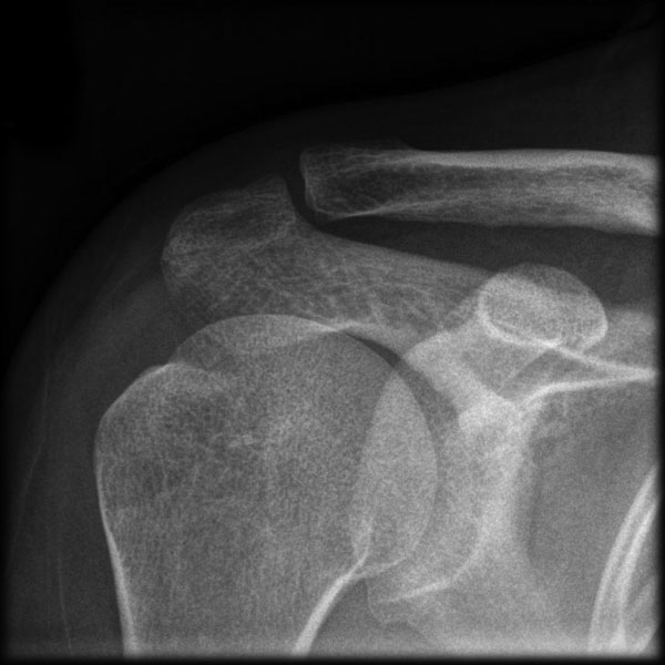

MRI is not always necessary for uncomplicated AC osteoarthritis. It becomes more useful when symptoms suggest a rotator cuff tear, labral involvement or another source of pain that X-rays cannot clarify.
| Element | Usefulness | Limitation |
|---|---|---|
| Pain on top of the shoulder | Guides the local examination | Does not identify the responsible tissue on its own. |
| Provocation tests | Reproduce certain symptoms | No single test confirms an AC source. |
| X-ray or MRI | Describes structures | Osteoarthritis is often asymptomatic. |
| Tracking meaningful movements | Assesses functional progress | A better image is not necessary for better function. |
The role of a diagnostic injection
When the diagnosis remains uncertain, a local anaesthetic injection into the AC joint may help determine whether this joint genuinely contributes to the pain. A clear, temporary reduction in pain after the injection strengthens the hypothesis of an acromioclavicular source.
This procedure belongs to an appropriately authorized medical professional. Its response may strengthen a hypothesis, but is neither absolute proof nor a reason to recommend the same procedure for every patient.
This information is particularly useful before considering surgery, because osteoarthritis visible on imaging can be entirely asymptomatic. [6]
Differential diagnosis
Pain on top of or at the front of the shoulder can also arise from other structures. The main possibilities include:
- rotator cuff tendinopathy or tear;
- subacromial pain;
- long head of biceps tendinopathy;
- adhesive capsulitis;
- glenohumeral osteoarthritis;
- referred pain from the neck;
- an old acromioclavicular sprain;
- distal clavicle osteolysis;
- less commonly, inflammatory arthritis or joint infection.
A complete shoulder and neck examination is therefore generally more useful than an assessment limited to the X-ray.
Conservative treatment
Treatment usually begins with non-surgical measures. A 2024 systematic review identified favourable outcomes with several approaches, while emphasizing variable study quality and the absence of one clearly superior strategy. [7]
Temporarily adapt irritating activities
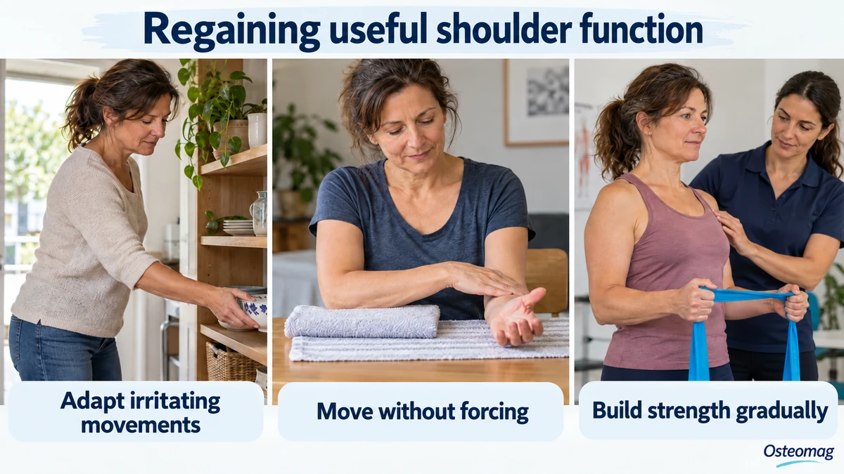
Temporarily reducing movements that strongly compress the joint may help, particularly repeated cross-body movements or certain strength-training exercises. This does not mean immobilizing the shoulder. The aim is to adjust loading while maintaining tolerable activity.
Exercise and rehabilitation
An exercise programme may target rotator cuff strength, scapular control, shoulder endurance and the ability to perform movements that usually provoke pain. The goal is not to ‘create space’ in the joint or erase imaging changes, but to improve functional capacity.
Evidence specific to physiotherapy for AC osteoarthritis remains less extensive than for other shoulder conditions. This supports a pragmatic approach: progressive loading, reducing highly provocative movements and gradual reintroduction according to the patient’s goals. [8]
Follow-up can focus on a meaningful task: carrying a light bag, dressing or reaching a shelf. Tolerance is compared across sessions and irritating tasks are gradually reintroduced. A flare-up does not require prolonged immobilization, but unusual pain or substantial loss of function warrants reassessment.
When is surgery considered?
Surgery is generally reserved for people whose pain remains substantial despite several months of conservative treatment and whose AC joint has been clearly identified as the source of symptoms.
The most common procedure is distal clavicle excision, also called the Mumford procedure. A small portion of the clavicle is removed to prevent painful contact with the acromion. This can be performed through open or arthroscopic surgery. [9]
Available studies do not show clear superiority of an open or arthroscopic technique in every case. The decision depends on the surgeon’s experience, other shoulder lesions and the clinical situation. AC joint arthrodesis is not the usual treatment for primary AC osteoarthritis. [10]
What is the role of osteopathy?
Osteopathy may be used as an adjunct once the diagnosis has been clarified. Manual treatment may seek to improve shoulder-girdle comfort, thoracic mobility or tolerance in nearby regions to make movement and exercise easier.
Manual care does not remove osteoarthritis, restore cartilage or put the joint back into place. The aim remains comfort and function; evidence specific to AC osteoarthritis does not justify promising lasting effectiveness from osteopathy alone.
Touch and suggested movements respect pain, medical context and the person’s goals. A very painful region, or one with warmth, redness and fever, needs medical advice first.
When should you seek prompt assessment?
Prompt assessment is indicated for a new deformity after trauma, sudden inability to raise the arm, marked redness with warmth or fever, substantial unexplained swelling, neurological weakness or severe pain after a fall.
Shoulder pain associated with feeling faint, chest pain, shortness of breath or atypical symptoms must not automatically be attributed to the AC joint.
Key points
Acromioclavicular osteoarthritis is extremely common on imaging and often asymptomatic. Diagnosing genuinely painful AC osteoarthritis must therefore depend on agreement between symptom location, clinical examination and imaging, sometimes supplemented by a diagnostic injection.
Most people begin with non-surgical care, including activity modification and individualized rehabilitation. Other options require medical discussion. Surgery remains reserved for selected persistent cases; manual treatment does not alter structural degeneration.
References
- [1] Vossen RJM, Verstift D, Van Etten-Jamaludin FS et al. Choice of Acromioclavicular Dislocation Treatment Should Not Be Influenced by Risk of Development of Acromioclavicular Osteoarthritis: A Systematic Review With Meta-Analysis. JBJS Rev. 2024;12(9):e24.00085. doi:10.2106/JBJS.RVW.24.00085.
- [2] Rossano A, Manohar N, Veenendaal WJ et al. Prevalence of acromioclavicular joint osteoarthritis in people not seeking care: A systematic review. J Orthop. 2022;32:85-91. doi:10.1016/j.jor.2022.05.009.
- [3] Ibounig T, Rämö L, Haas R et al. Imaging abnormalities of the acromioclavicular joint and subacromial space are common in asymptomatic shoulders: a systematic review. J Orthop Surg Res. 2025;20(1):7. doi:10.1186/s13018-024-05378-4.
- [4] Cadogan A, McNair P, Laslett M et al. Shoulder pain in primary care: diagnostic accuracy of clinical examination tests for non-traumatic acromioclavicular joint pain. BMC Musculoskelet Disord. 2013;14:156. doi:10.1186/1471-2474-14-156.
- [5] Krill MK, Rosas S, Kwon K et al. A concise evidence-based physical examination for diagnosis of acromioclavicular joint pathology: a systematic review. Phys Sportsmed. 2018;46(1):98-104. doi:10.1080/00913847.2018.1413920.
- [6] Walton J, Mahajan S, Paxinos A et al. Diagnostic values of tests for acromioclavicular joint pain. J Bone Joint Surg Am. 2004;86(4):807-12. doi:10.2106/00004623-200404000-00021.
- [7] Welch M, Rankin S, How Saw Keng M et al. A systematic review of the treatment of primary acromioclavicular joint osteoarthritis. Shoulder Elbow. 2024;16(2):129-144. doi:10.1177/17585732231157090.
- [8] Farrell G, Watson L, Devan H. Current evidence for nonpharmacological interventions and criteria for surgical management of persistent acromioclavicular joint osteoarthritis: A systematic review. Shoulder Elbow. 2019;11(6):395-410. doi:10.1177/1758573219840673.
- [9] Docimo S, Kornitsky D, Futterman B et al. Surgical treatment for acromioclavicular joint osteoarthritis: patient selection, surgical options, complications, and outcome. Curr Rev Musculoskelet Med. 2008;1(2):154-60. doi:10.1007/s12178-008-9024-5.
- [10] Rabalais RD, McCarty E. Surgical treatment of symptomatic acromioclavicular joint problems: a systematic review. Clin Orthop Relat Res. 2007;455:30-7. doi:10.1097/BLO.0b013e31802f5450.