Understanding a small sensory branch without confusing all ulnar neuropathies
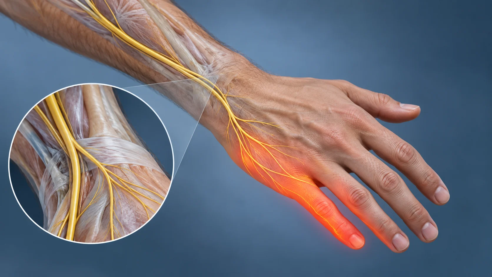
Numbness along the little-finger side of the back of the hand may suggest involvement of the dorsal cutaneous branch of the ulnar nerve. This small branch is superficial and vulnerable to certain injuries. Isolated involvement remains rare: symptom location offers a clue, but is not enough to establish a diagnosis. The history, neurological examination and, in some situations, additional investigations must be consistent. [1]
The value of this diagnosis is to distinguish a local sensory lesion from a problem affecting the ulnar nerve higher in the forearm or at the elbow, involvement at the wrist or another cause. In particular, new hand weakness must not automatically be explained by this cutaneous branch alone.
Where does this nerve branch run?
The ulnar nerve runs through the upper limb and contains motor and sensory fibers. Its dorsal cutaneous branch generally separates in the distal forearm, before the main nerve passes through Guyon’s canal at the wrist. It becomes superficial, curves around the ulnar border and branches toward the ulnar side of the back of the hand. Its course lies near tendinous, bony and subcutaneous structures in this region. [3][4]
It is not a single cable that is identical in everyone. Dissection studies describe several branches and variability in their course near the ulnar styloid. The 2019 anatomical study could not define a universally safe zone for every surgical procedure in this area. These findings are primarily relevant to preventing surgical injury; they do not provide a diagnostic method based on palpation alone. [3][4]
The usual territory includes part of the back of the hand toward the little finger and dorsal regions of the ulnar fingers. Distribution varies and may overlap other sensory territories. The fingertips also involve digital branches: an entire finger should not be colored on a diagram and then used to infer the lesion level with certainty. A clinical sensory map must be compared with the rest of the examination. [1][10]
Which symptoms may draw attention?
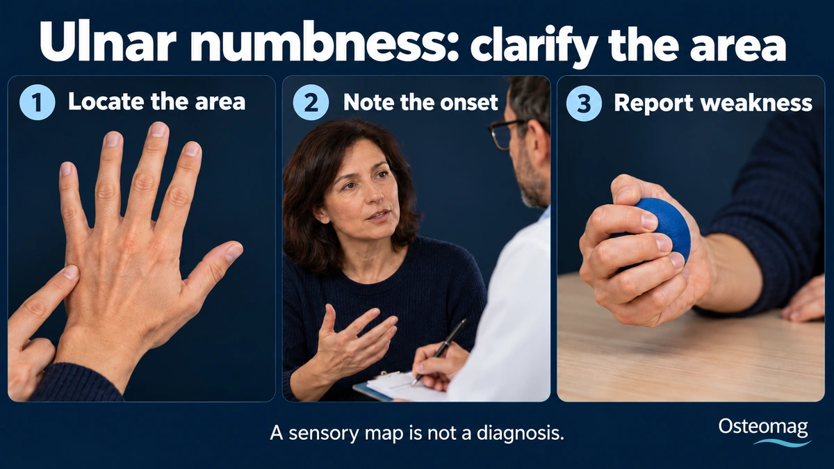
Possible manifestations include tingling, a padded sensation, reduced sensation, burning or pain with contact on the ulnar back of the hand. Some people notice more discomfort when a sleeve or bracelet brushes the area, or when leaning on the side of the wrist. These circumstances should be reported; they are not a stand-alone diagnostic test.
Truly isolated involvement of this branch is sensory. It does not directly explain paralysis of hand muscles. Functional difficulty may relate to pain, but objective loss of strength, progressive clumsiness or muscle wasting requires investigation for more extensive involvement. Distinguishing discomfort, apprehension and neurological weakness belongs to the clinical examination. [1][10]
Why can this nerve be injured?
Its superficial position exposes it to cuts, impacts, traction or repeated local pressure. A procedure on the ulnar side of the wrist may also affect it, directly or in a scarred environment. Previous surgery, wounds, fractures or immobilization should be explored carefully. A nearby scar does not, however, prove that the nerve has been severed or adheres to surrounding tissues. [1][3]
In a retrospective series of 14 patients referred for electrodiagnostic testing, 13 had a traumatic context, including eight after a healthcare procedure. Reduced pinprick sensation was noted in 11 patients, and the sensory response could not be recorded in 13. These figures describe a small selected group: they measure neither the frequency of the problem in the population nor the risk of a particular operation. [1]
The cause may remain uncertain. Continuing assessment is then preferable to asserting that fascial tension explains the symptom. A local abnormality, more proximal compression and other neuropathies may require different approaches.
What the examination aims to distinguish
The clinician clarifies onset, progression, distribution, triggering factors and history. They compare sensation in both hands, examine strength and look for other neurological signs. Examination may include the wrist, forearm, elbow and, depending on the presentation, the cervical region. An isolated provoked sign does not replace this overall assessment. [1][10]
With involvement confined to Guyon’s canal, sensation on the ulnar back of the hand is usually preserved because the dorsal branch has already separated upstream. More proximal ulnar nerve involvement may combine more extensive sensory and motor manifestations. Anatomical variations and complex presentations exist: this reasoning localizes a hypothesis, not a certainty. [2][10]
| Hypothesis | Possible clues | Point requiring attention |
|---|---|---|
| Isolated dorsal cutaneous branch | Sensory discomfort on the ulnar back of the hand | No motor deficit explained by this branch alone |
| More proximal ulnar nerve | Wider territory, sometimes weakness | Examine the elbow and motor functions |
| Involvement in Guyon’s canal | Variable palmar or motor manifestations | Ulnar back of the hand usually spared |
| Another neurological or local cause | Atypical distribution, tendon pain or associated symptoms | Do not force the presentation into a single diagnosis |
What are nerve conduction studies for?
Electrodiagnostic testing can examine the dorsal branch’s sensory response and compare it with other nerves or segments. When indicated, it helps clarify the location and nature of involvement. Historical work on this branch demonstrated its usefulness in localizing ulnar neuropathies. [5] Testing is performed and interpreted by a trained professional alongside clinical information.
Results also depend on technique and biological factors. A study in asymptomatic subjects highlights, in particular, the importance of temperature and standardized conditions to avoid errors. [6] A weak or absent response is therefore not interpreted in isolation, and a reassuring result does not necessarily explain all symptoms. Further investigations depend on progression and context.
Can ultrasound help?
High-resolution ultrasound can visualize a small nerve’s course and its relationships with neighboring tissues. An anatomical study supplemented by observations in healthy volunteers showed that this branch could be followed and that its course varied. This was not a treatment trial and does not establish diagnostic accuracy across all pathological situations. [7]
In 2024, a study of 25 healthy volunteers used ultrasound to locate the branch’s separation point and examine electrical stimulation conditions. It proposes technical landmarks for conduction testing; it does not validate universal screening or osteopathic treatment. [2] More broadly, ultrasound and electrodiagnostic testing provide complementary anatomical and functional information. Their indications remain individualized. [8][9]
When should prompt assessment be sought?
New or progressive loss of strength, worsening clumsiness, rapidly spreading numbness or muscle wasting warrant medical assessment. After a cut, significant trauma or recent surgery, a new sensory deficit should be reported to the relevant team without waiting for manual care to make it disappear.
A cold, pale or bluish hand, significant pain after trauma or a wound with a deficit requires urgent assessment. Sudden arm symptoms associated with facial changes, speech difficulty or one-sided weakness are also an emergency. These presentations go beyond a small isolated cutaneous neuropathy and must not be interpreted as a simple local tension problem.
Which protective measures can be discussed?
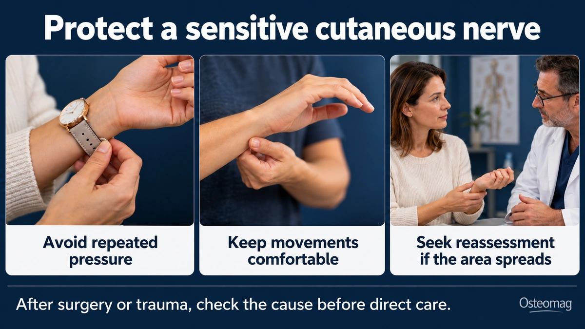
Once the cause has been assessed, reducing repeated pressure at the same spot may help: an overly tight accessory, prolonged leaning or irritating equipment. Adjustments depend on the person’s actual activity, not a blanket ban on using the hand. Comfortable movements may be maintained within limits specified by the responsible professional.
After surgery or trauma, protection and return-to-activity instructions take priority. Deep pressure on a very sensitive area and repeated tensioning intended to “free” the nerve without a diagnosis should be avoided. A flare-up or sustained spread of symptoms requires review of the strategy, not an automatic increase in treatment intensity.
What role can osteopathy and rehabilitation play?
Support may address comfort, permitted mobility and activity adjustments after the problem has been clarified. It does not replace neurological or surgical assessment when needed. None of the studies cited here demonstrates that an osteopathic technique repairs a lesion of this branch or restores impaired conduction.
Any rehabilitation is developed according to the cause, progression, sensitivity and functional goals. Following a useful task and relevant neurological signs is preferable to seeking a daily sensation of “release.” Recovery time cannot be inferred from a location on the skin: compression, irritation and more significant injury do not necessarily have the same prognosis.
Key points
The dorsal cutaneous branch of the ulnar nerve may explain a particular sensory territory, but must not become the automatic label for every instance of numbness along the side of the hand. Variable anatomy, a complete examination and appropriately indicated investigations help localize the problem. Weakness, worsening or onset after a wound or operation changes the priority: have the cause assessed before proposing local care.
References
- [1] Shields LB, Iyer VG, Zhang YP et al. Causes of Dorsal Cutaneous Branch of the Ulnar Nerve Neuropathy Among Patients Undergoing Electrodiagnostic Studies: A Series of 14 Patients. Cureus. 2023;15(4):e38162. doi:10.7759/cureus.38162.
- [2] Park E, Chang SY, Park HJ et al. Dorsal ulnar cutaneous nerve conduction study based on nerve ultrasound. Sci Rep. 2024;14(1):20422. doi:10.1038/s41598-024-71661-3.
- [3] Uerpairojkit C, Kittithamvongs P, Puthiwara D et al. Surgical anatomy of the dorsal cutaneous branch of the ulnar nerve and its clinical significance in surgery at the ulnar side of the wrist. J Hand Surg Eur Vol. 2019;44(3):263-268. doi:10.1177/1753193418815800.
- [4] Root CG, London DA, Schroeder NS et al. Anatomical relationships and branching patterns of the dorsal cutaneous branch of the ulnar nerve. J Hand Surg Am. 2013;38(6):1131-6. doi:10.1016/j.jhsa.2013.03.016.
- [5] Kim DJ, Kalantri A, Guha S et al. Dorsal cutaneous ulnar nerve conduction. Diagnostic aid in ulnar neuropathy. Arch Neurol. 1981;38(5):321-2. doi:10.1001/archneur.1981.00510050087021.
- [6] Young SH, Kalantri A. Dorsal ulnar cutaneous nerve conduction studies in an asymptomatic population. Arch Phys Med Rehabil. 2000;81(9):1171-2. doi:10.1053/apmr.2000.7163.
- [7] Le Corroller T, Bauones S, Acid S et al. Anatomical study of the dorsal cutaneous branch of the ulnar nerve using ultrasound. Eur Radiol. 2013;23(8):2246-51. doi:10.1007/s00330-013-2832-z.
- [8] Beekman R, Visser LH. High-resolution sonography of the peripheral nervous system — a review of the literature. Eur J Neurol. 2004;11(5):305-14. doi:10.1111/j.1468-1331.2004.00773.x.
- [9] Cartwright MS, Walker FO. Neuromuscular ultrasound in common entrapment neuropathies. Muscle Nerve. 2013;48(5):696-704. doi:10.1002/mus.23900.
- [10] Landau ME, Campbell WW. Clinical features and electrodiagnosis of ulnar neuropathies. Phys Med Rehabil Clin N Am. 2013;24(1):49-66. doi:10.1016/j.pmr.2012.08.019.