

Introduction
Osteoarthritis of the base of the thumb, a condition that unfolds gradually and often accompanies the aging process, manifests as a degeneration of the cartilage at the joint where the thumb meets the wrist. This particular form of osteoarthritis, also known as basal joint arthritis or carpometacarpal (CMC) arthritis, tends to affect individuals, particularly women, over the age of 40. The thumb’s basal joint, crucial for pinching and gripping activities, undergoes wear and tear over time, leading to the breakdown of cartilage and the exposure of underlying bone. This degeneration can result in a range of symptoms, including pain, swelling, and stiffness at the base of the thumb. The affected joint may lose its normal range of motion, impacting daily activities that involve fine motor skills. As the condition progresses, individuals may notice the development of bony nodules or spurs around the joint, contributing to deformities and further restricting movement. Osteoarthritis of the base of the thumb often presents challenges in performing routine tasks such as opening jars, turning keys, or even writing. The pain associated with this condition can be persistent and may be exacerbated by activities that involve gripping or pinching.
Factors such as genetic predisposition, joint instability, and prior trauma to the thumb joint can contribute to the development of basal joint arthritis. Additionally, repetitive use of the thumb in certain occupations or activities may increase the risk of developing osteoarthritis at the base of the thumb. Diagnosis typically involves a thorough examination by a healthcare professional, including a review of medical history, physical assessment, and possibly imaging studies such as X-rays to visualize the extent of joint degeneration. While there is no cure for osteoarthritis, various management strategies aim to alleviate symptoms and improve function. Non-surgical interventions may include lifestyle modifications, splinting, and pain management. In cases where conservative measures prove insufficient, surgical options like joint reconstruction or joint replacement may be considered. Physical therapy plays a crucial role in maintaining joint mobility and strength. Collaborative efforts between healthcare providers, including osteopaths, hand therapists, and orthopedic specialists, are integral in tailoring a comprehensive care plan for individuals grappling with osteoarthritis of the base of the thumb. Education about joint protection and adaptive strategies further empowers individuals to navigate daily activities with greater ease. As a chronic condition, proactive management and a holistic approach to care are paramount, ensuring that individuals with osteoarthritis of the base of the thumb can lead fulfilling lives while effectively addressing the challenges posed by this degenerative joint condition.
Anatomy of the Thumb Joint
The anatomy of the thumb is uniquely designed to provide the hand with remarkable dexterity, grip strength, and the ability to perform intricate movements. The joint most involved in osteoarthritis of the thumb is the carpometacarpal (CMC) joint, also referred to as the basal joint. Understanding the structure of this joint and how it functions is essential to grasp the impact that osteoarthritis can have on daily activities.
Carpometacarpal Joint (CMC)
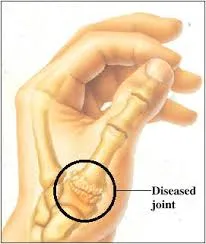
The CMC joint is a saddle-shaped joint located at the base of the thumb, where the first metacarpal bone meets the trapezium bone of the wrist. This unique shape allows the thumb to move in several planes, contributing to its ability to rotate, extend, flex, and oppose the other fingers. This joint is particularly critical for functions that require pinching and gripping, such as holding a pen or grasping a jar.
The CMC joint’s structure allows it to bear significant loads. However, because of its mobility and frequent use, it is also vulnerable to degeneration, particularly in people who frequently perform repetitive tasks that require pinching, twisting, or gripping. Over time, the cartilage that cushions the bones in this joint may wear down, leading to osteoarthritis, one of the most common degenerative conditions affecting the hand.
Ligaments and Muscles Supporting the Thumb
Several ligaments surround the CMC joint, providing stability during thumb movements. These ligaments work to ensure that the bones remain properly aligned, even under stress. Key ligaments include the anterior oblique ligament, which is often the first to deteriorate in individuals with osteoarthritis. Ligament laxity can lead to joint instability, causing the bones in the joint to shift out of place, contributing to deformities like subluxation (partial dislocation) and bone spurs.
Muscles such as the abductor pollicis longus and the opponens pollicis control thumb movements and work alongside the joint’s ligaments. These muscles enable the thumb to perform complex tasks, such as opposition (bringing the thumb toward the other fingers) and gripping. The loss of cartilage in the CMC joint can cause pain during these movements, weakening the surrounding muscles and limiting hand function over time.
Other Key Joints of the Thumb
In addition to the CMC joint, the thumb is supported by two other important joints: the metacarpophalangeal (MCP) joint and the interphalangeal (IP) joint. The MCP joint connects the first metacarpal bone to the proximal phalanx (first bone in the thumb). This hinge-like joint allows the thumb to bend and straighten. The IP joint, located between the two bones in the thumb, further enhances its range of motion.
Though osteoarthritis most commonly affects the CMC joint, it can also occur in the MCP and IP joints. Degeneration in these areas often contributes to the overall stiffness and discomfort people experience in their hands.
Blood Supply and Nerve Innervation
The thumb is richly supplied with blood through branches of the radial artery, ensuring that the bones, muscles, and ligaments receive adequate nutrients and oxygen. The nerves responsible for sensation and motor control of the thumb include the median and radial nerves. As osteoarthritis progresses, joint inflammation can sometimes compress these nerves, leading to tingling or numbness in the thumb and affecting fine motor skills.
Understanding the complex anatomy of the thumb highlights why osteoarthritis at the base of the thumb can be so debilitating. The intricate balance of bones, ligaments, muscles, and nerves must work seamlessly for the thumb to function properly. When osteoarthritis disrupts this balance, it leads to pain, loss of motion, and weakness, severely impacting hand function and overall quality of life.
Causes of osteoarthritis of the base of the thumb
The development of osteoarthritis at the base of the thumb, a condition marked by the gradual degradation of cartilage in the joint connecting the thumb to the wrist, stems from a combination of intrinsic and extrinsic factors. While osteoarthritis is often associated with the aging process, several key contributors can hasten or exacerbate its onset. Genetic predisposition emerges as a significant factor, as individuals with a family history of osteoarthritis may be more susceptible to developing this condition at the base of the thumb. Joint instability, another intrinsic factor, plays a crucial role, as variations in the anatomy or alignment of the thumb joint can predispose it to increased stress and wear. Trauma, such as fractures or dislocations involving the thumb, can disrupt the delicate balance of the joint and contribute to the accelerated breakdown of cartilage. Repetitive use of the thumb, particularly in certain occupations or activities that involve frequent gripping or pinching, constitutes an extrinsic factor that can heighten the risk of osteoarthritis. Women, especially those over the age of 40, are more commonly affected, with hormonal changes potentially playing a role in the development of basal joint arthritis. The wear and tear on the thumb’s basal joint over time, aggravated by these various factors, lead to the degeneration of cartilage and the subsequent exposure of underlying bone. This degeneration manifests clinically through a range of symptoms, including pain, swelling, and stiffness at the base of the thumb. As the condition progresses, the joint may undergo structural changes, with the formation of bony nodules or spurs contributing to deformities and further restricting mobility. Diagnosis typically involves a comprehensive evaluation by a healthcare professional, encompassing a detailed medical history, physical examination, and imaging studies such as X-rays to assess the extent of joint degeneration. Understanding the multifaceted nature of the causes of osteoarthritis at the base of the thumb is essential for developing effective management and prevention strategies. While certain risk factors like genetic predisposition cannot be modified, lifestyle modifications, ergonomic adjustments, and joint protection strategies can mitigate the impact of extrinsic factors such as repetitive use. Proactive measures, including maintaining a healthy weight and engaging in exercises that promote joint strength and flexibility, play a crucial role in preventing or delaying the onset of osteoarthritis. Collaborative efforts between healthcare providers, including orthopedic specialists, hand therapists, and osteopaths, are instrumental in tailoring individualized approaches to address the unique causes and manifestations of osteoarthritis at the base of the thumb, empowering individuals to manage and mitigate the impact of this degenerative joint condition.
- Normal age-related wear and tear: Osteoarthritis is often associated with aging. Over time, the cartilage that covers the ends of bones can wear away, which can cause the bones to rub together when you move, causing osteoarthritis.
- Genetic predisposition: Some people may have a genetic predisposition to developing joint problems, including osteoarthritis. If members of your family have been affected by this condition, you may be at increased risk.
- Repetitive activities: Certain activities or occupations that involve repetitive movements or excessive use of the thumb and wrist may increase the risk of developing osteoarthritis at the base of the thumb. This may include frequent manual tasks or specific sporting activities.
- Joint trauma: Previous injuries to the thumb, such as sprains or fractures, can increase the risk of developing osteoarthritis. Past joint damage can alter the structure of the joint, contributing to cartilage deterioration.
- Joint instability: Joint instability, often due to displacement or ligamentous laxity, can promote the development of osteoarthritis. An unstable joint is less able to distribute the load evenly during movements.
- Chronic Inflammation: Chronic inflammatory conditions, such as rheumatoid arthritis, may increase the risk of osteoarthritis of the base of the thumb. Persistent inflammation can damage the cartilage and lead to degenerative changes in the joint.
- Obesity: Being overweight or obese can put increased pressure on joints, including the one at the base of the thumb, which can accelerate cartilage wear.
Risk Factors for Thumb Osteoarthritis
Osteoarthritis of the thumb, particularly in the carpometacarpal (CMC) joint, is a degenerative condition influenced by several risk factors. While aging is the most well-known contributor, other factors—such as gender, genetics, occupation, and lifestyle—also play significant roles in the onset and progression of this condition. By understanding these risk factors, individuals can take proactive steps to manage or prevent the development of thumb osteoarthritis.
Age
As with many forms of osteoarthritis, the risk of developing thumb osteoarthritis increases with age. Cartilage naturally wears down over time, and by the age of 40 or 50, the cumulative effects of everyday stress on the thumb joint become more apparent. The process of cartilage degeneration is gradual, but over decades of use, the cartilage that cushions the bones in the thumb joint may erode, leading to bone-on-bone contact. This causes pain, inflammation, and joint stiffness, typical symptoms of osteoarthritis.
Gender
Women are disproportionately affected by thumb osteoarthritis, especially after the age of 40. This gender disparity may be due, in part, to hormonal changes that occur during menopause. Estrogen, a hormone that helps protect cartilage, declines during menopause, potentially contributing to an increased risk of joint degeneration. Studies have also suggested that women may have looser ligaments and a greater degree of joint flexibility compared to men, which could contribute to joint instability and wear.
Genetic Predisposition
Genetics also plays a significant role in the development of osteoarthritis. Individuals with a family history of joint disorders or osteoarthritis are more likely to develop the condition themselves. Certain inherited traits, such as joint shape or the strength of connective tissues, may predispose individuals to cartilage breakdown, particularly in weight-bearing or frequently used joints like the thumb’s CMC joint.
Joint Instability
Anatomical differences or variations in the thumb’s structure can lead to joint instability, which in turn increases the risk of osteoarthritis. When the ligaments around the thumb are unable to maintain proper alignment and support, the joint experiences abnormal stress. Over time, this additional stress wears down the cartilage and accelerates joint degeneration. Individuals with hypermobility (excessive joint movement) are particularly vulnerable to joint instability, and they may develop osteoarthritis at a younger age than the general population.
Previous Joint Trauma
A history of injury to the thumb, such as fractures, dislocations, or severe sprains, can predispose an individual to osteoarthritis later in life. Joint trauma can damage the cartilage or alter the normal alignment of the joint, making it more susceptible to wear and tear. Even when initial injuries heal, they may leave lasting effects that weaken the joint and contribute to long-term degeneration.
Repetitive Use and Occupational Hazards
Occupations or activities that require repetitive thumb movements, such as pinching, gripping, or twisting, increase the risk of thumb osteoarthritis. Jobs that involve manual labor, such as carpentry or sewing, place a significant strain on the CMC joint. Likewise, athletes who participate in sports requiring repetitive thumb use, like tennis or rock climbing, are more likely to develop joint problems over time.
Obesity
Excess body weight places additional stress on the joints, particularly in the lower body. While the thumb does not bear the body’s weight directly, obesity is still considered a risk factor for osteoarthritis because of the overall increase in systemic inflammation. Inflammation is a known contributor to cartilage breakdown, and individuals with obesity may be more prone to developing osteoarthritis in various joints, including the thumb.
Chronic Inflammation
Conditions that cause chronic inflammation, such as rheumatoid arthritis, can also elevate the risk of osteoarthritis. Prolonged inflammation in the joints leads to cartilage damage and accelerates degeneration. For individuals with rheumatoid arthritis or other inflammatory conditions, the risk of developing secondary osteoarthritis in the thumb is higher, and they may experience a more rapid progression of symptoms.
By recognizing and addressing these risk factors, individuals can take steps to protect their thumb joints and slow the progression of osteoarthritis. Preventative strategies, including weight management, injury prevention, and avoiding repetitive strain, are essential in reducing the likelihood of developing this painful condition.
Symptoms of osteoarthritis of the base of the thumb
Osteoarthritis of the base of the thumb manifests through a spectrum of symptoms, collectively impacting the functionality and comfort of the affected hand. Central to these symptoms is pain, often the initial indication of joint degeneration. Individuals with this condition commonly experience pain localized at the base of the thumb, particularly during activities that involve gripping or pinching. The pain can range from a dull ache to sharp discomfort, influencing daily tasks and diminishing the overall quality of life. Swelling, another hallmark symptom, accompanies the inflammatory response triggered by the degeneration of cartilage. The affected joint may exhibit visible swelling, contributing to a sense of fullness or tightness around the thumb’s base. Stiffness emerges as a pervasive symptom, especially after periods of inactivity or upon waking. Reduced flexibility and limited range of motion in the thumb joint become apparent, hindering fine motor skills and impeding activities that demand precise hand movements. As osteoarthritis progresses, the joint may undergo structural changes, leading to the development of bony nodules or spurs. These palpable, often visible, abnormalities can contribute to deformities in the thumb, altering its appearance and further restricting mobility.
Functional limitations extend beyond pain and stiffness, with weakness in the affected hand becoming increasingly noticeable. Grip strength diminishes, and individuals may struggle with tasks requiring a firm hold or sustained pressure. The combined impact of pain, swelling, stiffness, and weakness can significantly disrupt daily activities, affecting the ability to perform routine tasks such as opening jars, turning keys, or writing. Moreover, the symptoms of osteoarthritis at the base of the thumb may influence overall hand function and dexterity, challenging individuals both physically and emotionally. The chronic nature of these symptoms underscores the importance of comprehensive management strategies. Conservative approaches, such as lifestyle modifications and joint protection techniques, aim to alleviate symptoms and improve function. Splinting may be employed to provide support and reduce stress on the affected joint, particularly during activities that trigger discomfort. Pain management strategies, including over-the-counter or prescription medications, may be recommended to enhance individuals’ overall comfort. Physical therapy plays a pivotal role in maintaining joint mobility and strength, equipping individuals with exercises to improve thumb function and lessen the impact of symptoms. Surgical interventions, such as joint reconstruction or joint replacement, may be considered in cases where conservative measures prove inadequate. Understanding and effectively addressing the symptoms of osteoarthritis at the base of the thumb necessitate a collaborative effort involving healthcare professionals, including orthopedic specialists, hand therapists, and osteopaths. Tailoring a comprehensive care plan to the unique needs of each individual ensures a holistic approach that empowers them to navigate the challenges posed by this degenerative joint condition while maximizing hand function and overall well-being.
- Pain: Pain is often the most pronounced symptom. It may be felt at the base of the thumb or wrist, and it may worsen with movement or use of the hand.
- Swelling: Osteoarthritis can lead to inflammation of the joints, which may manifest as localized swelling.
- Stiffness: People with osteoarthritis of the base of the thumb may feel stiffness in the hand, especially when waking up or after a period of inactivity.
- Reduced strength: Due to pain and joint degeneration, grip strength may decrease, which can make it difficult to perform certain manual activities.
- Joint deformity: As osteoarthritis progresses, structural changes may occur, eventually leading to joint deformity.
- Crackle: Some patients may feel or hear a rubbing or crackling sound when they move their thumb.
- Limitation of movement: Osteoarthritis can limit the range of movement of the thumb, which can affect the ability to grasp objects or perform daily activities.
Stages of Thumb Osteoarthritis
Osteoarthritis of the thumb, particularly at the base or carpometacarpal (CMC) joint, progresses through several stages. Each stage is marked by increasing degeneration of the joint, leading to worsening symptoms and functional limitations. Understanding these stages can help individuals recognize their condition’s severity and work with healthcare providers to determine appropriate treatment options.
Stage 1: Early Wear and Tear
In the first stage of thumb osteoarthritis, there may be minimal symptoms, and cartilage deterioration has only just begun. During this early phase, the cartilage covering the bones in the CMC joint starts to thin, but there is still sufficient cushioning to prevent the bones from rubbing together directly. As a result, individuals might not experience significant pain or discomfort. Some may feel mild soreness or stiffness after activities that involve repetitive thumb use, such as gripping, pinching, or typing.
At this stage, X-rays may show very mild narrowing of the joint space, but the overall joint structure remains intact. Many individuals are unaware that they have osteoarthritis at this point, and the condition is often diagnosed incidentally during imaging for other hand or wrist concerns.
Stage 2: Increased Cartilage Loss
As osteoarthritis progresses to stage 2, the cartilage in the thumb joint becomes more noticeably worn. This stage is characterized by increased pain, particularly during activities that put pressure on the thumb. Individuals may experience discomfort while holding objects, twisting doorknobs, or gripping tools. Swelling and inflammation around the joint may become more apparent, and stiffness often occurs, especially in the morning or after periods of inactivity.
X-rays at this stage will typically show a narrowing of the joint space as the cartilage continues to deteriorate. Small bony outgrowths, known as osteophytes or bone spurs, may begin to form around the joint. These spurs develop as the body attempts to repair the damaged joint, but they often contribute to additional discomfort and limit joint mobility.
Stage 3: Significant Joint Damage
By stage 3, the cartilage in the CMC joint has significantly eroded, leading to more persistent symptoms. Pain is no longer limited to periods of activity and may become constant, even at rest. The thumb joint may appear visibly enlarged or deformed due to the formation of bony nodules. Swelling and inflammation may cause a sensation of tightness around the joint, and gripping strength may weaken considerably.
Individuals in stage 3 often find daily tasks, such as opening jars, writing, or buttoning clothing, increasingly difficult. Limited range of motion and increased joint stiffness are common. X-rays will reveal advanced joint damage, with substantial cartilage loss, reduced joint space, and prominent bone spurs.
At this point, conservative treatments like splinting, physical therapy, and anti-inflammatory medications may offer temporary relief but are often insufficient to manage the symptoms. Some individuals may start considering surgical options for more long-term relief.
Stage 4: End-Stage Osteoarthritis
In stage 4, the CMC joint is severely damaged, and the bones may be in direct contact with one another due to the complete loss of cartilage. The constant friction between the bones causes chronic pain, severe stiffness, and significant limitations in thumb movement. Deformities of the joint are usually prominent at this stage, with noticeable swelling, bony growths, and potentially visible changes in the thumb’s alignment.
Individuals with end-stage osteoarthritis often experience severe difficulties in performing basic hand functions. The pain is persistent and debilitating, greatly impacting the ability to perform tasks that require grip, pinch, or fine motor skills. At this point, non-surgical treatments are generally ineffective, and most individuals will require surgical intervention to restore function and alleviate pain.
X-rays in stage 4 show nearly complete joint destruction, with bone-on-bone contact, extensive bone spurs, and joint deformity. Joint replacement or fusion surgery may be recommended to relieve pain and restore some level of hand function.
Treatment by Stage
Understanding the stage of thumb osteoarthritis is crucial for determining the most effective treatment approach. In the early stages, conservative treatments such as lifestyle modifications, joint protection, physical therapy, and splinting can help slow the disease’s progression and alleviate symptoms. In the later stages, when joint damage is more severe, surgical options like joint reconstruction, joint replacement, or fusion may be necessary to provide long-term relief and improve hand function.
Recognizing the symptoms and stages of thumb osteoarthritis early can empower individuals to seek timely medical intervention, potentially delaying the need for more invasive treatments and preserving their quality of life.
Test for thumb osteoarthritis
Test 1
- Hold the base of the thumb and move the thumb.
- There is osteoarthritis if there is pain and movement causes a grinding sound called crepitus.
Test 2
- Touch the tip of the thumb with the tip of the index finger.
- It should all form an O.
- If the shape looks more like a D, this indicates osteoarthritis of the thumb. This “O” shape indicates the absence of osteoarthritis

Treatment of thumb osteoarthritis
he treatment of thumb osteoarthritis aims to alleviate symptoms, improve joint function, and enhance overall hand mobility. A multifaceted approach involving both non-surgical and surgical interventions is often tailored to the individual’s specific needs and the severity of the condition.
Non-surgical interventions are typically considered as the first line of treatment for thumb osteoarthritis:
- Lifestyle Modifications: Simple lifestyle changes can be beneficial, such as avoiding activities that exacerbate symptoms and incorporating joint-friendly habits in daily tasks.
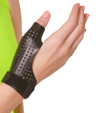
- Splinting: Wearing a splint or brace can provide support to the thumb joint, reducing stress and promoting proper alignment. This can be particularly helpful during activities that may strain the joint.
- Pain Management: Over-the-counter pain relievers like acetaminophen or nonsteroidal anti-inflammatory drugs (NSAIDs) may help manage pain and inflammation. However, their prolonged use should be monitored under the guidance of a healthcare professional.
- Physical Therapy: Targeted exercises and physical therapy can enhance joint flexibility, strengthen surrounding muscles, and improve overall hand function. A therapist may design a tailored exercise program to address specific limitations.
- Topical Treatments: Creams or patches containing anti-inflammatory medications may provide localized relief to the affected area.
- Assistive Devices: Using assistive devices, such as ergonomic tools or utensils with larger handles, can reduce strain on the thumb joint during daily activities.
For cases where conservative measures are insufficient or the condition is more advanced, surgical options may be considered:
- Joint Reconstruction: Surgical procedures, such as ligament reconstruction or tendon interposition, aim to stabilize the joint, improve alignment, and reduce pain.
- Joint Replacement: In cases of severe joint degeneration, joint replacement surgery may be recommended. This involves removing the damaged joint surfaces and replacing them with artificial components.
- Arthroscopy: Minimally invasive procedures, like arthroscopy, may be employed to assess and treat joint abnormalities. This involves inserting a small camera into the joint to guide surgical interventions.
- Fusion (Arthrodesis): In certain situations, fusing the bones in the joint may be considered to provide stability and relieve pain. However, this limits joint motion.
The choice of treatment depends on various factors, including the patient’s overall health, the extent of joint damage, and individual preferences. A collaborative approach involving healthcare professionals such as orthopedic specialists, hand therapists, and osteopaths is crucial for developing a comprehensive and personalized treatment plan. Regular follow-ups and ongoing management strategies, including lifestyle modifications and continued physical therapy, are essential to optimize long-term outcomes for individuals dealing with thumb osteoarthritis.
Radiographic sign of osteoarthritis of the thumb
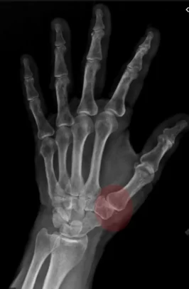
Radiographic assessment plays a crucial role in identifying and confirming osteoarthritis of the thumb, providing valuable insights into the structural changes within the affected joint. One distinctive radiographic sign indicative of thumb osteoarthritis is the presence of joint space narrowing. As the cartilage at the base of the thumb degenerates, the space between the bones in the joint diminishes. This narrowing is observable on X-rays and indicates the loss of the protective cushioning between the bones, contributing to the characteristic symptoms of pain, stiffness, and reduced range of motion.
In addition to joint space narrowing, osteophyte formation is a notable radiographic feature. Osteophytes, also known as bone spurs, are bony outgrowths that develop in response to the ongoing degeneration of cartilage. These spurs may appear along the edges of the bones within the thumb joint, contributing to the structural changes and potentially causing further discomfort and restricted movement.
Another radiographic sign often seen in thumb osteoarthritis is subchondral sclerosis. This term refers to an increased density or hardening of the bone just beneath the cartilage surface. As osteoarthritis progresses, the bone undergoes changes, and the subchondral sclerosis becomes apparent on X-rays. This sclerosis is reflective of the alterations in the bone structure associated with the degenerative process occurring within the thumb joint.
Furthermore, cyst formation may be observed on radiographs of individuals with thumb osteoarthritis. These cysts, often termed geodes or subchondral cysts, represent fluid-filled sacs that develop within the bone. They can be a consequence of the ongoing degeneration and changes in the bone and may contribute to the radiographic picture of osteoarthritis.
Radiographic signs provide a comprehensive view of the structural alterations occurring in the thumb joint affected by osteoarthritis. While joint space narrowing, osteophyte formation, subchondral sclerosis, and cysts are key indicators, the interpretation of X-rays should always be done in conjunction with a thorough clinical assessment. The combination of radiographic findings and clinical symptoms aids healthcare professionals, including orthopedic specialists and radiologists, in making accurate diagnoses and formulating effective management plans for individuals dealing with osteoarthritis of the thumb.
- Reduced joint space: Osteoarthritis causes progressive breakdown of the cartilage that covers the joint surfaces. When cartilage thins, the joint space between bones decreases on x-rays.
- Osteophytes: Also called “parrot beaks,” these are bony growths that form around joints in response to cartilage deterioration. Osteophytes may be visible on x-rays and contribute to joint deformity.
- Subchondral sclerosis: This is a densification or thickening of the bone beneath the cartilage in response to increased stress on the joint from loss of cartilage.
- Joint erosions: Signs of erosion or wear of the joint surface may be detected on x-rays.
- Joint deformity: Osteoarthritis can cause structural changes and deformity of the joint, which may also be visible on x-ray images.
Prevention Strategies for Thumb Osteoarthritis
While thumb osteoarthritis is a common degenerative condition, especially as individuals age, there are several proactive measures that can be taken to prevent or delay its onset. Prevention strategies largely focus on minimizing joint stress, maintaining healthy lifestyle habits, and reducing risk factors such as obesity and repetitive strain on the thumb joint. By adopting these strategies, individuals can reduce the likelihood of developing thumb osteoarthritis and protect the long-term health of their joints.
1. Ergonomic Adjustments
One of the most effective ways to prevent thumb osteoarthritis is by making ergonomic adjustments in daily tasks. For individuals who perform repetitive hand movements, such as typing, gripping tools, or using smartphones, using ergonomic tools can reduce strain on the thumb joint. Specially designed keyboards, larger-handled utensils, and tools with padded grips can help distribute pressure more evenly across the hand, reducing stress on the thumb.
In addition, individuals should aim to take regular breaks from repetitive tasks to allow their joints to rest and recover. Stretching exercises during these breaks can also help maintain flexibility in the thumb and reduce stiffness.
2. Joint Protection Techniques
Learning and practicing joint protection techniques is crucial for reducing the wear and tear on the thumb joint. For example, using the whole hand rather than just the thumb to grip or hold objects can distribute the load more evenly across multiple joints. When carrying heavy objects, it’s advisable to use both hands and avoid placing excessive pressure on the thumb alone.
Individuals should also be mindful of their posture and hand positioning during daily activities. Keeping the thumb in a neutral, relaxed position can help reduce unnecessary strain. When gripping or pinching is necessary, performing the movement slowly and gently, rather than forcefully, can prevent joint damage.
3. Regular Exercise and Strengthening
Maintaining strong, flexible muscles around the thumb joint can help protect it from degeneration. Regular hand exercises, such as thumb stretches, resistance training, and range-of-motion exercises, improve muscle strength and joint flexibility. Strengthening the muscles that support the thumb joint helps to stabilize the joint and reduce the risk of injury.
Exercise also improves circulation, which can aid in keeping the joint healthy by delivering nutrients and oxygen to the cartilage. In addition to specific hand exercises, maintaining an overall active lifestyle through regular aerobic exercise helps control body weight and reduces the stress on joints.
4. Healthy Weight Management
Obesity is a significant risk factor for osteoarthritis, including that of the thumb. Excess body weight contributes to increased systemic inflammation, which accelerates joint degeneration. Maintaining a healthy weight through a balanced diet and regular physical activity can reduce the inflammatory burden on joints and slow the development of osteoarthritis.
A diet rich in anti-inflammatory foods, such as fruits, vegetables, whole grains, and fatty fish, can further protect against joint damage. Omega-3 fatty acids, in particular, are known for their anti-inflammatory properties and may help reduce cartilage breakdown.
5. Avoiding Overuse
Overuse of the thumb joint is a common contributor to osteoarthritis. Individuals who work in occupations or participate in hobbies that require frequent thumb use should be aware of the risks associated with repetitive stress. Activities that involve excessive gripping, pinching, or twisting can place significant strain on the CMC joint, leading to cartilage wear over time.
To prevent overuse, individuals should rotate tasks when possible, allowing the thumb joint to rest periodically. Using assistive devices, such as jar openers or grip aids, can also help reduce the mechanical stress on the joint.
6. Early Intervention
Recognizing early signs of thumb pain or stiffness and seeking prompt medical advice can help prevent the progression of osteoarthritis. Early intervention strategies, such as physical therapy, splinting, or anti-inflammatory treatments, can alleviate symptoms and delay further joint damage. Regular check-ups with a healthcare professional, especially for individuals with a family history of osteoarthritis, can help monitor joint health and implement preventative measures as needed.
Living with Thumb Osteoarthritis
Living with thumb osteoarthritis presents challenges that affect both physical abilities and emotional well-being. As the thumb plays a crucial role in nearly every hand movement, osteoarthritis at the base of the thumb (carpometacarpal joint) can significantly impact daily activities, work, and leisure. However, with a proactive management plan and lifestyle adaptations, individuals can continue to live fulfilling lives despite the condition.
Adapting Daily Activities
One of the most immediate impacts of thumb osteoarthritis is the difficulty in performing common tasks such as opening jars, turning doorknobs, holding a pen, or even using a smartphone. These activities, which involve gripping, pinching, and twisting motions, place stress on the thumb joint, leading to pain and stiffness. Adapting these daily activities can help reduce strain on the joint and alleviate symptoms.
Ergonomic tools and assistive devices can make a significant difference in reducing the impact on the thumb. For example, jar openers with larger handles, key turners, and pen grips are designed to ease the pressure on the CMC joint. Additionally, using both hands to perform tasks that require significant thumb pressure can help distribute the load more evenly and reduce joint wear.
Emotional and Psychological Impact
Living with chronic pain and limitations can also take a toll on mental health. The inability to perform tasks that were once simple can lead to frustration, feelings of helplessness, and anxiety. For individuals who rely on their hands for their profession—such as musicians, artists, or manual laborers—the condition can be particularly distressing.
It’s important to acknowledge the emotional side of living with osteoarthritis and to seek support when needed. This can include talking to a therapist, joining a support group, or discussing concerns with healthcare providers. Building a strong support network of family, friends, and healthcare professionals can help individuals cope with the psychological challenges of the condition.
Maintaining Mobility and Strength
Maintaining joint mobility and strength is essential to managing thumb osteoarthritis. Physical therapy can play a vital role in this, as therapists can guide patients through exercises designed to improve range of motion, strengthen muscles around the thumb joint, and reduce pain. Simple hand exercises, like stretching the thumb and performing resistance exercises, can be done at home to maintain flexibility and hand function.
In addition to hand exercises, staying physically active through low-impact activities such as swimming, walking, or yoga can improve overall joint health. Regular exercise helps keep the body’s weight in check, reduces inflammation, and maintains muscle strength, all of which contribute to slowing the progression of osteoarthritis.
Pain Management
Pain management is another key aspect of living with thumb osteoarthritis. Over-the-counter pain relievers such as acetaminophen or NSAIDs can be helpful in reducing pain and inflammation. Topical treatments, including creams or patches with anti-inflammatory ingredients, can provide targeted relief for joint pain.
Some individuals may benefit from periodic corticosteroid injections into the thumb joint to alleviate inflammation and pain. While injections are not a long-term solution, they can provide temporary relief during particularly painful flare-ups.
For more advanced cases, medical devices such as thumb braces or splints can stabilize the thumb joint and prevent excessive motion. This reduces pain during activities and provides support for weakened ligaments.
Long-Term Outlook
Thumb osteoarthritis is a progressive condition, meaning that it typically worsens over time. However, the progression can be slow, and with proper management, individuals can maintain hand function and quality of life. Surgery may be necessary in severe cases when conservative treatments no longer provide relief, but many individuals manage the condition successfully for years with a combination of lifestyle modifications, therapy, and pain management.
Staying proactive in managing symptoms, remaining informed about treatment options, and working closely with healthcare providers are crucial steps in living well with thumb osteoarthritis. The right approach to care can make a significant difference in maintaining independence and continuing to engage in meaningful activities.
Frequently Asked Questions (FAQs
Here are answers to some common questions people have about thumb osteoarthritis, its treatment, and management.
1. What is thumb osteoarthritis, and how does it affect me?
Thumb osteoarthritis is a degenerative condition that affects the carpometacarpal (CMC) joint at the base of the thumb. As the cartilage in this joint wears down, the bones can rub against each other, causing pain, swelling, stiffness, and loss of function. This condition can make it difficult to perform tasks that involve gripping, pinching, or twisting.
2. What are the early signs of thumb osteoarthritis?
Early signs of thumb osteoarthritis often include pain at the base of the thumb, especially during or after activities that involve gripping or pinching, such as opening jars or holding objects. You may also notice stiffness, particularly in the morning or after periods of rest, and mild swelling around the joint. If you feel a grinding sensation when moving your thumb, this can also be an early symptom.
3. How is thumb osteoarthritis diagnosed?
Thumb osteoarthritis is typically diagnosed through a physical examination by a healthcare provider, who will assess the range of motion, tenderness, and swelling in the thumb. X-rays may be used to confirm the diagnosis by revealing joint space narrowing, bone spurs, or other signs of cartilage wear. In some cases, advanced imaging techniques like MRI can provide further details about the condition.
4. Can thumb osteoarthritis be cured?
There is no cure for osteoarthritis, including thumb osteoarthritis. However, there are many treatment options available to manage symptoms and slow the progression of the disease. Early intervention with conservative treatments—such as physical therapy, splinting, and pain management—can help maintain function and reduce discomfort.
5. What are the best treatments for thumb osteoarthritis?
The best treatment depends on the severity of your symptoms. Early-stage thumb osteoarthritis can often be managed with lifestyle modifications, physical therapy, splinting, and over-the-counter pain medications like NSAIDs. In more advanced cases, corticosteroid injections, joint reconstruction, or joint replacement surgery may be recommended. Working closely with a healthcare provider to tailor treatment to your specific needs is essential.
6. How can I prevent thumb osteoarthritis from getting worse?
To prevent thumb osteoarthritis from worsening, it’s important to avoid activities that put excessive strain on the thumb joint. Using ergonomic tools, taking regular breaks from repetitive tasks, and practicing joint protection techniques can help. Engaging in exercises that strengthen the muscles around the thumb and maintaining a healthy weight can also support joint health and reduce the risk of further damage.
7. Will I need surgery for thumb osteoarthritis?
Surgery is not usually the first option for treating thumb osteoarthritis. It is generally reserved for cases where conservative treatments have failed to provide relief and the condition has significantly progressed. Surgical options include joint reconstruction, joint replacement, or fusion of the joint, depending on the severity of the joint damage and the individual’s needs. Your doctor will help determine if and when surgery might be necessary.
8. Can I continue working with thumb osteoarthritis?
Many people with thumb osteoarthritis are able to continue working, especially if they adapt their work environment and habits. Using ergonomic tools, modifying tasks to reduce strain on the thumb, and following a physical therapy regimen can help manage symptoms and maintain function. For individuals with jobs that require frequent hand use, splints or braces can offer additional support.
9. How long does it take to recover from thumb surgery?
Recovery time after surgery for thumb osteoarthritis varies depending on the type of procedure performed. Joint reconstruction or replacement surgery typically requires several weeks to months of healing, followed by physical therapy to restore strength and mobility. Full recovery may take up to six months, but pain relief and improved function are usually noticeable within a few weeks post-surgery.
10. Can thumb osteoarthritis affect other joints?
While thumb osteoarthritis specifically affects the CMC joint, osteoarthritis can occur in other joints of the hand, including the metacarpophalangeal (MCP) joint or the interphalangeal (IP) joint. In addition, osteoarthritis commonly affects other weight-bearing joints such as the knees, hips, and spine. It is important to maintain overall joint health to reduce the risk of osteoarthritis developing in other areas.
Conclusion
Thumb osteoarthritis, particularly at the base of the thumb or carpometacarpal (CMC) joint, is a progressive and degenerative condition that significantly affects hand function and quality of life. It develops as cartilage wears down over time, leading to pain, stiffness, and a loss of mobility that can make even simple tasks challenging. Factors such as aging, genetics, joint instability, and repetitive thumb use contribute to the onset and progression of this condition. Recognizing the symptoms early, understanding the underlying causes, and seeking timely intervention are crucial to managing the disease.
Effective management of thumb osteoarthritis requires a combination of lifestyle modifications, joint protection techniques, and both non-surgical and surgical treatments. Conservative measures such as splinting, physical therapy, and pain management can offer substantial relief, especially in the early stages of the condition. For individuals with advanced osteoarthritis, surgical options like joint reconstruction or replacement may become necessary to restore function and alleviate pain.
Furthermore, preventive strategies—including ergonomic adjustments, maintaining a healthy weight, and engaging in regular hand exercises—are essential to protecting the thumb joint and slowing the disease’s progression. Supplements like OsteoMag, which contain magnesium and other essential nutrients, may offer additional support in maintaining joint health and reducing inflammation.
Living with thumb osteoarthritis can be challenging both physically and emotionally, but a proactive approach that incorporates medical advice, physical therapy, pain management, and lifestyle changes can greatly enhance the ability to manage the condition. With proper care and tailored treatment plans, individuals can maintain hand function, reduce pain, and continue to engage in daily activities with greater ease and comfort.
By staying informed about the condition and working closely with healthcare professionals, individuals can navigate the complexities of thumb osteoarthritis and improve their long-term outcomes, ensuring a higher quality of life despite the challenges posed by this degenerative joint disease.
References/Bibliography
Providing a comprehensive list of credible references and resources is essential for readers who want to explore thumb osteoarthritis more deeply. Below are some key sources that can be included to support the information discussed in the text, including academic studies, clinical guidelines, and authoritative websites on osteoarthritis, joint health, and treatment options.
1. Clinical Guidelines and Research Studies
These sources provide evidence-based information about the diagnosis, management, and treatment of thumb osteoarthritis:
- American College of Rheumatology (ACR) Guidelines for the Management of Osteoarthritis of the Hand
This guideline outlines best practices for the non-surgical and surgical management of hand osteoarthritis, including specific recommendations for thumb osteoarthritis. The guidelines offer evidence-based strategies for pain management, physical therapy, and when to consider surgical interventions.
Source: American College of Rheumatology - Cooper, C., & Javaid, M. K. (2017). Osteoarthritis: A Clinical Review
This clinical review published in BMJ offers a detailed overview of osteoarthritis, including risk factors, stages, and available treatment options. The review provides insights into how osteoarthritis affects various joints, including the thumb, and discusses emerging research in joint health.
Source: BMJ, Volume 356, Article i2373. - Zhang, W., et al. (2010). OARSI Recommendations for the Management of Hand Osteoarthritis
The Osteoarthritis Research Society International (OARSI) offers guidelines specifically for managing osteoarthritis in the hand, which includes the thumb CMC joint. The recommendations cover non-pharmacological, pharmacological, and surgical options.
Source: Osteoarthritis and Cartilage, 18(9), 1206-1214.
2. Books on Osteoarthritis and Joint Health
These comprehensive resources provide in-depth knowledge about osteoarthritis, joint anatomy, and treatments:
- “Osteoarthritis: Diagnosis and Medical/Surgical Management” by Roland W. Moskowitz
This textbook provides an extensive look at osteoarthritis, from its pathophysiology to medical and surgical interventions. The book is an invaluable resource for healthcare providers and patients seeking to understand the progression and management of osteoarthritis, including that of the thumb. - “Physical Therapy for Osteoarthritis” by Timothy E. Sell, PT, PhD
This book focuses on the role of physical therapy in managing osteoarthritis. It provides an array of exercises, stretches, and mobility techniques designed to maintain joint health and function, particularly in the hands and thumbs.
3. Medical Journals and Research Articles
These peer-reviewed journals offer the latest research findings on osteoarthritis:
- “Thumb Carpometacarpal Osteoarthritis: A Review of the Current Literature” by Christina Chung, MD, and Joseph E. Rabinovich, MD
This article provides an overview of the causes, symptoms, and stages of thumb CMC osteoarthritis. The authors also review current treatment approaches, including non-surgical and surgical methods.
Source: Journal of Hand Surgery, 43(2), 133-140. - “Magnesium’s Role in Bone and Joint Health” by Li, X., et al. (2019)
This study explores the impact of magnesium on cartilage health and joint stability, offering insights into how magnesium supplements, such as OsteoMag, may benefit those with osteoarthritis.
Source: Journal of Bone and Mineral Research, 34(5), 850-860.
4. Online Resources for Patients and Healthcare Providers
These websites provide trusted information about thumb osteoarthritis, its management, and the latest treatments:
- Arthritis Foundation
The Arthritis Foundation is a leading source of information about osteoarthritis, providing resources for patients, caregivers, and healthcare providers. The website offers detailed sections on hand osteoarthritis, including treatment options, pain management tips, and lifestyle adaptations.
Source: www.arthritis.org - Mayo Clinic
The Mayo Clinic’s comprehensive guide to osteoarthritis includes detailed descriptions of the symptoms, diagnosis, and treatment of osteoarthritis affecting different joints, including the thumb. Their resources are known for being accessible to patients while grounded in scientific research.
Source: www.mayoclinic.org - National Institute of Arthritis and Musculoskeletal and Skin Diseases (NIAMS)
NIAMS is a branch of the National Institutes of Health (NIH) that offers up-to-date research findings and educational materials on osteoarthritis and joint health. The site is particularly helpful for patients seeking evidence-based information about treatment advancements.
Source: www.niams.nih.gov
5. Videos and Multimedia
For individuals who prefer learning through visual or interactive methods, these video resources provide comprehensive educational material on thumb osteoarthritis:
- “Understanding Thumb Arthritis: Causes and Treatments” by Johns Hopkins Medicine
This video series from Johns Hopkins Medicine features orthopedic specialists explaining the causes, stages, and treatments for thumb osteoarthritis. The series is highly informative and includes visuals that make understanding the anatomy and progression of the disease easier for viewers.
Source: Johns Hopkins YouTube Channel - “Hand Therapy for Thumb Arthritis” by Occupational Therapy Videos
This video provides guided hand therapy exercises aimed at improving thumb function and reducing pain caused by osteoarthritis. It’s an excellent resource for those looking to integrate physical therapy into their daily routine.
Source: Occupational Therapy YouTube Channel
6. Patient Testimonial Sources
Patient experiences offer valuable insights into what it’s like to live with thumb osteoarthritis and how treatments like OsteoMag can impact joint health. Including testimonials from reputable sources can provide a real-life perspective:


