When a tendon produces a painful click on the inside of the knee
Hearing the knee click while climbing stairs does not necessarily indicate an injury. The sound becomes more informative when it always occurs in the same place, reproduces familiar pain and limits an activity. Possible causes include snapping pes anserinus, also called snapping pes syndrome. A tendon abruptly changes position as the knee bends or straightens. This phenomenon has been described in small series. It remains unusual and should not become the automatic explanation for every click on the inner side of the knee [1, 2].
The practical question is which structure moves and whether that movement truly explains the difficulty. A tendon seen on ultrasound may glide without causing pain. Conversely, medial knee pain may arise from a meniscus, a bursa or another tissue. Examination during movement, considered alongside the symptom history, helps connect these findings. An image obtained at rest does not necessarily reveal what happens during the snap [2, 3].
Three tendons converge towards the tibia
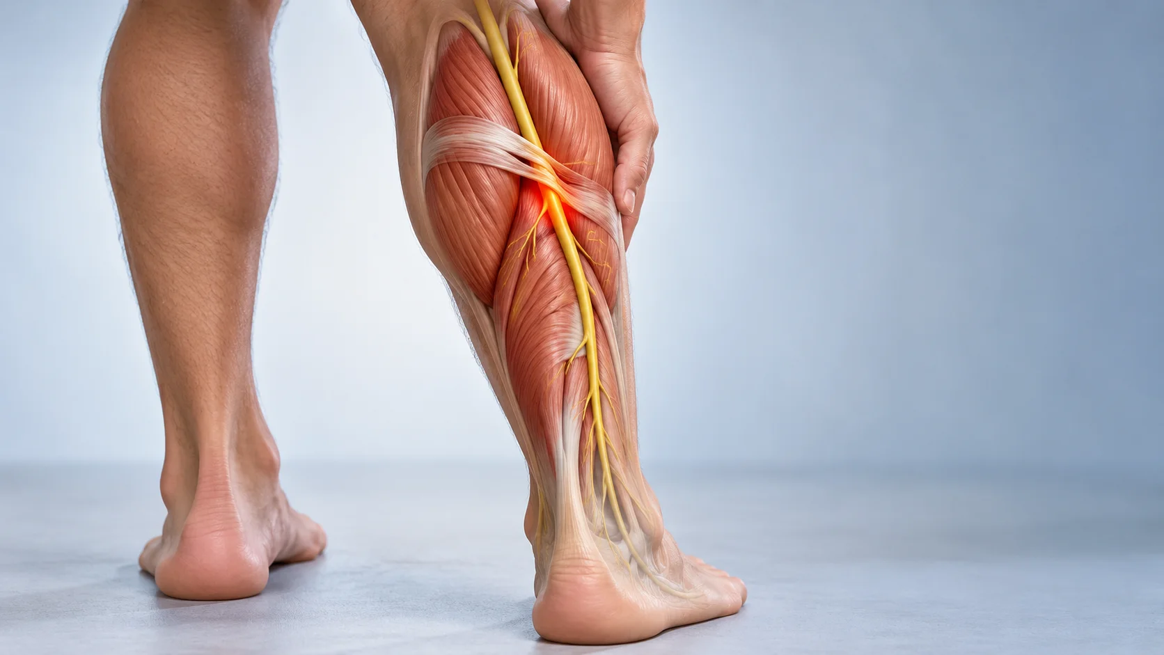
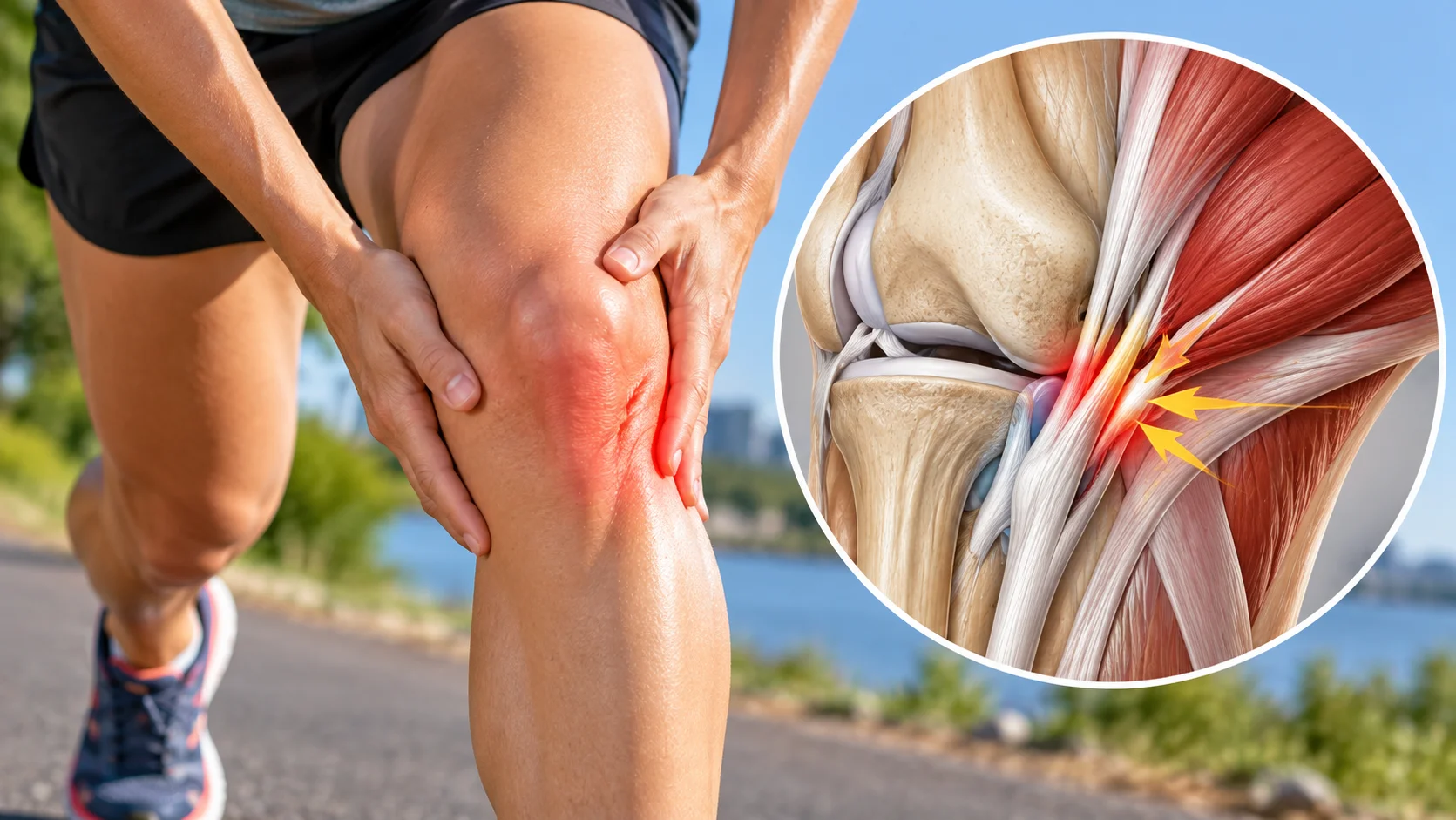
The pes anserinus consists of the sartorius, gracilis and semitendinosus tendons, which attach to the anteromedial tibia below the knee joint line. They originate in different regions of the thigh and contribute to knee flexion and control of the lower limb. Sartorius lies more superficially; gracilis and semitendinosus run in deeper layers. A bursa facilitates gliding between the tendons and neighbouring tissues.
These structures naturally move as the knee angle changes. In published cases, gracilis or semitendinosus may abruptly cross a bony prominence or a neighbouring structure. The sensation resembles a cord moving from one side of an obstacle to the other, rather than an entire joint dislocating. The exact course varies. The involved tendon must be identified instead of assuming an identical mechanism in every patient [2, 3, 12].
Semimembranosus, which neighbours semitendinosus, is not one of the three pes anserinus tendons. Nevertheless, a report describes snapping involving these two tendons in a triathlete. This anatomical distinction matters: “snapping on the inside of the knee” covers several situations, some extending beyond the pes anserinus in its strict anatomical sense. Approximate localisation is therefore insufficient for selecting an intervention [6].
Describe the symptom before naming it
The history clarifies how long the click has been present, whether it followed an injury or surgery and which activities trigger it. Snapping during the final degrees of straightening differs from catching in deep flexion. Distinguishing the sound heard, movement felt beneath the skin and pain is useful. These experiences may coincide, but their association should be checked rather than assumed [1, 2].
Cycling, running, squatting or standing from sitting may reveal the phenomenon. This does not prove that these activities damaged the tendon. They may simply repeat the angle at which it changes position. Someone with a painless click, preserved movement and unrestricted activities has different needs from someone avoiding stairs because of repeated pain.
The practitioner observes a few comfortable movements and attempts to reproduce the usual symptom without repeated irritating tests. Palpation may identify superficial displacement but cannot always distinguish neighbouring tendons precisely. A click observed once without matching pain remains a limited clue. Comparing the other knee adds context without turning every difference into a problem [1, 2].
Several causes of medial pain resemble one another
The following table offers assessment clues, not a self-diagnosis tool. Two problems may coexist. Localised pain, swelling or catching is not exclusive to one tissue. Identifying a tendon displacement that matches the symptom is particularly useful for avoiding attribution of superficial snapping to an incidental meniscal abnormality found on imaging [2].
| Possible explanation | Possible findings | What to check |
|---|---|---|
| Snapping pes anserinus tendon | Superficial movement, sometimes palpable, at a particular point in bending or straightening | Observe the tendon during the symptom; a click alone is insufficient. |
| Meniscal involvement | Joint-line pain, sometimes swelling, catching or locking | Symptoms overlap; investigate a cause within the joint. |
| Pes anserine bursitis | Tenderness below the medial joint line, often during activity | A painful bursa does not necessarily cause a tendon to snap. |
| Medial collateral ligament involvement | Pain after a lateral force, sometimes a feeling of instability | Consider the injury history and ligament examination. |
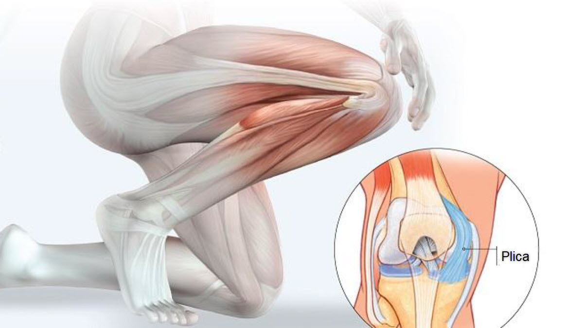
| Plica or patellofemoral pain | More anterior catching or pain depending on the task | The click and pain should match anatomically. |
| Bony prominence | A hard mass or unusual contour, particularly in a young person | Radiographs may be needed; do not assume simple tension. |
Dynamic ultrasound shows the tendon crossing
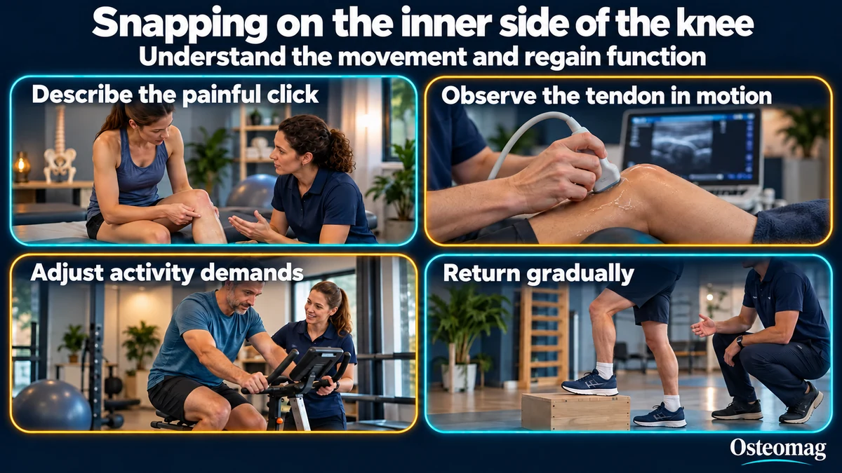
Dynamic ultrasound follows tissues while the person reproduces the movement. The probe is placed over the relevant region, and the examiner observes the tendons during flexion and extension. Its value lies in seeing the displacement at the same moment as the usual click and pain. Publications have documented snapping of gracilis, semitendinosus or their combination in this way [2, 3, 7].
The examination depends on the operator’s experience and the ability to reproduce the phenomenon. A movement occurring only under load may be difficult to reproduce lying down. Probe pressure can also alter gliding. Findings must therefore be interpreted in relation to the person’s actual circumstances. Ultrasound provides dynamic information; it does not automatically make every unusual movement causal.
Radiographs answer a different question: is there a bony prominence, a mass or a post-surgical feature? MRI may help when an intra-articular cause or another tissue injury is suspected. However, normal MRI at rest does not exclude snapping. A dancer described in the literature had an observable phenomenon despite normal MRI, illustrating the difference between static imaging and movement [5].
After surgery and in children
Snapping has been reported after total and unicompartmental knee replacement. These rare observations do not establish an individual’s risk. They show why a new painful click after surgery should be considered in the operative context: relationships between tendons and nearby structures may have changed. Contacting the team following the knee is preferable to independently starting intensive stretching [8, 9].
In children and adolescents, a hard mass near the proximal tibia warrants assessment. A small series described five young people with pes anserine symptoms associated with an osteochondroma; only one had snapping. Other reports concern tibial spurs irritating the bursa. These publications do not mean that every click in a young person comes from a bone tumour. They justify checking a palpable prominence or unusual persistent pain [10, 11].
Adapting activity without demanding complete silence
When symptoms do not suggest an urgent situation, support may begin by temporarily reducing the most irritating movement. This may mean shortening a session, reducing resistance or choosing a more comfortable range. The objective is to maintain tolerated activities while avoiding daily repetition of clearly painful clicking. Complete rest is not a universal programme, and immediate disappearance of the sound is not the only measure of progress.
On a bicycle, a modest change in resistance or position may be tested with a competent professional. For stairs, a lower step or additional support may help temporarily. The response is evaluated by whether pain decreases and the task becomes easier. These are individual adjustments; the small available studies do not establish that a particular setting corrects tendon movement in every patient [1, 2].
Building a return guided by tolerance
Progressive strengthening may support the overall capacity of the lower limb. Depending on needs, it may include knee flexion against light resistance, bridges, step-ups or hip exercises. Selection depends on the movement response and objectives. Practice begins within a tolerated range, followed by gradual increases in one variable, such as repetitions or resistance. No robust trial establishes a specific dose for this rare syndrome.
Stretching is not required simply because a tendon snaps. It may be considered when a relevant mobility restriction exists, provided it does not strongly reproduce pain. The idea of pulling harder on the muscle to move a tendon into the “right place” is too simplistic. An anatomical variation, bony prominence or particular tendon course does not automatically disappear with greater flexibility.
Follow-up focuses on a task chosen with the patient: comfortable stair numbers, cycling duration or running distance. Discomfort during activity and recovery afterwards are recorded. Improvement means important activities become accessible again and symptoms are less disruptive. Residual painless noise does not necessarily require stopping progression. Increasing pain or locking, however, calls for reassessment.
What can specific treatments actually achieve?
The literature mainly consists of isolated cases and very small series. One publication describes four patients; another reports three procedures on medial hamstring tendons. Favourable outcomes after targeted intervention have been reported, but these observations cannot robustly compare options or predict another person’s result. They do not validate a protocol applicable to everyone [1, 4].
Surgical discussion may be appropriate when the mechanism is documented, pain remains limiting and suitable management has been insufficient. Reported procedures depend on the responsible tendon or prominence. A case of selective gracilis surgery highlights the value of prior ultrasound identification; it does not establish superiority in every situation. Expected benefit, functional consequences and uncertainty should be discussed individually [7].
The role of osteopathy
An osteopathic approach may help understand provoking tasks, examine comfortable movement and support adaptations consistent with the patient’s goals. Observing the hip and ankle may be useful if changing their contribution actually changes the task. It does not establish that pelvic asymmetry caused the snapping.
There is no strong clinical evidence that osteopathy permanently changes the course of the tendon responsible for this syndrome. Improved comfort does not demonstrate a changed attachment. The relationship between structure and function encourages precise observation, checking effects during activity and recognising the limits of touch. Referral remains necessary when imaging or specialist advice is indicated.
When to seek prompt assessment
Significant trauma, inability to bear weight, a very hot and swollen knee, fever, true locking, an enlarging mass or rapid deterioration requires prompt assessment. These signs extend beyond the explanation of simple tendon snapping.
Scientific references
- [1] Bollen and Arvinte. Snapping pes syndrome: a report of four cases. 2008.
- [2] Shapiro et al. Snapping Pes Anserinus and the Diagnostic Utility of Dynamic Ultrasound. 2017.
- [3] Karataglis et al. Snapping knee syndrome in an athlete caused by the semitendinosus and gracilis tendons. 2008.
- [4] Geeslin and LaPrade. Surgical treatment of snapping medial hamstring tendons. 2010.
- [5] Guney et al. Semitendinosus snapping: analysis of movement, electromyographic activities, muscle strength and endurance, motor control and joint position sense. 2013.
- [6] von Dercks et al. Snapping knee syndrome caused by semitendinosus and semimembranosus tendons. 2016.
- [7] Akagawa et al. Snapping Pes Syndrome Caused by the Gracilis Tendon: Successful Selective Surgery with Specific Diagnosis by Ultrasonography. 2020.
- [8] Tensho et al. Snapping pes syndrome after total knee arthroplasty. 2014.
- [9] Inui et al. Snapping Pes Syndrome after Unicompartmental Knee Arthroplasty. 2016.
- [10] Sakamoto and Matsuda. Pes Anserinus Syndrome Caused by Osteochondroma in Paediatrics: A Case Series Study. 2017.
- [11] Tiwari et al. Pes Anserinus Bursitis due to Tibial Spurs in Children. 2017.
- [12] Bae and Kwon. Snapping knee caused by the gracilis and semitendinosus tendon. 1997.