
Interpret the lower-back curve alongside symptoms and function
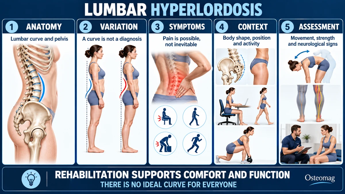
Lumbar hyperlordosis describes a lumbar curve considered more pronounced than expected in a particular context. Lordosis itself is normal: it contributes to the shape of the spine. A visible arch does not automatically indicate disease, muscle weakness or the cause of pain. Assessment needs to consider body shape, position and capacity. [1]
Expressions such as “an overly hollow back” or “a misplaced pelvis” can create unnecessary worry. There is no single ideal angle for the whole population. Someone with a pronounced curve may be comfortable and active, while someone with less curvature may have pain. Care therefore focuses on symptoms and function while looking for situations that warrant investigation.
Anatomy: understanding the lumbar curve
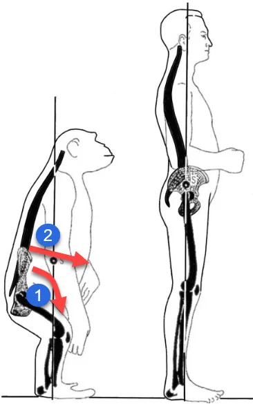
The five lumbar vertebrae, discs and sacrum contribute to the curve seen from the side. Anatomical features of the pelvis influence the spine’s sagittal organisation. Pelvic incidence is a morphological parameter; it should not be confused with pelvic tilt, which changes with position. [2]
Facet joints, capsules, ligaments and muscles contribute to movement. The abdominals, paraspinal muscles, hip muscles and diaphragm participate in different tasks. Their activity depends on the movement and load. A person’s outline cannot establish that a muscle is too short, too strong or insufficiently active.
Nerve roots pass through the foramina between the vertebrae. Localised low back pain differs from radicular pain, which may be accompanied by tingling or weakness in the leg. A pronounced curve alone does not establish nerve compression. [3]
Measuring lordosis: why the numbers vary
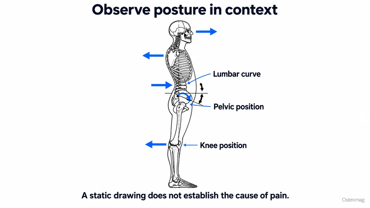
The measured angle depends on the vertebral landmarks, measurement method, position and individual characteristics. A photograph, side-view observation or skin-surface measurement does not provide the same information as an X-ray. Results obtained with different methods are not directly interchangeable. [1] [2]
A systematic review found variation in lordosis and mobility with age and sex. It highlights the importance of context when interpreting a value. A single dividing line between normal and pathological would therefore be too simplistic. [1]
Imaging is not justified merely to check whether someone is standing straight enough. When medically indicated, the clinician interprets overall spinal balance alongside symptoms. An isolated measurement provides neither a certain prognosis nor an automatic exercise prescription.
Hyperlordosis and low back pain: association is not causation
The meta-analysis by Chun and colleagues does not support the simplistic idea that every increase in lordosis causes low back pain. In some comparisons, people with low back pain instead had reduced lordosis. Studies remain heterogeneous and cannot explain each person’s pain from an angle. [4]
A review of cohort studies examined several physical characteristics before low back pain developed. The reported associations concern groups and have limitations; they are not a reliable tool for predicting an individual’s pain or imposing postural correction. [5]
Research comparing posture in people with and without low back pain also needs cautious interpretation. An average difference does not show that changing posture will eliminate symptoms. People may change how they hold themselves when in pain, further complicating interpretation. [6]
In practice, assessment looks for what matters to the person: an irritating movement, reduced capacity, difficulty recovering or a specific condition. An angle cannot replace this clinical investigation.
Mechanisms and contributing factors
The arch may vary with pelvic position, hip movement, thoracic posture and the task being performed. Someone may arch more while standing than sitting, or during an activity that requires looking upwards. This does not prove a permanent defect.
Limited hip movement or a particular movement strategy may increase reliance on lumbar extension during some tasks. A professional can test a variation to see whether it improves comfort. This is a practical strategy, not proof that the pelvis was previously in the wrong position.
Pregnancy, growth, spinal deformity or spondylolisthesis may also alter the profile. These situations cannot be diagnosed from body shape alone. Persistent pain in an adolescent athlete, particularly during repeated extension activities, deserves an appropriate assessment rather than a quick conclusion about posture. [3] [7]
Symptoms: what deserves attention?
Hyperlordosis can be entirely painless. People who seek care sometimes report discomfort with prolonged standing, extension movements or the end of a demanding day. These symptoms are not specific and can occur with very different spinal curves.
Radiating pain, numbness or loss of strength calls for assessment of nerve structures and consideration of other diagnoses. Hip pain may also be felt around the lower back. The location, onset and circumstances of pain guide assessment more than the impression of an exaggerated arch.
Disliking the appearance of the back in a mirror does not establish a disorder. An aesthetic concern should be distinguished from a medical limitation. Without pain, reduced capacity or unusual signs, a longstanding stable curve does not automatically require treatment.
Clinical examination: look beyond the side profile
History and functional assessment
The practitioner asks about symptom duration, previous health problems, injury, radiating pain and limited activities. Goals may include standing longer, returning to sport or carrying a child. These guide the measures used to follow progress.
Examination assesses lumbar and hip movements, walking and tolerance of particular tasks. Depending on symptoms, it includes strength, sensation, reflexes and nerve tests. Standing observation is useful, but it captures one moment rather than the back’s entire function. [7]
Test adaptations without prescribing one posture
The professional may suggest support, a different working height or a variation in movement. If the task becomes more comfortable, this helps build the programme. It does not mean that the initial posture was dangerous.
Pain on palpation cannot identify the responsible tissue with certainty or establish a structural abnormality. A coherent assessment relies on several converging findings and how they change over time.
Differential diagnosis
| Possibility to consider | Useful findings |
|---|---|
| Non-specific low back pain | Variable symptoms without specific signs of serious disease. |
| Radicular pain | Compatible radiation, with possible sensory or motor findings. |
| Spondylolysis or spondylolisthesis | Clinical context, particularly persistent extension pain in a young athlete. |
| Hip condition | Symptoms linked to hip movement, restriction or groin pain. |
| Inflammatory disease, fracture or infection | History and accompanying signs requiring medical assessment. |
Non-specific low back pain remains a clinical diagnosis after assessment. This does not mean the pain is imaginary; it means that it often cannot confidently be attributed to one structure. An explanation focused only on the arch risks missing another cause. [3]
When is imaging appropriate?
NICE advises against routine imaging for low back pain without a particular suspicion. In specialist care, it is considered when the result could change decisions. An X-ray may investigate certain deformities or bony lesions; MRI addresses other questions, including neurological or inflammatory ones. [8]
A meta-analysis did not find improved outcomes from routine immediate imaging in people without signs of serious disease. A radiological lordosis measurement should therefore not become compulsory before beginning to move. [9]
Degenerative findings are also common in people without pain. Their discovery needs a contextual explanation, rather than a narrative portraying the back as damaged or destined to deteriorate. [10]
Warning signs and medical referral
New urinary retention, loss of sensation around the perineum or rapidly progressive weakness requires urgent assessment. Pain after major trauma, or a fall in someone at risk of fracture, also needs investigation. [7] [11]
Fever, feeling unwell, suspected infection or a cancer history with new unexplained pain warrants medical advice. Warning signs are interpreted alongside the history: isolated night pain or a visible curve does not establish a serious diagnosis. [11]
A child or adolescent with persistent pain, a recent change in profile or limited sporting participation should be assessed. In adults, deterioration, neurological signs or failure to improve with appropriate care leads to reconsideration of the working explanation.
Rehabilitation: improve activity tolerance
Choose exercises for a task
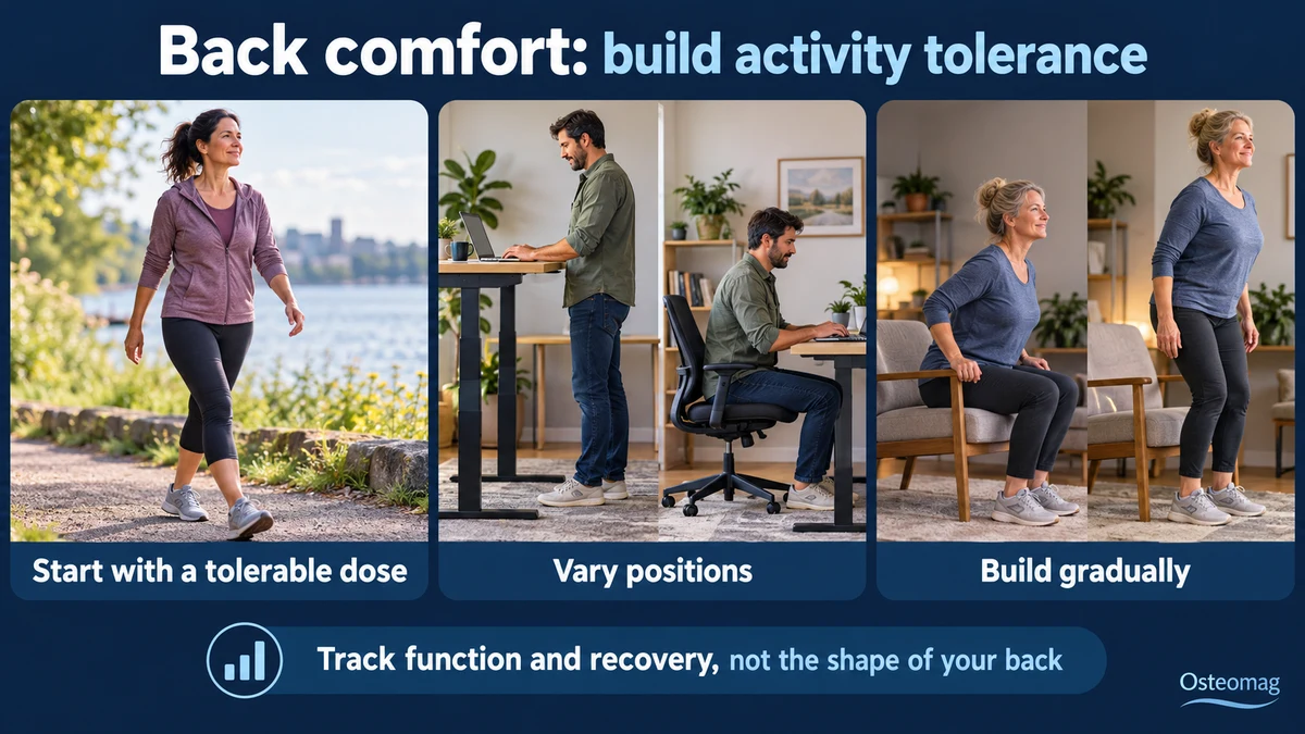
The programme may include walking, trunk and hip strengthening, mobility exercises and gradual practice of difficult movements. The Cochrane review supports exercise for chronic low back pain, with variable benefits. It does not demonstrate that lordosis must decrease to obtain a worthwhile result. [12]
Someone uncomfortable while standing may try walking breaks, alternating weight-bearing or changing working height. Someone uncomfortable during a sporting movement may temporarily reduce volume and then rebuild tolerance. Adaptations should remain practical and compatible with personal goals.
There is no need to keep the abdominals contracted continuously or hold the pelvis in a prescribed position. Such instructions may cause fatigue and excessive monitoring of movement. A control exercise can be useful at times; it should not turn the whole day into permanent correction.
Progression and monitoring
The starting point depends on current capacity. A few repetitions or a short walk may be enough initially. Increase one variable at a time, such as duration, load or difficulty. Lasting aggravation calls for adjustment, while new weakness calls for medical advice.
Follow-up focuses on activities: standing, bending, working and sleeping. Improvement without a visible change in the curve is still a valid result. WHO favours individualised care for chronic primary low back pain according to needs and context. [13]
Prepare for a sustainable return
Choosing two or three important activities helps set realistic goals. Record their duration, the breaks needed and the next-day response. If a flare occurs, temporarily returning to a tolerable dose helps activity resume without abandoning all movement. The programme should remain simple enough to continue in daily life. Gains in confidence, endurance and participation matter alongside reduced pain. If capacity declines despite these adjustments, clinical reassessment can review the diagnosis and the relevance of the next steps.
The realistic role of osteopathy
An osteopath may help explain variation in lumbar shape, identify limiting tasks and recognise signs that require referral. They can support adjustments and collaborate with other professionals. The osteopath does not make the medical diagnosis of an underlying spinal condition.
Evidence on low back pain does not show that osteopathic care returns the pelvis to an ideal position or that such a change is necessary for recovery. Any care chosen for comfort should be judged by observable effects, without replacing exercise, independence or medically indicated investigation.
Key points
A pronounced arch is not proof of disease. Assessment looks for symptoms, limitations and warning signs. Rehabilitation aims to improve the ability to live and move without imposing one shape of back on everyone.
Scientific references
- [1] Arshad R, et al. Effect of age and sex on lumbar lordosis and the range of motion: systematic review and meta-analysis. J Biomech. 2019. Read the source.
- [2] Roussouly P, et al. Classification of the normal variation in the sagittal alignment of the human lumbar spine and pelvis in the standing position. Spine. 2005;30:346–353. Read the source.
- [3] Hartvigsen J, et al. What low back pain is and why we need to pay attention. Lancet. 2018. Read the source.
- [4] Chun SW, et al. The relationships between low back pain and lumbar lordosis. Spine J. 2017. Clinical association analysis. Read the source.
- [5] Sadler SG, et al. Restriction in lateral bending range of motion, lumbar lordosis, and hamstring flexibility predicts the development of low back pain: systematic review of prospective cohort studies. 2017. Read the source.
- [6] Sugavanam T, et al. Postural asymmetry in low back pain: systematic review and meta-analysis of observational studies. Disabil Rehabil. 2025 (online 2024). Read the source.
- [7] George SZ, et al. Interventions for the management of acute and chronic low back pain: revision 2021. JOSPT. 2021. Read the source.
- [8] NICE. Low back pain and sciatica in over 16s: assessment and management. NG59. Recommendations. Read the source.
- [9] Chou R, et al. Imaging strategies for low-back pain: systematic review and meta-analysis. Lancet. 2009. Read the source.
- [10] Brinjikji W, et al. Systematic literature review of imaging features of spinal degeneration in asymptomatic populations. AJNR. 2015. Read the source.
- [11] Downie A, et al. Red flags to screen for malignancy and fracture in patients with low back pain: systematic review. BMJ. 2013. Read the source.
- [12] Hayden JA, et al. Exercise therapy for chronic low back pain. Cochrane. 2021. Read the source.
- [13] World Health Organization. WHO guideline for non-surgical management of chronic primary low back pain. 2023. Read the source.